Rejection of the Kidney Allograft N Engl J


 ligands of the toll-like")


")


 cells drive the cellular immune")
 cells § express the transcription factor forkhead box")


, with mononuclear cells crossing")


 due to")


- Slides: 46
Rejection of the Kidney Allograft N Engl J Med 2010; 363: 1451 -62. R 3 박세윤
CLINICAL FEATURES OF ALLOGRAFT REJECTION
Introduction Duration Hyperacute Pathophysiologic change Cellularintestitial Acute Vascular Late acute Antibodyendothelial Chronic Severity Extent of histologic inflammatio n and injury Presence or absence of renal dysfunction Acute rejection Subclini cal rejection
Introduction Response to treatment • Presence of glucocorticoid resistance • Absence of glucocorticoid resistance Immunologic mechanisms • Adaptive immune system response • Innate immune system response Cell-mediated immune response Humoral immune response
The innate immune system § Injured tissues express. . 1) ligands of the toll-like receptor system - DAMP (damage-associated molecular-pattern molecules) 2) Other innate danger molecules § Toll-like receptors - detect pathogens - sence the presence of foreign-tissue molecules - can produce factors the cause the maturation and activation of dendritic cells : important role in promoting acute rejection !
The innate immune system § Complement system - produces C 3 a and C 5 a - directly activate intragraft T cells and antigen-presenting cells § MICA (MHC class I peptide-related sequence A) antigens - endothelial surfaces - activate natural killer cells and CD 8 T cells - there is an association between poor graft outcomes and sensitization to the highly polymorphic MICA antigens
ANTIBODY-MEDIATED REJECTION HLA molecules Endothelial-cell antigens ABO blood-group antigens on endothelial cells and red cells
Antibodies against blood-group antigens § ABO-incompatible kidneys successfully transplanted : with use of an experimental protocol entails perioperative removal of antibodies from the recipient by means of plasmapheresis or immunoadsorption § After they have been removed, anti-blood-groop antibodies can rise to pretreatment levels after transplantation -> adhere to the microvasculature activate complement but they generally do not injure the endothelium ‘accommodation’ but mechanism is unknown
Hyperacute Rejection § Rejection of the renal graft that occurs almost immediately after release of the vascular cross clamps is classified as hyperacute. § The kidney appears flaccid and mottled : reflecting the deposition of antibodies against HLA antigens expressed on the endothelium of the glomeruli and microvasculature § Activation of the classic complement cascade within the graft, endothelial necrosis, platelet deposition, local coagulation
Acute Antibody-Mediated Rejection § Antibody-mediated rejection often begins within days after trasnplantation (or within weeks, if antilymphocyte antibody therapy was given) § The main feature is rapid graft dysfunction due to inflammation § Agonistic angiotensin II type 1 (AT 1)–receptor antibodies § associated with corticosteroid-resistant vascular rejection accompanied by malignant hypertension § pathogenic role remains unclear
antibodies against donor antigens bind to antigens expressed on endothelial cells in the graft vessel (Panel A).
Acute Antibody-Mediated Rejection § Promote platelet aggregation § Von Willebrand factor § P-selectin § Cause leukocytes to adhere to glomeruli (glomerulitis) or to dilated peritubular capillaries (margination) § Interleukin-1α § Interleukin-8 § Chemokine (C-C motif) ligand 2 (CCL 2) § Chemoattractants C 3 a and C 5 a
Acute Antibody-Mediated Rejection § C 4 d, a marker of classic complement activation, is frequently found in peritubular capillaries § C 5 b triggers the assembly of the membrane-attack complex (C 5 b–C 9) § causes localized endothelial necrosis and apoptosis § detachment of endothelial cells from the basement membrane § Microthrombi, with hemorrhage and arterial-wall necrosis and infartion, occur in severe cases
mononuclear cells adhere to the endothelium of the glomeruli (Panel B, arrows; periodic acid–Schiff stain) and the peritubular capillaries (shown at higher magnification in Panel C, arrows;
This process is accompanied by C 4 d deposition in the glomeruli and peritubular capillaries (Panel D, arrows; C 4 d immunohistochemical stain) and in the peritubular capillaries between ghost outlines of the renal tubules (Panel E, arrows; C 4 d immunofluorescent stain).
Acute Antibody-Mediated Rejection § Early diagnosis and treatment are essential for salvaging grafts § Treatments include removal of antibodies § § § plamapheresis immunoadsorption high-dose pulses of glucocorticoids intravenous immune globulin antiproliferative agents
Acute Antibody-Mediated Rejection § Supplementary therapies § rituximab § antilymphocyte antibody § New, investigational agents § Eculizumab (a monoclonal antibody that inhibits the cleavage of C 5) § Bortezomib (a proteasome inhibitor that can inhibit plasma cells)
T-CELL-MEDIATED REJECTION
Antigen Presentation Immature dendritic cells Transplant ed organ dornor antigen Recipient’s draining lymph nodes and spleen These antigens mature into APCs These T cells differenciate into various subgroups The APCs then home to lymphoid organs, where they activate the recipient’s T
The Major Histocompatibility Complex § HLA class I - HLA-A. -B, -C § HLA class II - DP, -DQ, -DR § HLA class III - Complement system, Cytokine, Heat shock proein
endogenous antigen -> proteosome -> class I MHC : CD 8+ lymphocytes exogenous antigen -> endosome -> class II MHC : CD 4+ lymphocytes
The Major Histocompatibility Complex MHC class II Internal protein (e. g. , viral proteins) -> cytotoxic CD 8 T cells Extracellular proteins (e. g. , bacterial proteins) -> CD 4 T cells HLA encode glycoproteins (MHC molecules) that enable the APCs to display fragments of antigens (peptides) to receptors on T cells.
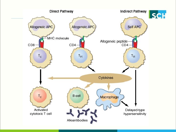
Recognition of Alloantigens by T Cells § Normally, only a small proportion of the T-cell population responds to a specific antigen (approximately 1 cell in 105 to 106 T cells). § In contrast, the responding proportion in transplantation is 1 to 10% § The recipient’s T lymphocytes can sense alloantigens § donor’s APCs (direct pathway) § recipient’s APCs (indirect pathway) § recipient’s APCs can also take up membrane
T-Cell Subgroups § CD 4 T cells produce inflammatory cytokines interferon-γ and interleukin-2 : drive a cellular response interleukin-4, interleukin-5, and interleukin-13, : produce a humoral response § CD 8 T cells mediate cytotoxicity, their effector functions overlap
T-Cell Subgroups § type 1 helper T (Th 1) cells drive the cellular immune response § type 2 helper T (Th 2) cells produce the humoral immune response § can reject grafts, using pathways that involve eosinophils
T-Cell Subgroups § Regulatory T (Treg) cells § express the transcription factor forkhead box P 3 (FOXP 3) § The number of Treg cells correlates with markers of T-cell rejection, including interstitial inflammation, tubulitis, and cytotoxic gene expression, but not with the graft outcome, suggesting that FOXP 3 -positive cells aid in stabilizing inflammation within the graft
Treg : Regulatory T cells – limit the rejection response t. Th 17 : type 17 helper T cells – mediate glucocorticoid-
Costimulation § T-cell activation requires signals other than those engendered by the MHC–peptide complex, termed costimulatory signals § T cells become anergic when presented with an antigen in the absence of these signals, and agents that block these signals are under development § CD 80 (B 7 -1) and CD 86 (B 7 -2) ; these two B 7 molecules are ligands for two T-cell– membrane receptors, CD 28 (stimulate) and CTLA-4 (inhibitory)
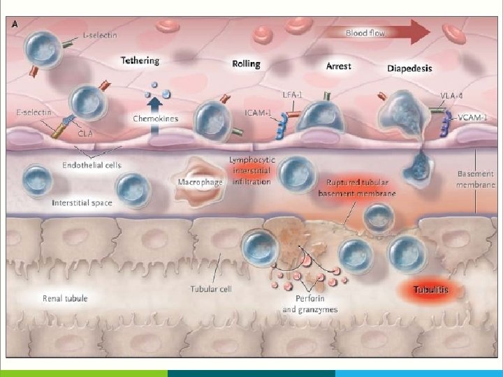
T-Cell Movement in The Allograft § T cells use adhesion molecules, including leukocyte-function–associated antigen 1 (LFA 1) to roll along and tether to endothelium, migrate across peritubular capillaries, and enter the graft § Fingolimod, a small molecule that blocks the egress of T cells from lymph nodes, and anti–LFA-1 agents block such T-cell movement, but until now they have had a limited clinical effect in transplantation
dense interstitial lymphocytic infiltration (Panel B, arrow; periodic acid–Schiff stain), with mononuclear cells crossing the tubular basement membrane (pink) into the renal tubules, resulting in tubulitis (Panel C, arrow; periodic acid–Schiff stain).
In acute vascular rejection, mononuclear cells adhere to the endothelium of small muscular arteries
Effector T Cells § CD 8 T cells release perforin, which perforates target-cell membranes, and granzymes A and B, which enter cells and induce caspase-mediated apoptosis. § The Fas ligand on cytotoxic T cells activates Fas, a receptor on cells of the graft, and this interaction also induces caspase-mediated apoptosis
Effector T Cells § CD 4 T cells can attack grafted cells expressing minor MHC antigens and can also secrete TNF-α and tumor necrosis factor β (TNF-β), which bind to TNF receptors on endothelial or tubular cells, causing them to undergo apoptosis § In animals, blockade of TNF by antibody or knockout of TNF-receptor genes prolongs allograft survival.
OTHER PATTERNS OF REJECTION Vascular rejection Late actue rejection Chronic rejection
Vascular Rejection § CD 4 and CD 8 T cells and macrophages invade the subendothelium and intima of muscular arteries by means of intercellular adhesion molecule 1 (ICAM-1) or vascular-cell adhesion molecules (VCAM) on activated endothelium and by means of chemokine (e. g. , CCL 4, CCL 5, and CXCL 8) gradients. § Experimental evidence suggests that anti-MHC antibodies, T-cell– mediated immunity to minor MHC antigens, Natural killer cells, and interferon-γ all play a role in the invasion of vessels.
Vascular Rejection § Vascular rejection is a severe condition that does not respond to glucocorticoid therapy and instead requires potent antilymphocyte- antibody therapy (muromonab-CD 3 [Orthoclone OKT 3, Ortho Biotech] or antithymocyte globulin).
Lacte Acute Rejection § Severe and difficult to reverse, with a high risk of subsequent graft loss § Its main features are active immune inflammation and chronic tubulointerstitial damage, which frequently involves graft-directed antibody.
Chronic Rejection § Chronic allograft rejection — ongoing immune injury to the graft — is due to a failure to maintain sufficient immunosuppression to control residual antigraft lymphocytes or antibodies § Its features include a progressive decline in renal function, Invasion of the renal parenchyma by T cells, and persistent infiltration of the interstitium by T cells and macrophages § Occasionally, one also sees smooth-muscle proliferation and hyperplasia in vessels, forming a neointima; focal destruction
Chronic Rejection § In chronic antibody-mediated rejection, undetected preexisting donor-specific antibodies or antibodies generated after transplantation deposit on the capillary endothelium § Endothelial injury to glomerular and peritubular capillaries causes cellular hypertrophy, subendothelial deposition of fibrillary material, expansion and duplication of the glomerular basement membrane, or mesangial- cell interposition
In chronic vascular rejection, neointimal thickening (Panel E, arrow; Masson trichrome stain) due to
antibody-mediated rejection results in transplant glomerulopathy, with thickened glomerular capillaries (Panel A, arrows; periodic acid–Schiff stain) and double contours (Panel B, arrows; Masson green and silver stain)
C 4 d in peritubular capillaries containing mononuclear cells (Panel C, arrows; C 4 d immunohistochemical stain) and flocculent subendothelial material below an activated endothelial cell of the glomerular capillary (Panel D, arrows; electron microscopy)