Acute rejection Acute rejection rates have steadily declined


![Am J Transplant 17[Suppl 1]: 21– 116, 2017](https://slidetodoc.com/presentation_image_h2/3fef189e278c11647e1a3384643163b7/image-3.jpg "Am J Transplant 17[Suppl 1]: 21– 116, 2017")
 • Banff 2017 guidelines: • • • Ia :")
: • All three criteria below required (Banff 2017 guidelines) A-Histologic")


, with or without rituximab,")


, with or without rituximab, was")


- Slides: 21
Acute rejection • Acute rejection rates have steadily declined from nearly 100% in the first era of organ transplantation to approximately 10% more recently • T-cell mediated = Steroid responsive= pre transplant donor-specific antibodies (DSA) = lymphocytic infiltrate of tubules (tubulitis) and larger vessels (vasculitis). • Ab mediated = Steroid non responsive = HLA A/B/DR mismatch (1) active tissue injury, (2) immuno-histologic evidence of peritubular capillary complement split-product C 4 d deposition (3) circulating DSA
Risk Factors of Rejection • Positive panel reactive antibody test ? • Repeat transplant? • Black race • Recipient age • Immunosuppression regimen and exposure
Am J Transplant 17[Suppl 1]: 21– 116, 2017
Acute T cell–mediated rejection (TCR) • Banff 2017 guidelines: • • • Ia : >25% Interstitial inflammation with moderate tubulitis (t 2) Ib : >25% Interstitial inflammation with severe tubulitis (t 3) IIa Mild-to-moderate intimal arteritis (v 1) IIb Severe intimal arteritis (v 2) III Transmural arteritis and/or fibrinoid necrosis
Acute antibody-mediated rejection (AMR): • All three criteria below required (Banff 2017 guidelines) A-Histologic evidence of tissue injury including one or more of the following: 1 - Microvascular inflammation 2 - Arteritis 3 - Thrombotic microangiopathy 4 - Acute tubular injury B- Evidence of current/recent Ab interaction with endothelium including 1 or more of the following: 1 - Positive C 4 d staining of peritubular capillaries 2 - Moderate microvascular inflammation 3 - Increased expression of gene transcripts in biopsy tissue strongly associated with AMR C-Serologic evidence of donor-specific antibodies (DSA) 1 - Positive C 4 d staining or presence of AMR-associated gene transcripts may substitute for DSA
• The revised 2013 Banff criteria for antibody mediated rejection diagnosis removed the requirement for C 4 d detection • Recent Banff consensus notes studies removed the requirement for documented circulating DSA in the setting of positive C 4 d staining and microvascular inflammation. Am J Transplant 18: 293– 307, 2018
Peritubular lymphocytic infiltration
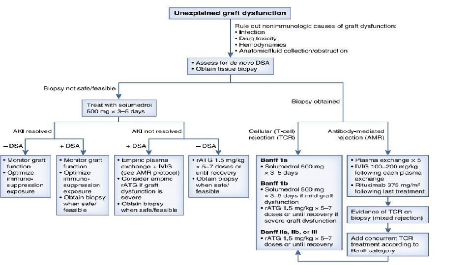
Acute Rejection Diagnosis • The gold standard for diagnosing acute rejection in kidney transplant recipients is tissue biopsy. • It should be pursued without delay in patients with graft dysfunction that is not explained by other nonimmunologic causes
Diagnosis • Numerous studies of urine and blood biomarkers, such as CXCL 9, CXCL 10, granzyme B, perforin, and Fas ligand, have generally shown mixed sensitivity and specificity for identifying acute rejection, differentiating T cell–mediated rejection from antibody-mediated rejection, and distinguishing immunologic injury from other forms of graft damage • Decreased urinary mi. R-210 levels have been associated with T cell– mediated rejection and subsequent 1 -year GFR decline • Donor-derived cellfree DNA (cf-DNA) profiling has been applied to the noninvasive diagnosis of antibody-mediated rejection
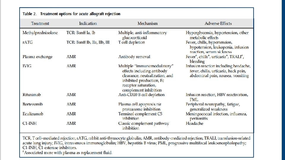
Acute Rejection Treatment • Treatment strategies differ between T cell–mediated rejection and antibody-mediated rejection • Any untreated clinical acute rejection episode will ultimately result in accelerated graft loss.
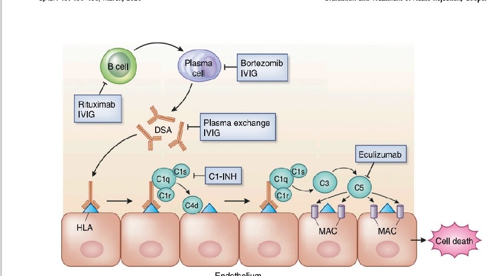
Antibodymediated rejection treatments • Removing antibody-producing B cells or plasma cells • Removing antibodies (DSA) • Inhibiting the subsequent complementregulated graft damage
Antibodymediated rejection treatments • Plasma exchange and intravenous Ig (IVIG), with or without rituximab, was the most commonly used strategy and is generally considered standard of care for antibodymediated rejection treatment • Daily or every other day plasma exchange consisting of 1. 5 plasma volume removal with each treatment followed by IVIG at 100– 200 mg/kg, with or without a single dose of rituximab at 3. 75 mg/m 2. Transplantation 102: 557– 568, 2018 • Treatment regimen consisting only of IVIG is inadequate for most cases
• T cell–mediated rejection involving lymphocytic infiltrate of the vasculature (Banff II and III lesions) generally requires T cell–depleting therapy, most commonly r. ATG. • Antibody therapy was superior to steroid therapy in reversing T cell– mediated rejection.
Antibody-Mediated Rejection • Several new therapeutic treatment options have been studied in recent years. • Treatments are directed at: 1. removing antibody-producing B cells or plasma cells 2. removing antibodies (DSA) 3. inhibiting the subsequent complement-regulated graft damage
Antibody-Mediated Rejection • Plasma exchange and intravenous Ig (IVIG), with or without rituximab, was the most commonly used strategy and is generally considered standard of care for antibody-mediated rejection treatment (CJASN 15: 430– 438, March, 2020 ) • Daily or every other day plasma exchange consisting of 1. 5 plasma volume removal with each treatment followed by IVIG at 100– 200 mg/kg, with or without a single dose of rituximab at 3. 75 mg/m 2.
Refractory Antibody-Mediated Rejection • 1 -Bortezomib, a proteasome inhibitor for treatment of multiple myeloma, has received special focus because of its ability to induce apoptosis in antibody-producing plasma cells. • There is minimal effect on DSA burden when used as a sole agent. • 2 - The humanized m. Ab eculizumab has been targeted in the prevention and treatment of antibody-mediated injury because of its mechanism of complement component C 5 inhibition. • Patients treated with combination splenectomy and eculizumab experienced no graft loss and minimal transplant glomerulopathy on protocol biopsy.
• 3 -A pilot study of the anti-IL 6 receptor antibody tocilizumab has shown promising results for patients with chronic antibody-mediated rejection (Am J Transplant 17: 2381– 2389, 2017) • 4 - C 1 -esterase inhibitors (C 1 -INH) have recently used for the treatment of acute antibody-mediated rejection. • C 1 -INH inhibits proximal enzymes in the classic complement pathway including C 1 q.
• There is significant reduction in acute rejection in patients treated with belatacept by adding tacrolimus. Adams et al. Am J Transplant 17: 2922– 2936, 2017