
ROLE OF ALLOGRAFT BIOPSY IN THE MANAGEMENT OF
ROLE OF ALLOGRAFT BIOPSY IN THE MANAGEMENT OF TRANSPLANTED PATIENT Parmjeet Randhawa Professor, Division of Transplantation Department of Pathology University of Pittsburgh
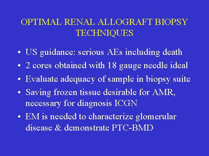
OPTIMAL RENAL ALLOGRAFT BIOPSY TECHNIQUES • • US guidance: serious AEs including death 2 cores obtained with 18 gauge needle ideal Evaluate adequacy of sample in biopsy suite Saving frozen tissue desirable for AMR, necessary for diagnosis ICGN • EM is needed to characterize glomerular disease & demonstrate PTC-BMD
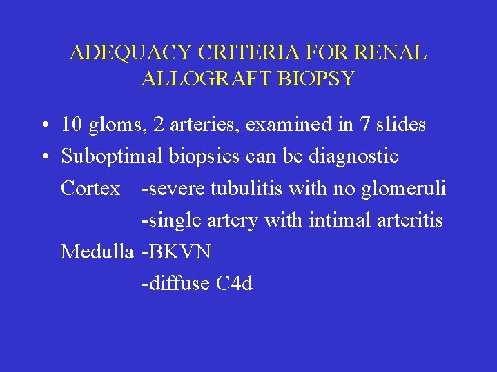
ADEQUACY CRITERIA FOR RENAL ALLOGRAFT BIOPSY • 10 gloms, 2 arteries, examined in 7 slides • Suboptimal biopsies can be diagnostic Cortex -severe tubulitis with no glomeruli -single artery with intimal arteritis Medulla -BKVN -diffuse C 4 d
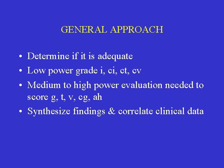
GENERAL APPROACH • Determine if it is adequate • Low power grade i, ct, cv • Medium to high power evaluation needed to score g, t, v, cg, ah • Synthesize findings & correlate clinical data
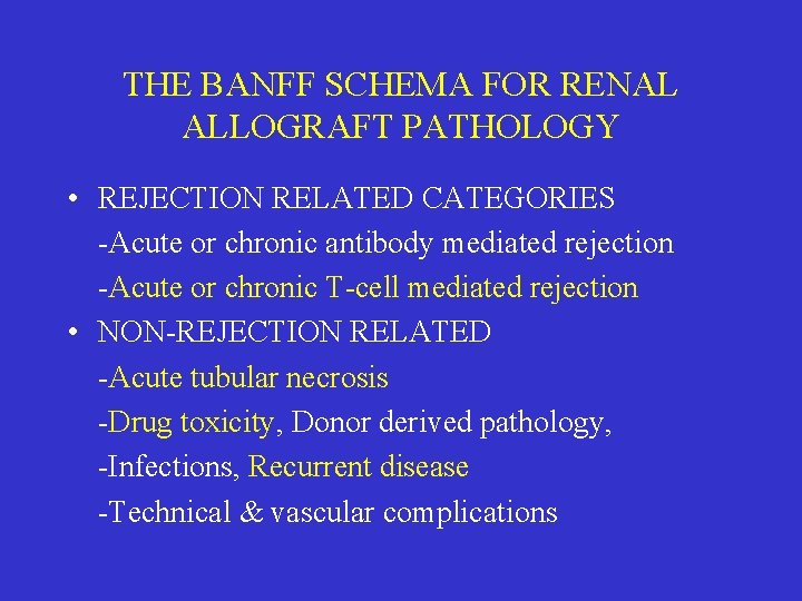
THE BANFF SCHEMA FOR RENAL ALLOGRAFT PATHOLOGY • REJECTION RELATED CATEGORIES -Acute or chronic antibody mediated rejection -Acute or chronic T-cell mediated rejection • NON-REJECTION RELATED -Acute tubular necrosis -Drug toxicity, Donor derived pathology, -Infections, Recurrent disease -Technical & vascular complications
ACUTE REJECTION • Acute T-cell mediated rejection • Acute cellular rejection
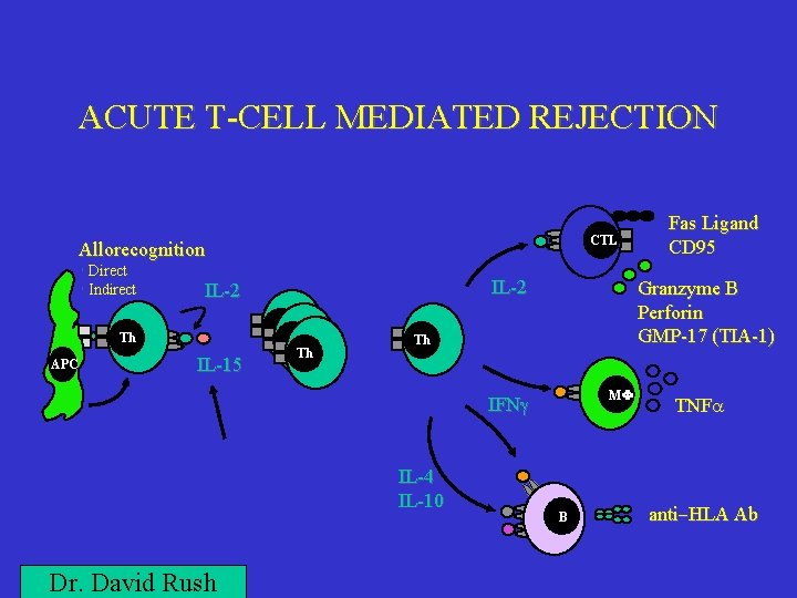
ACUTE T-CELL MEDIATED REJECTION CTL Allorecognition • Direct • Indirect Th APC IL-2 IL-15 Th Th Th Granzyme B Perforin GMP-17 (TIA-1) Th M IFNg IL-4 IL-10 Dr. David Rush Fas Ligand CD 95 B TNFa anti-HLA Ab
ACUTE T-CELL MEDIATED REJECTION • An alloimmune reaction mediated by cell mediated immunity • Subclinical with normal creatinine • Laboratory evidence of graft dysfunction • Severe cases may show fever, graft tenderness, leukocytosis and eosinophilia
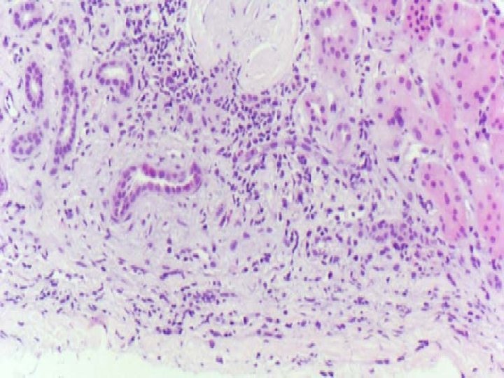
HISTOLOGIC CRITERIA FOR DIAGNOSIS OF ACUTE T-CELL MEDIATED REJECTION • Predominantly mononuclear infiltrate • Presence of parenchymal damage • Occurrence of subendothelial inflammation in venules, arteries
GRADING OF ACUTE TCMR-1 • • Severity of inflammation Intensity of tubulitis Disruption of tubular architecture Presence of arteritis
GRADING OF INTERSTITIAL INFLAMMATION • Limit assessment to cortex unless medulla alone sampled • Ignore areas in continuity with capsule • <10% area is assigned grade 0 • Grades 1, 2, 3 are 10 -25, 26 -50, >50% • Area is evaluated not the intensity
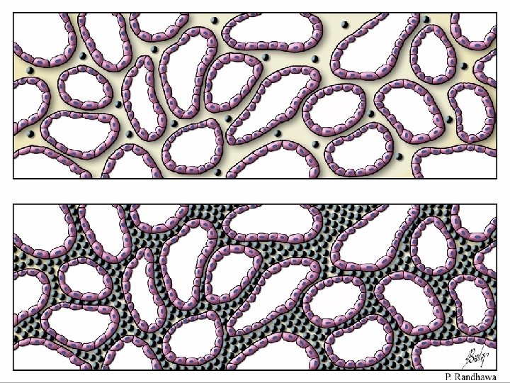
INFLAMMATION IN AREAS OF SCARRING
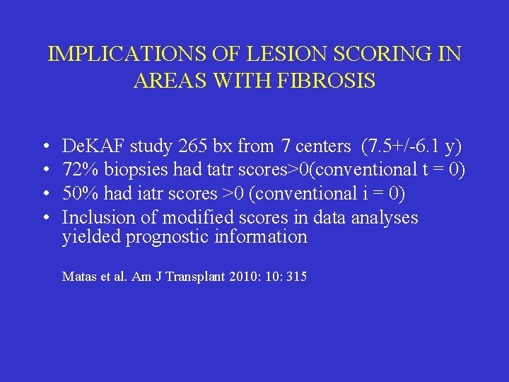
IMPLICATIONS OF LESION SCORING IN AREAS WITH FIBROSIS • • De. KAF study 265 bx from 7 centers (7. 5+/-6. 1 y) 72% biopsies had tatr scores>0(conventional t = 0) 50% had iatr scores >0 (conventional i = 0) Inclusion of modified scores in data analyses yielded prognostic information Matas et al. Am J Transplant 2010: 315
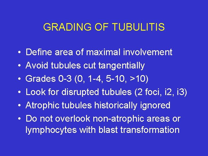
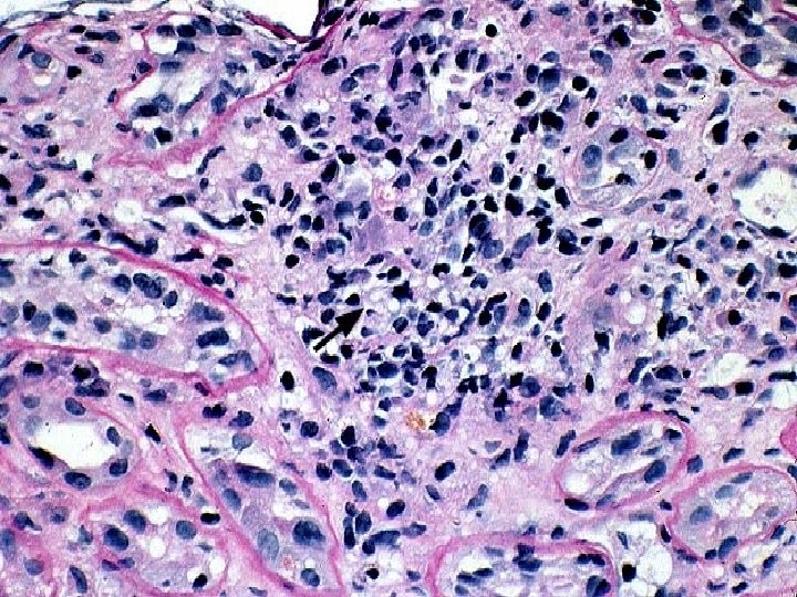
GRADING OF TUBULITIS • • • Define area of maximal involvement Avoid tubules cut tangentially Grades 0 -3 (0, 1 -4, 5 -10, >10) Look for disrupted tubules (2 foci, i 2, i 3) Atrophic tubules historically ignored Do not overlook non-atrophic areas or lymphocytes with blast transformation
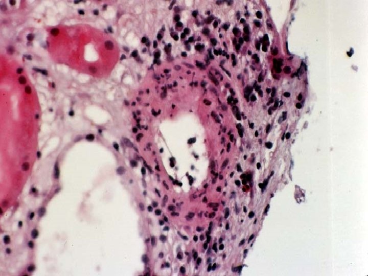
GRADING OF INTIMAL ARTERITIS • Look closely if interstitial hemorrhage, glomerulitis, or PTC inflammation • Caveat added to report if < 4 arteries • Grade v 1 (<25%) (mild to moderate) • Grade v 2 (>25%) (severe) • Grade v 3 fibrinoid change, necrosis, transmural i (transmural intimal arteritis)
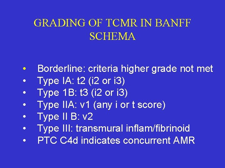
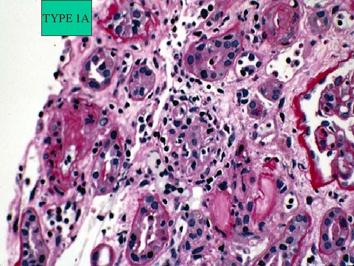
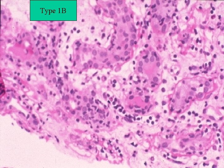
GRADING OF TCMR IN BANFF SCHEMA • • Borderline: criteria higher grade not met Type IA: t 2 (i 2 or i 3) Type 1 B: t 3 (i 2 or i 3) Type IIA: v 1 (any i or t score) Type II B: v 2 Type III: transmural inflam/fibrinoid PTC C 4 d indicates concurrent AMR
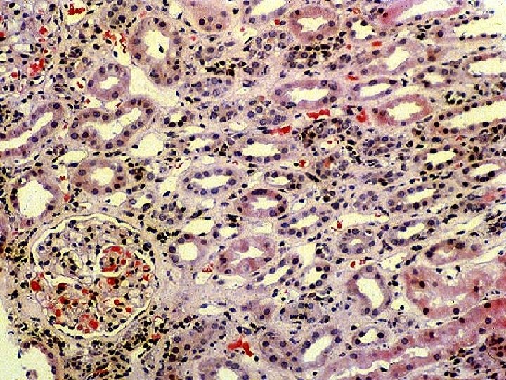
BORDERLINE CHANGES SUSPICIOUS FOR ACUTE REJECTION • • • Looks like rejection but criteria I, II not met i 1 with any t grade OR t 1 with any i Most often t 1 tubulitis and i 1 inflammation For biopsies with i 2, 3 look for t 2, t 3, v 1 Theoretically i 1, t 2, t 3 & i 2, i 3, t 1 allowed Some cases evolving or treated higher AR
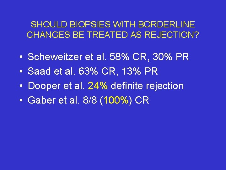
SHOULD BIOPSIES WITH BORDERLINE CHANGES BE TREATED AS REJECTION? • • Scheweitzer et al. 58% CR, 30% PR Saad et al. 63% CR, 13% PR Dooper et al. 24% definite rejection Gaber et al. 8/8 (100%) CR
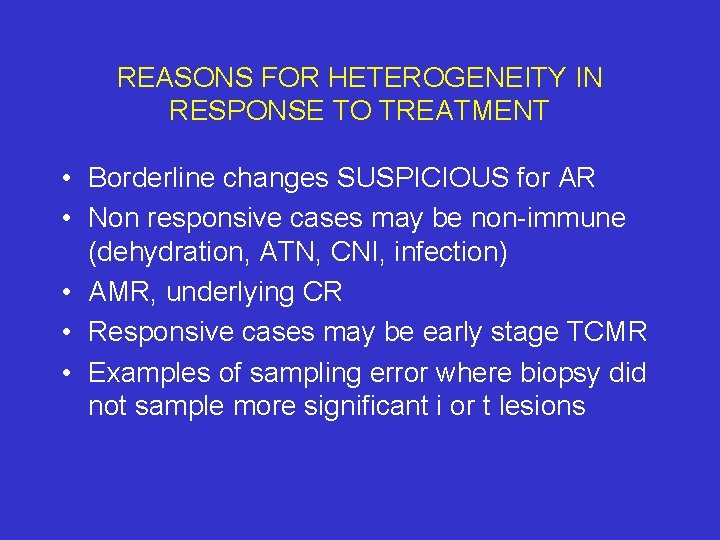
REASONS FOR HETEROGENEITY IN RESPONSE TO TREATMENT • Borderline changes SUSPICIOUS for AR • Non responsive cases may be non-immune (dehydration, ATN, CNI, infection) • AMR, underlying CR • Responsive cases may be early stage TCMR • Examples of sampling error where biopsy did not sample more significant i or t lesions
TYPE 1 A
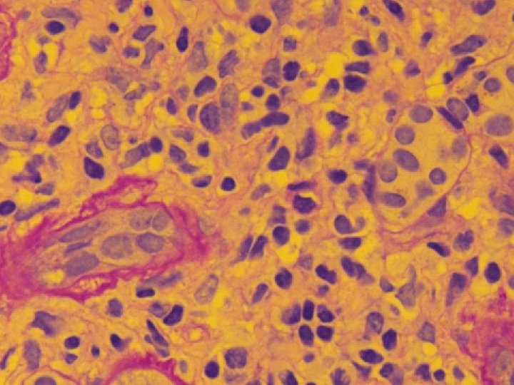
Type 1 B
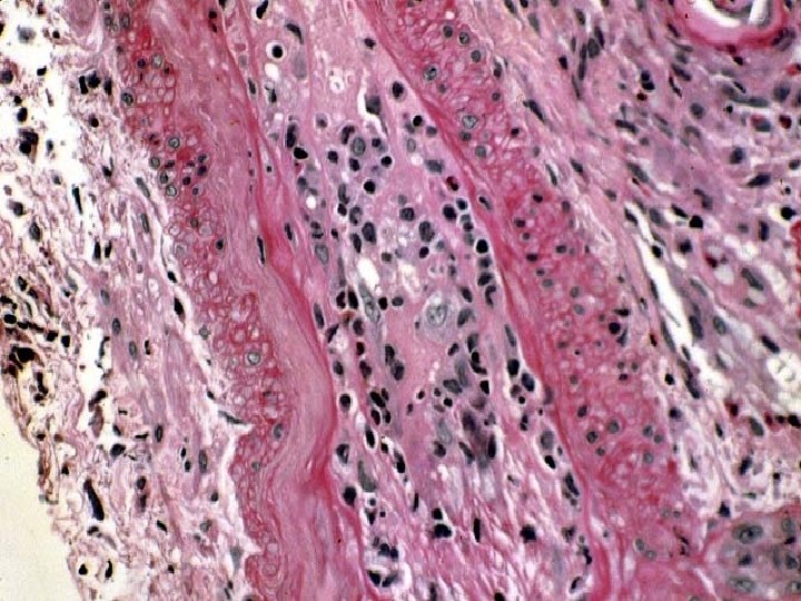
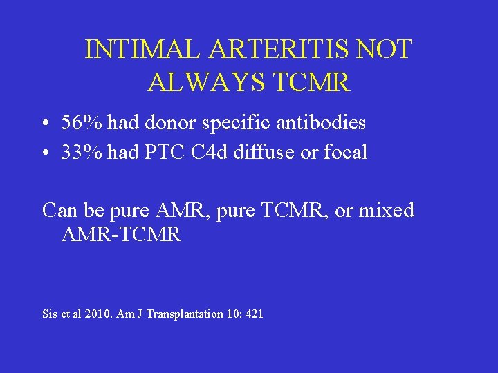
INTIMAL ARTERITIS NOT ALWAYS TCMR • 56% had donor specific antibodies • 33% had PTC C 4 d diffuse or focal Can be pure AMR, pure TCMR, or mixed AMR-TCMR Sis et al 2010. Am J Transplantation 10: 421
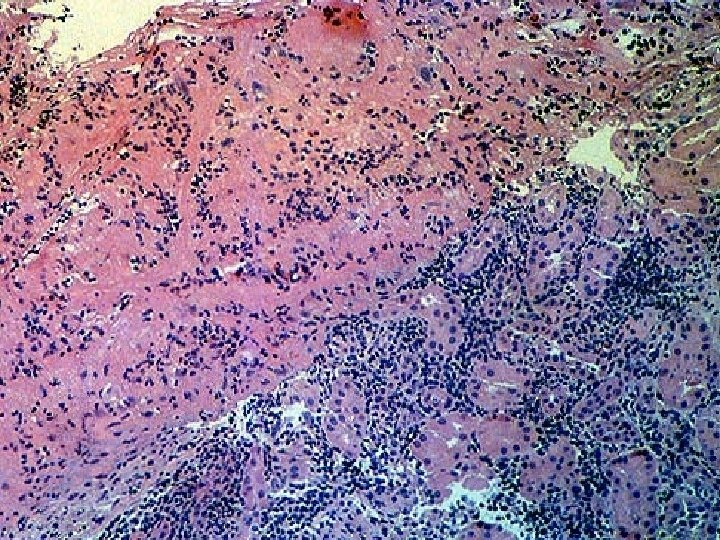
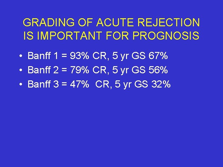
GRADING OF ACUTE REJECTION IS IMPORTANT FOR PROGNOSIS • Banff 1 = 93% CR, 5 yr GS 67% • Banff 2 = 79% CR, 5 yr GS 56% • Banff 3 = 47% CR, 5 yr GS 32%
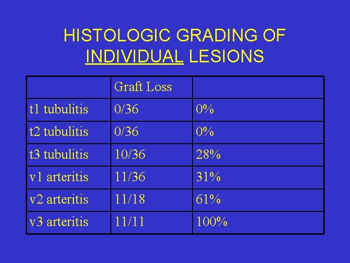
HISTOLOGIC GRADING OF INDIVIDUAL LESIONS Graft Loss t 1 tubulitis 0/36 0% t 2 tubulitis 0/36 0% t 3 tubulitis 10/36 28% v 1 arteritis 11/36 31% v 2 arteritis 11/18 61% v 3 arteritis 11/11 100%
IMMUNOHISTOCHEMICAL STUDIES IN ACUTE REJECTION • • • Not necessary for Dx (except C 4 d) May occasionally help d/d PTLD vs AR Intraglomerular CD 68 worse prognosis CD 20 not correlation AMR or response CD 4/CD 8/CD 68: stage of evolution, patient selection, immunosuppression.
MOLECULAR DIAGNOSIS TCMR • • MDx potentially valuable non-invasive tool Dx TCMR: sensitivity & specificities of 70 -90% Disagreements in an average of 20% Reflect sampling issues, arbitrary grading thresholds & low frequency diagnoses • Provides information that is complementary


. • ACUTE TUBULAR NECROSIS • ACUTE TUBULAR INJURY • ACUTE KIDNEY INJURY
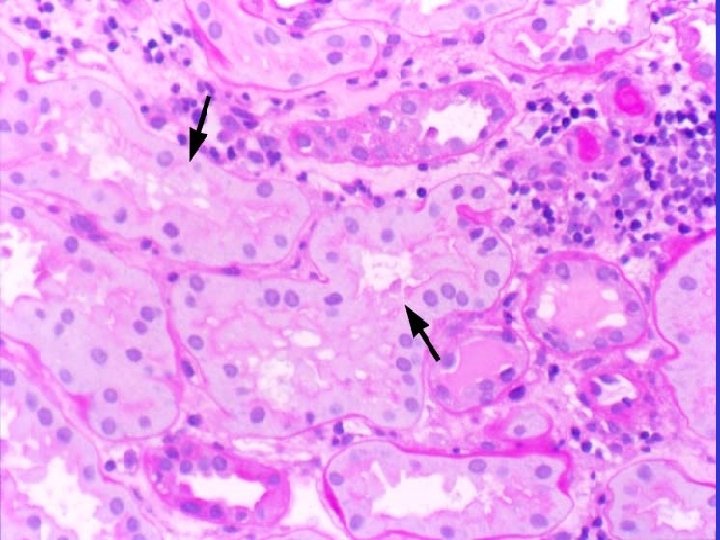
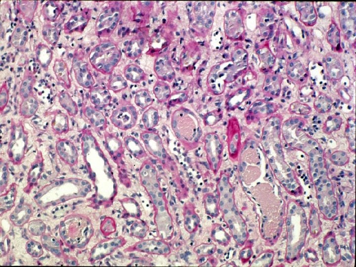
CALCINEURIN INHIBITOR TOXICITY 1. Cyclosporine 2. Tacrolimus
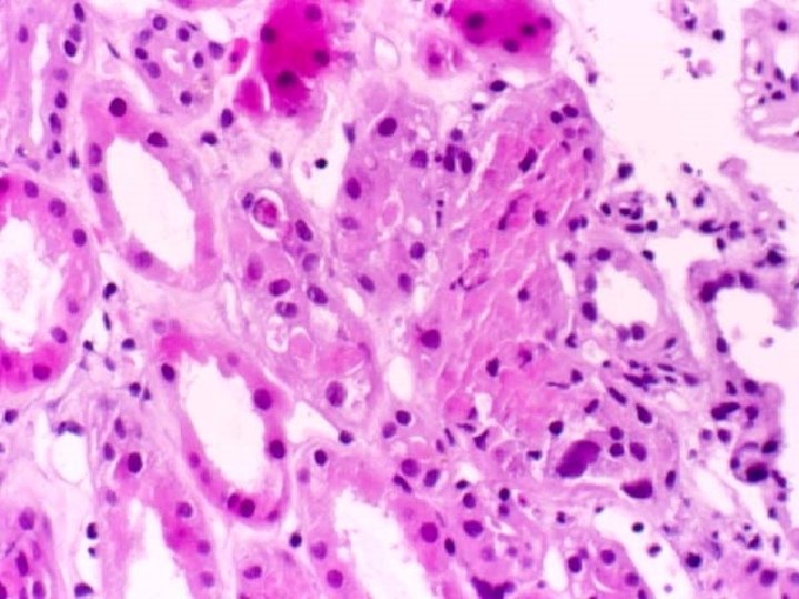
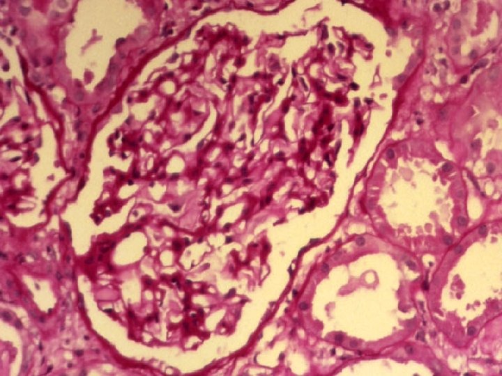
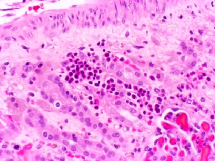
CALCINEURIN INHIBITOR CAN CAUSE FUNCTIONAL TOXICITY • • • Elevation in serum creatinine Blood levels Tac/Csa may be elevated Biopsy shows no specific pathology Reduction of dosage restores serum creat Graft dysfunction attributed to vasospasm
CALCINEURIN INHIBITORS CAN CONTRIBUTE TO ATI • CNI induced AKI confirmed in animals • Clinically, prolonged cases of DGF can recover once calcineurin inhibitors switched • Likely mechanism is reversal of drug induced vasoconstriction
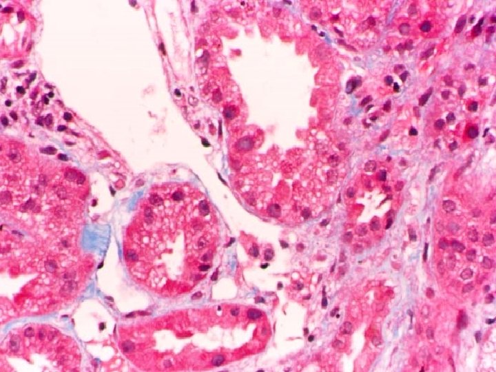
TUBULAR VACUOLIZATION IS NOT SPECIFIC FOR CNI • Use of plasma expanders (dextran, mannitol), radiocontrast media, IVIG preps containing sucrose as a stabilizer, hyperosmolar sucrose infusions • Frequently seen in context of ACR • Occurs in donor biopsies • Patients maintained on Azathioprine • Attributed to CNI by exclusion
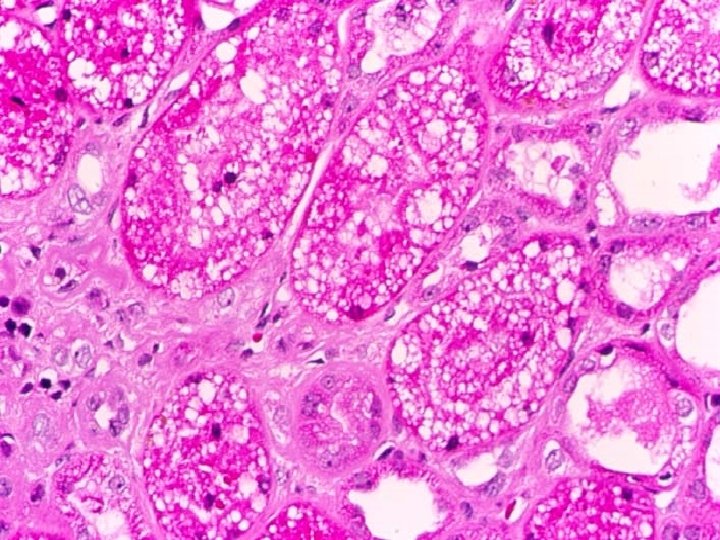
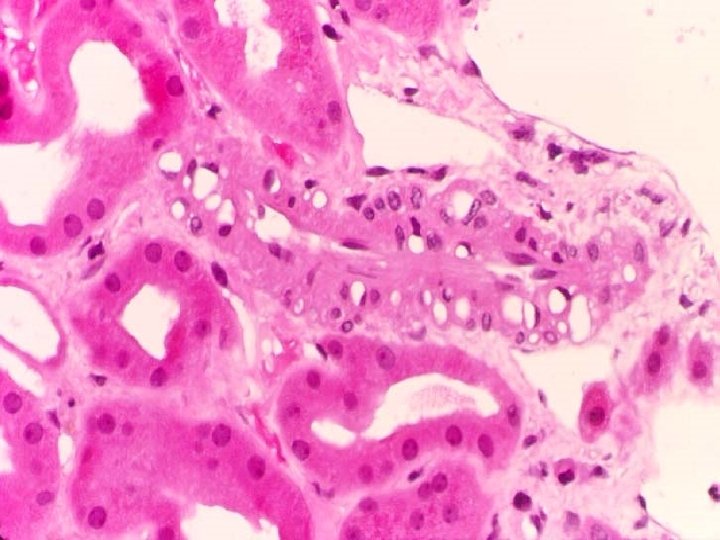
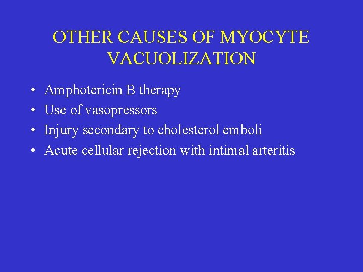
OTHER CAUSES OF MYOCYTE VACUOLIZATION • • Amphotericin B therapy Use of vasopressors Injury secondary to cholesterol emboli Acute cellular rejection with intimal arteritis
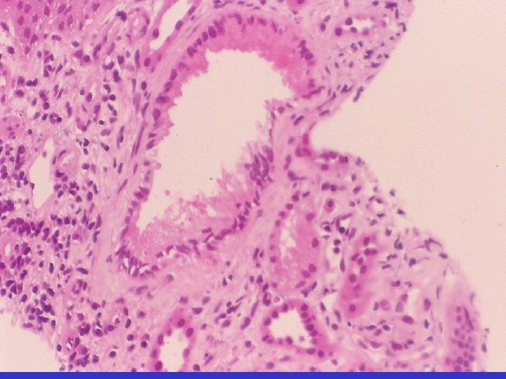
GIANT MITOCHONDRIA • Association with CNI confirmed in rats • Lesion of limited diagnostic utility, since it is uncommon, not readily recognized, & may needs EM to exclude lysosomes • Not specific: described in azathioprine Rx, glomerulonephritis, acute tubular necrosis

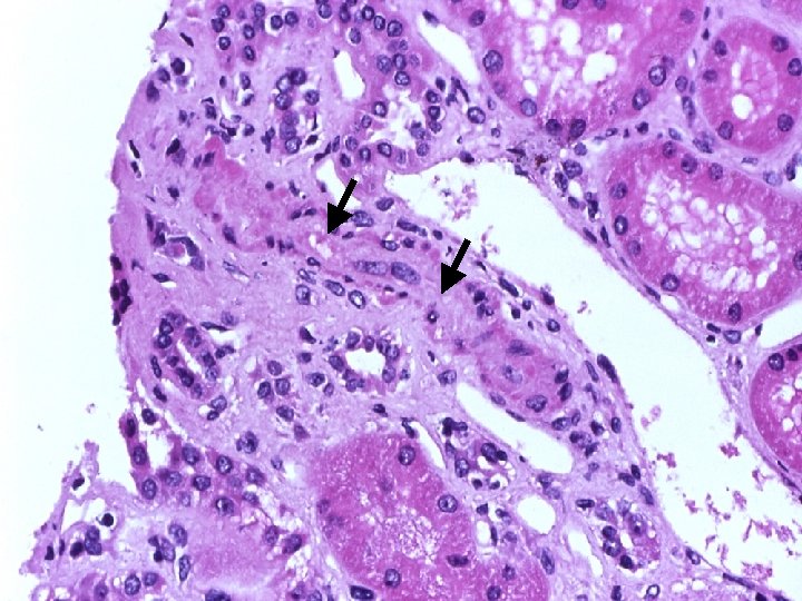
. THROMBOTIC MICROANGIOPATHY
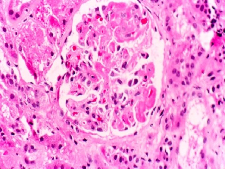
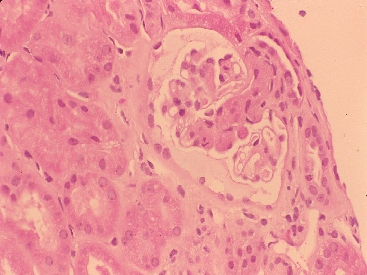
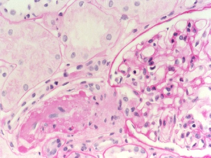
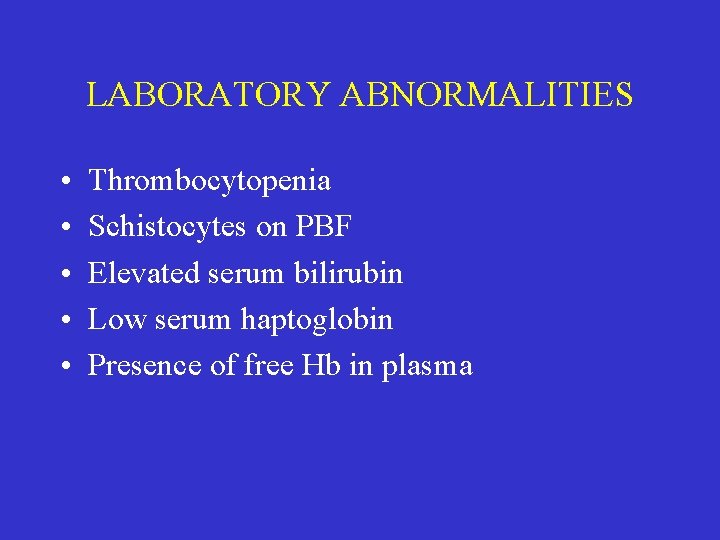
LABORATORY ABNORMALITIES • • • Thrombocytopenia Schistocytes on PBF Elevated serum bilirubin Low serum haptoglobin Presence of free Hb in plasma
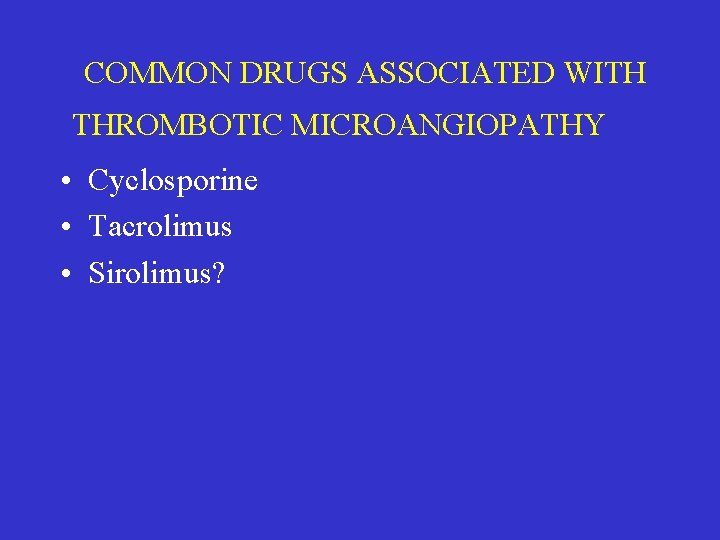
COMMON DRUGS ASSOCIATED WITH THROMBOTIC MICROANGIOPATHY • Cyclosporine • Tacrolimus • Sirolimus?
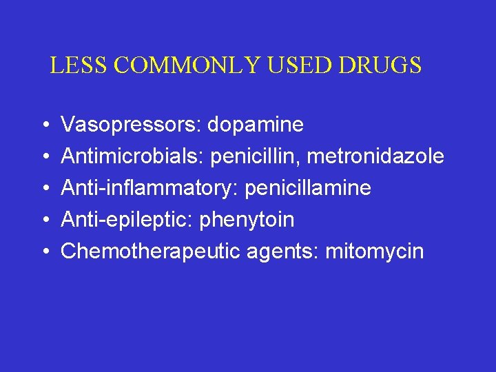
LESS COMMONLY USED DRUGS • • • Vasopressors: dopamine Antimicrobials: penicillin, metronidazole Anti-inflammatory: penicillamine Anti-epileptic: phenytoin Chemotherapeutic agents: mitomycin
THROMBOTIC MICROANGIOPATHY IS MAY BE OF IMMUNE ORIGIN • • Antibody mediated rejection T-cell mediated ac rej with intimal arteritis. Ac rejection treated with OKT 3 Autoimmune disorders with circulating autoab (ACA, APA, ANCA)
INFECTIONS ARE IN IMPORTANT CAUSE OF TMP • • • Gastroenteritis (E. coli, Shigella) CMV infection (damage to endothelium) HCV (anti-cardiolipins) Parvovirus infection (targets endothelium) HIV infection (TTP syndrome)
RECURRENT GENETIC HUS • Recurrent rate depends on mutation • 50% for CFH, CFI (inhibitors complement) -liver-kid transplant may be curative • Not expected MCP/CD 46 (present in donor kidney, will catalyse breakdown of C 3 b, C 4 b normally) -extrarenal activation of complement -chimerism in glomerular capillaries -
OTHER BIOCHEMICAL DEFECTS ASSOCIATED TMP • • • Deficiency of anti-thrombin III Protein C Protein S Factor V mutation (resistance to Protein C) Homocysteinemia (B 6, B 12 deficiency)
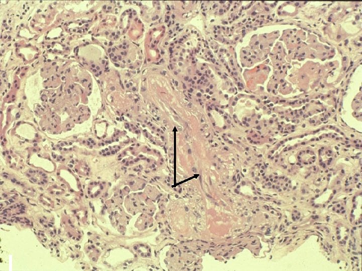
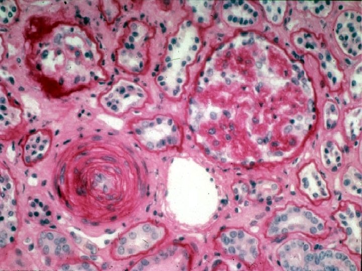
CNI ASSOCIATED ACUTE ARTERIOLOPATHY • Intimal edema and fibrin • Necrosis of myocytes leaves behind empty basement membranes bags • Filled up by knot like protein deposits • Knots fuse to form ring of pearls
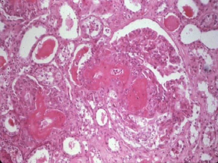
ARTERIOLONECROSIS • Fibrinoid change in arteriolar wall • Collapse of distal glomeruli • Healing by onion skin change
CHRONIC GRAFT DYSFUNCTION • • Evolves slowly Usually begins > 3 m post-tx Typically progressive Rate of decline, may be punctuated by periods of stability
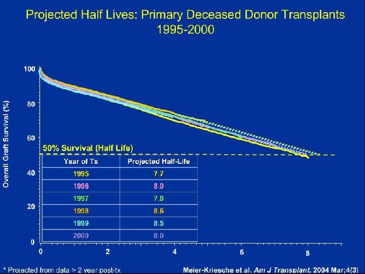
MAGNITUDE OF PROBLEM
CAUSES OF CHRONIC GRAFT DYSFUNCTION • REJECTION RELATED CATEGORIES -Chronic active T-cell mediated rejection -Cronic active antibody mediated rejection • NON-REJECTION RELATED CATEGORIES -Calcineurin inhibitor toxicity -Glomerulonephritis -Recurrence of original disease -Donor disease (age, hypertension) -RA stenosis, reflux nephropathy, recurrent UTI/BKN -Technical & vascular complications
CHRONIC REJECTION (BANFF 1991) No distinction made between chronic active TCMR or chronic AMR • Chronic Transplant Glomerulopathy • New onset arteriosclerosis (not preexisting donor disease)
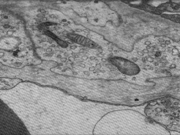
CHRONIC TRANSPLANT GLOMERULOPATHY • Reduplication of GBM • Subendothelial translucent material • Mesangial interposition (tram track MST) Suggested increase in capillary wall thickness of >2 fold, at least 3 capillary loops Ivanyi 2001; Mod Pathol 14: 1200
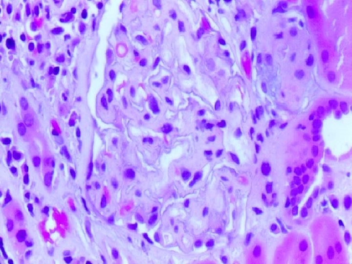
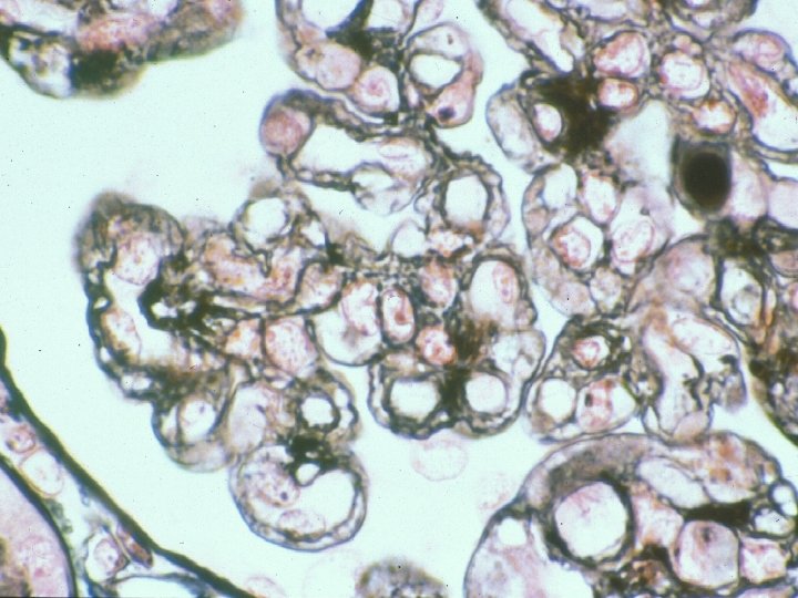
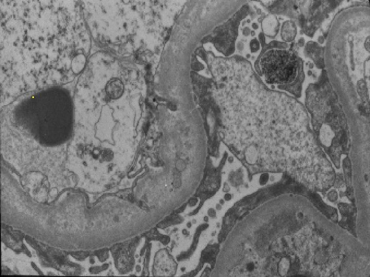
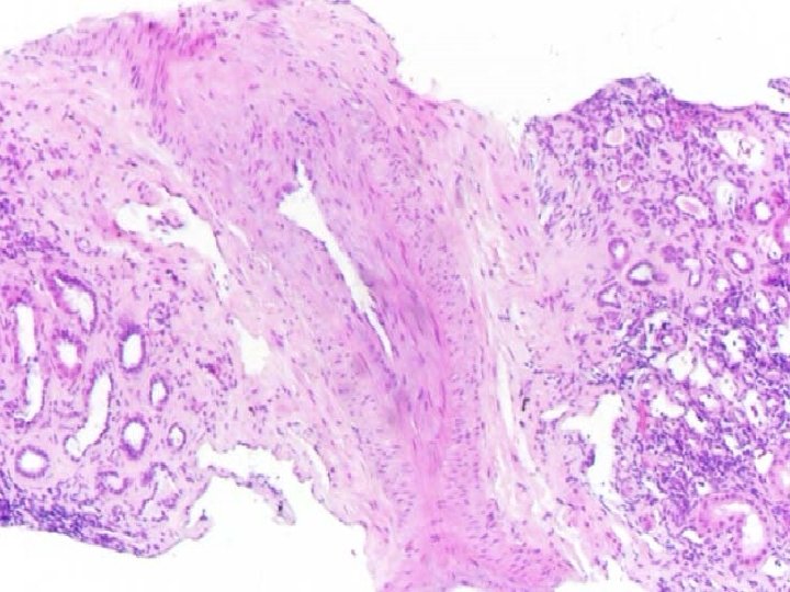
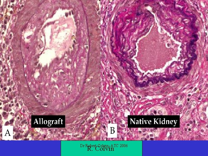
DIAGNOSTIC CRITERIA FOR CHRONIC REJECTION (BANFF 1991) • Chronic Transplant Glomerulopathy • New onset arteriosclerosis (exclude preexisting donor disease) - intimal fibrosis without elastosis - fibroelastosis may develop after prolonged graft dysfunction
Dr Robert Colvin, ATC 2006 R. Colvin
DIFFERENCES BETWEEN GRAFT AND ENDEMIC ATHEROSCLEROSIS 1. 2. 3. 4. Extent of involvement (diffuse vs proximal) Type of luminal compromise distinctive Calcification is late & less common in GAS. Differences in biochemical composition (Apo -A, E, biglycans vs Apo-B, decorin) Differences are relative & not absolute
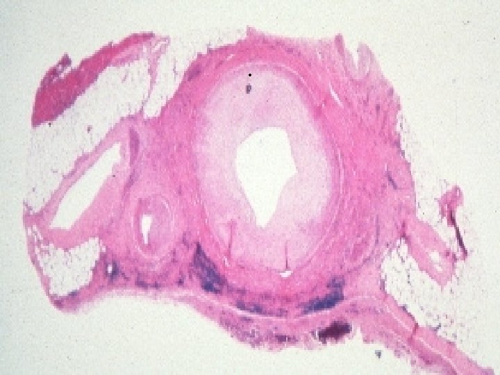
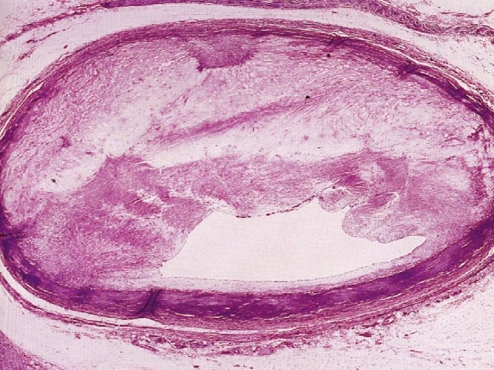
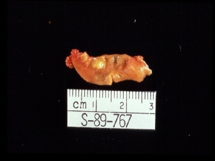
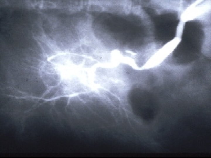
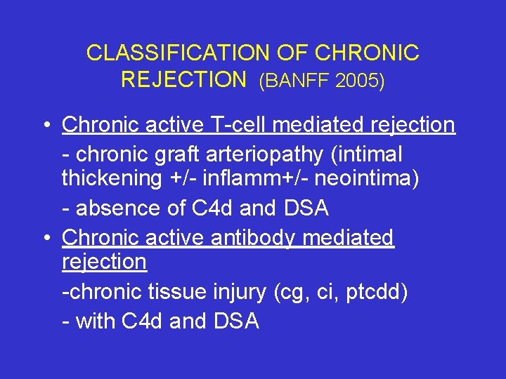
CLASSIFICATION OF CHRONIC REJECTION (BANFF 2005) • Chronic active T-cell mediated rejection - chronic graft arteriopathy (intimal thickening +/- inflamm+/- neointima) - absence of C 4 d and DSA • Chronic active antibody mediated rejection -chronic tissue injury (cg, ci, ptcdd) - with C 4 d and DSA
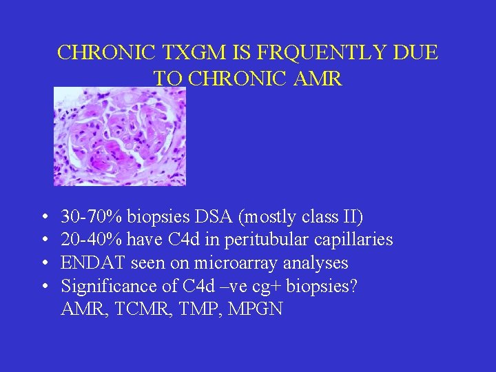
CHRONIC TXGM IS FRQUENTLY DUE TO CHRONIC AMR • • 30 -70% biopsies DSA (mostly class II) 20 -40% have C 4 d in peritubular capillaries ENDAT seen on microarray analyses Significance of C 4 d –ve cg+ biopsies? AMR, TCMR, TMP, MPGN
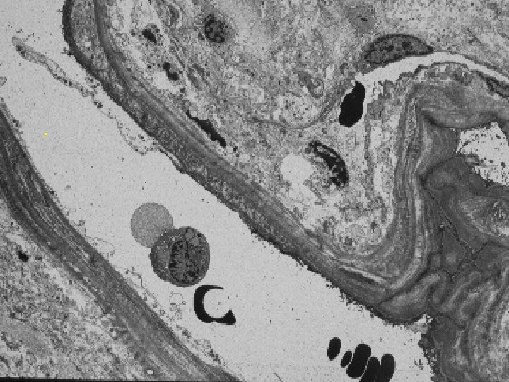
REDUPLICATION OF PERITUBULAR CAPILLARY BASEMENT MEMBRANES • • Needs EM: single PTC with 7 layers 3 PTC’s with 5 -6 layers Duplication >60 -75% of circumference Thus defined, lesion will only rarely occur in other diseases: TMP, reflux, Phenacetin kidney (repeated endothelial injury)
CHRONIC CALCINEURIN INHIBITOR TOXICITY • Cyclosporine • Tacrolimus
PATTERNS OF TISSUE INJURY IN CHRONIC CNI TOXICITY • • Arteriolar lesions Striped Interstitial fibrosis/tubular atrophy Chronic phase thrombotic microangiopathy Ischemic glomerulopathy Focal segmental glomerulosclerosis Juxtaglomerular hyperplasia Tubular vacuolization and calcification
ARTERIOLAR LESIONS • 1 -3 layers smooth muscle • No complete internal elastic lamina • Diameter < 1/3 rd of a glomerulus
ARTERIOLAR HYALINOSIS • • Deposition of glassy material Initially in endothelial layer Followed by circumferential extension Medial involvement
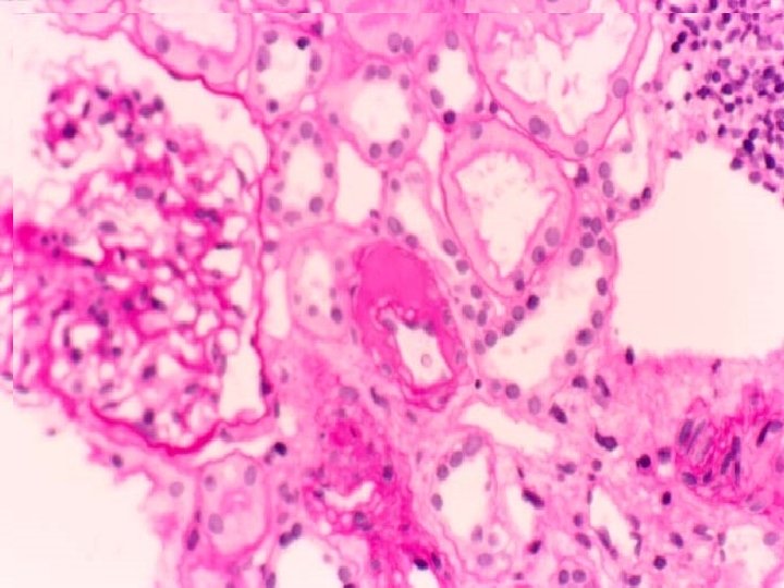
ARTERIOLAR HYALINOSIS • • • Donor disease Diabetes mellitus Hypertension PSS Chronic thrombotic microangiopathy Radiation
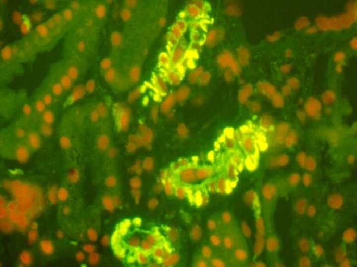
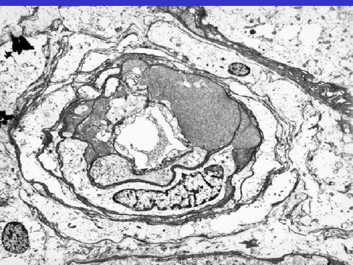

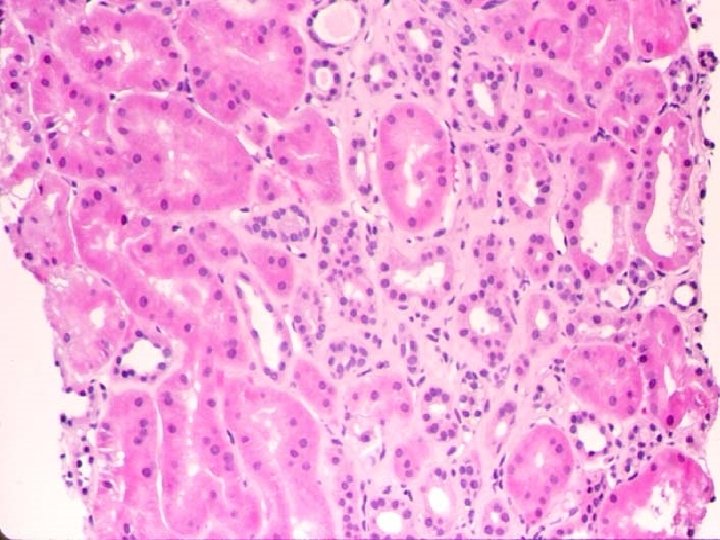
. INTERSTITIAL FIBROSIS IN CALCINEURIN INHIBITOR TOXICITY
PATTERNS OF FIBROSIS • Striped fibrosis: alternating areas of atrophic AND normal or even hypertrophic renal parenchyma • Diffuse interstitial fibrosis: more extensive and uniformly distributed collagen deposition
GLOMERULONEPHRITIS IN THE ALLOGRAFT KIDNEY • Donor transmitted glomerulonephritis • De-novo glomerulonephritis • Recurrence of original disease Morphologic spectrum similar to native kidneys
DONOR TRANSMITTED GLOMERULONEPHRITIS • Ig. A nephropathy • SLE • MPGN
DE-NOVO GLOMERULONEPHRITIS IN THE ALLOGRAFT KIDNEY • • • Work up infection, neoplasms, autoimmune Review medication (captopril, hydralazine) Elicit past h/o horse ATG for TCMR Recently C 4 d + cases MGN recognized Most frequently, however, no cause found
DE-NOVO ANTI-GLOMERULAR BASEMENT MEMBRANE DISEASE • • • Seen in patients with Alport’s (<15%) Exposure to Goodpasture’s antigen Often subclinical but may cause graft loss Histology: proliferative or crescentic GN ELISA, IF confirmation anti. GBMab
RECURRENT GLOMERULONEPHRITIS • 3 rd most frequent cause of graft loss at 10 y (1=chr rej, 2=death with functional graft) • Accounts for 8. 4% of all lost grafts overall • Range 5. 9 -12. 0 %, highest if male sex, primary disease FSGS or MGN Briganti et al 2002; NEJM 347: 103
RECURRENT GLOMERULAR DISEASE • • • Focal segmental glomerulosclerosis 3. 4 y Membranoproliferative GN 3. 6 y Membranous nephropathy 3. 3 y Ig. A nephropathy 4. 4 y Life of avg CRTX 13 y, LRD 21 y Hariharan et al 1999; Transplantation 68: 635
RATES OF GRAFT FAILURE • • • Focal segmental glomerulosclerosis 65% Membranoproliferative GN 66% Membranous nephropathy 44% Ig. A nephropathy 41% Diabetes mellitus 53% HUS/TTP 63% Hariharan et al 1999; Transplantation 68: 635
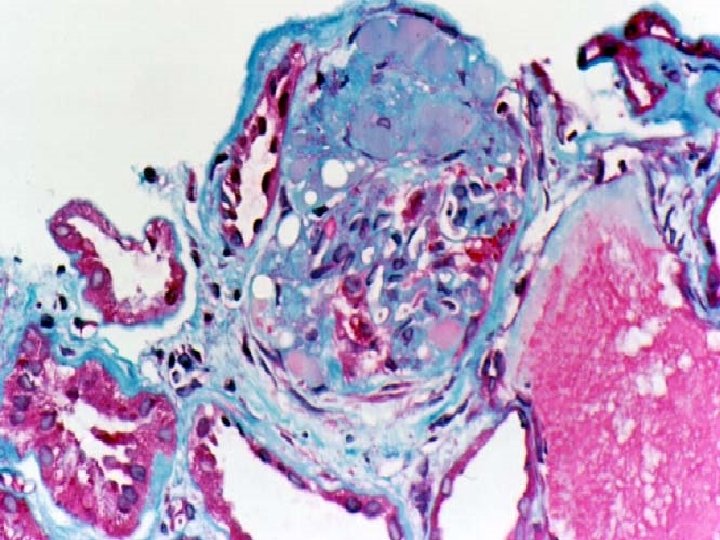
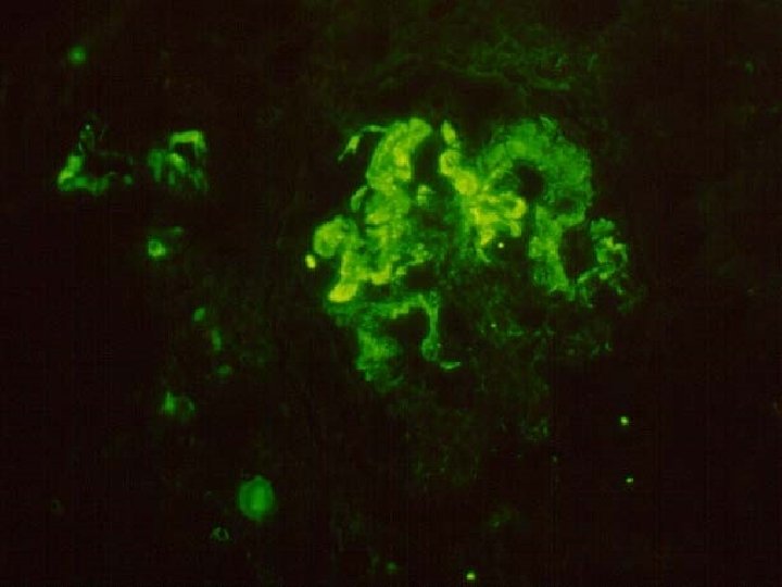
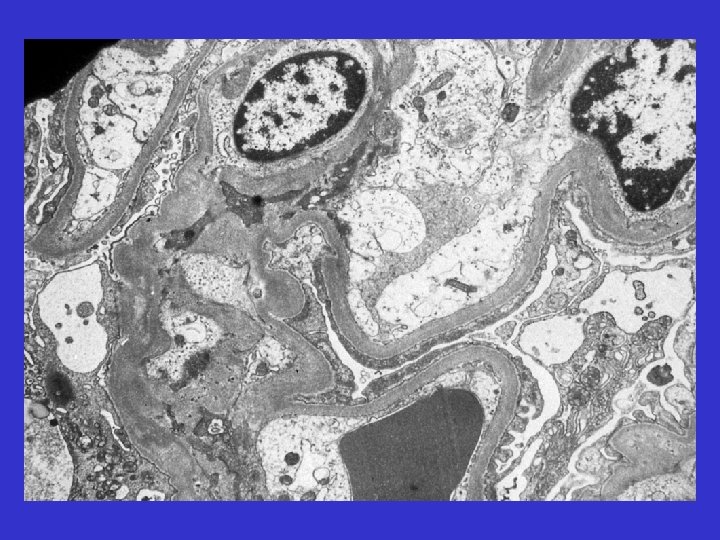
MANAGEMENT OF PATIENTS TRANSPLANTED FOR FSGS • • • Early dx & intervention best prognosis Ur. prot/creat ratio OD to disch, wx 4, mx 12, Ratio > 0. 5 consider 24 hr measurement Puria >2 g/day consider biopsy confirmation Plasmapheresis removes permeability factor
DE NOVO FOCAL SEGMENTAL GLOMERULOSCLEROSIS • FSGS in patient tx for another cause • Common finding in ISTA • Most cases months to years after Transplant -Ischemic glomerulopathy secondary to ah -Hyperfiltration injury, a compensatory response to loss renal mass
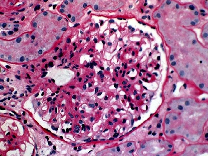
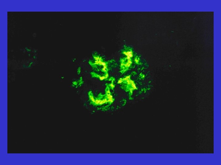
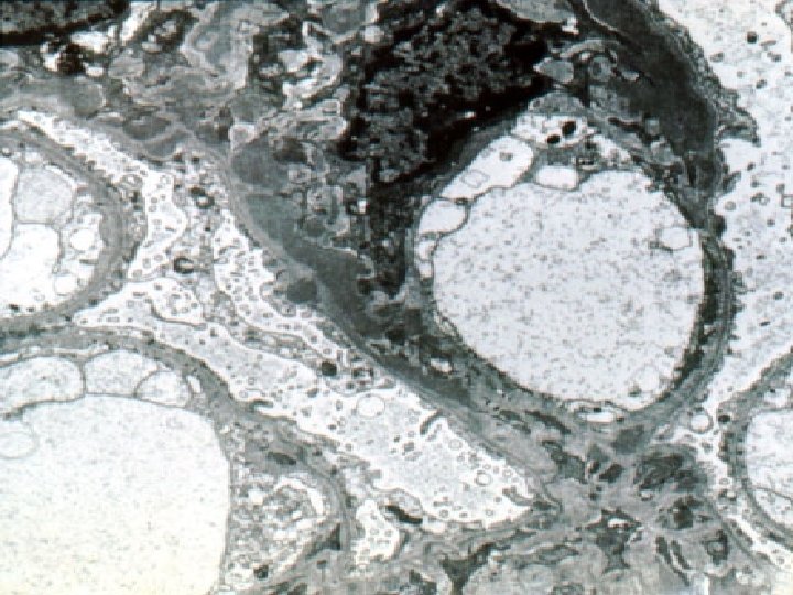
RECURRENT METABOLIC DISEASES • • • Diabetes mellitus Amyloidosis LCDD Lipoprotein glomerulopathy Fabry’s disease
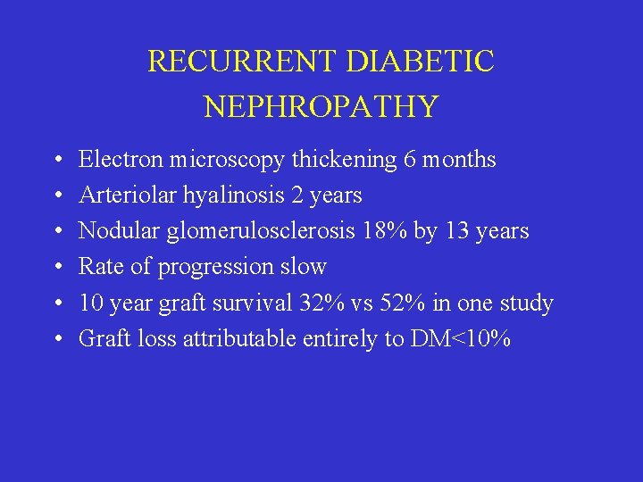
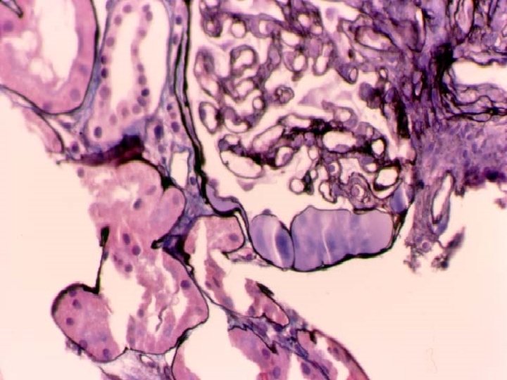
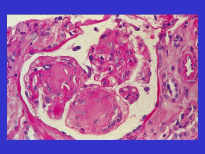
RECURRENT DIABETIC NEPHROPATHY • • • Electron microscopy thickening 6 months Arteriolar hyalinosis 2 years Nodular glomerulosclerosis 18% by 13 years Rate of progression slow 10 year graft survival 32% vs 52% in one study Graft loss attributable entirely to DM<10%
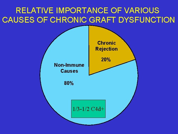
RELATIVE IMPORTANCE OF VARIOUS CAUSES OF CHRONIC GRAFT DYSFUNCTION Chronic Rejection Non-Immune Causes 20% 80% 1/3 -1/2 C 4 d+
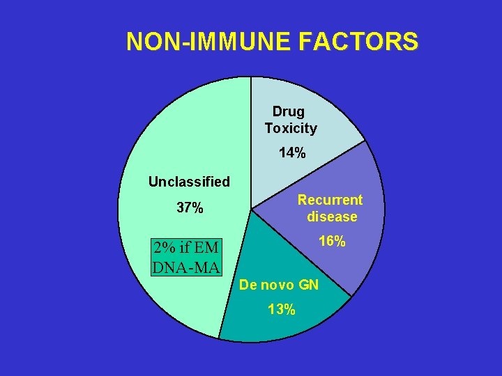
NON-IMMUNE FACTORS Drug Toxicity 14% Unclassified Recurrent disease 37% 16% 2% if EM DNA-MA De novo GN 13%
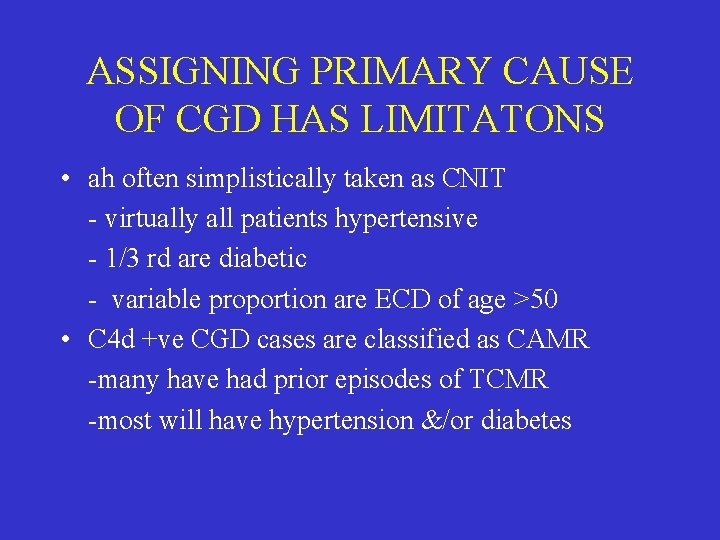
ASSIGNING PRIMARY CAUSE OF CGD HAS LIMITATONS • ah often simplistically taken as CNIT - virtually all patients hypertensive - 1/3 rd are diabetic - variable proportion are ECD of age >50 • C 4 d +ve CGD cases are classified as CAMR -many have had prior episodes of TCMR -most will have hypertension &/or diabetes
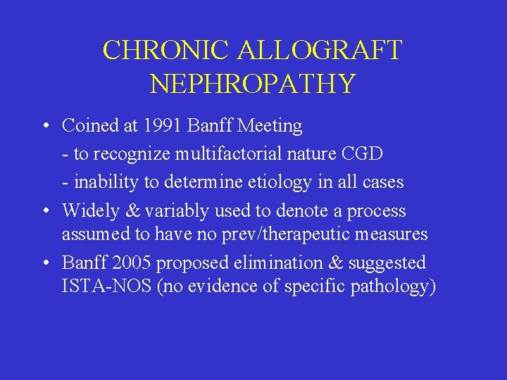
CHRONIC ALLOGRAFT NEPHROPATHY • Coined at 1991 Banff Meeting - to recognize multifactorial nature CGD - inability to determine etiology in all cases • Widely & variably used to denote a process assumed to have no prev/therapeutic measures • Banff 2005 proposed elimination & suggested ISTA-NOS (no evidence of specific pathology)
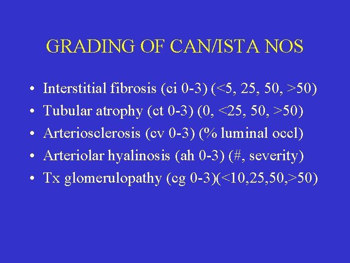
GRADING OF CAN/ISTA NOS • • • Interstitial fibrosis (ci 0 -3) (<5, 25, 50, >50) Tubular atrophy (ct 0 -3) (0, <25, 50, >50) Arteriosclerosis (cv 0 -3) (% luminal occl) Arteriolar hyalinosis (ah 0 -3) (#, severity) Tx glomerulopathy (cg 0 -3)(<10, 25, 50, >50)
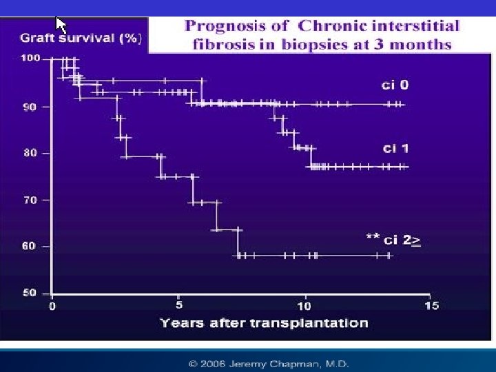
PROGNOSTIC IMPORTANCE OF HISTOLOGIC GRADING
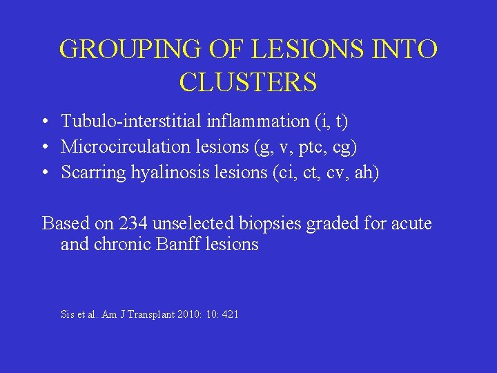
GROUPING OF LESIONS INTO CLUSTERS • Tubulo-interstitial inflammation (i, t) • Microcirculation lesions (g, v, ptc, cg) • Scarring hyalinosis lesions (ci, ct, cv, ah) Based on 234 unselected biopsies graded for acute and chronic Banff lesions Sis et al. Am J Transplant 2010: 421
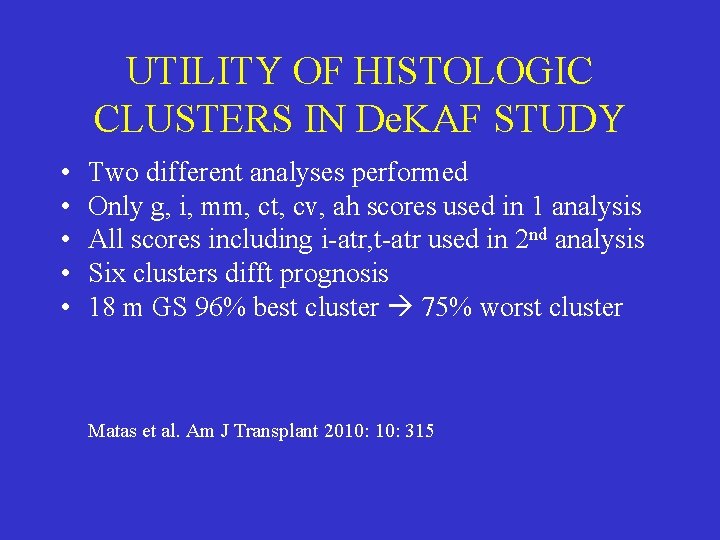
UTILITY OF HISTOLOGIC CLUSTERS IN De. KAF STUDY • • • Two different analyses performed Only g, i, mm, ct, cv, ah scores used in 1 analysis All scores including i-atr, t-atr used in 2 nd analysis Six clusters difft prognosis 18 m GS 96% best cluster 75% worst cluster Matas et al. Am J Transplant 2010: 315

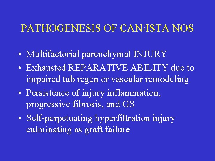
PATHOGENESIS OF CAN/ISTA NOS • Multifactorial parenchymal INJURY • Exhausted REPARATIVE ABILITY due to impaired tub regen or vascular remodeling • Persistence of injury inflammation, progressive fibrosis, and GS • Self-perpetuating hyperfiltration injury culminating as graft failure
CAUSES OF RENAL ALLOGRAFT DYSFUNCTION • REJECTION RELATED CATEGORIES -Acute & chronic Ab mediated rejection -Acute & chronic T-cell mediated rejection • NON-REJECTION RELATED CATEGORIES -ATN, CNIT, Infections, GN, Recurrent native disease, donor derived pathology
RENAL BIOPSY REPORTS SHOULD USE STANDARDIZED TERMINOLOGY Example of non-standardized report: Allograft kidney, needle biopsy: • Interstitial inflammation and tubulitis c/w ac acute rejection (? TCMR, AMR, ? grade) • Patchy interstitial fibrosis & tubular atrophy
A STANDARDIZED BIOPSY REPORT • • Diagnosis: acute T-cell mediated rejection Grade severity: Banff grade 1 (g 0, i 2, t 3, v 0) Worse tubulitis compared to prior biopsy Chronicity: severe interstitial fibrosis (cg 2, ci 3, ct 3, cv 2) We use standardized Banff scoring templates saves time & facilitates large databases


- Slides: 134