Acute Rejection in Kidney Transplantation Valrie HAGE MD


- Slides: 27
Acute Rejection in Kidney Transplantation Valérie HAGE MD, MPH Second IPNA Teaching course Habtoor 2019
Introduction • Major cause of allograft dysfunction • Some kidneys do not regain function even with maximal antirejection therapy. • Can have a negative impact on long-term graft survival
Definitions • Acute deterioration in allograft + specific pathologic changes. • Two principal histologic forms: - Active (acute) antibody-mediated rejection (ABMR) - Acute T cell-mediated (cellular) rejection (TCMR) • Subclinical rejection = histologic evidence of acute rejection on biopsy without an elevation in the serum creatinine
ABMR TCMR • Binding of circulating antibodies to donor • T cells that react to donor alloantigens on graft endothelial cells (HLA/ histocompatibility antigens ABO/ non HLA) PRIMARY LESIONS • Severe vasculitis, glomerulitis with neutrophils in the GC and PTCs • Interstitial infiltration with mononuclear cells • Presence of linear staining for C 4 d • Tubulitis and interstitial mononuclear cell inflammation
ABMR B. Nankivell: N Engl J Med, 2010
TCMR B. Nankivell: N Engl J Med, 2010
Clinical features • Most episodes of acute rejection occur within the first six months after transplantation • Rejection >12 months typically from noncompliance overaggressive reduction in immunosuppression. • Most patients are asymptomatic • Occasionally: present with fever, oliguria, graft pain and/or tenderness
Radiographic manifestations Nonspecific exclude other causes of acute kidney injury Ultrasonography: - increased graft size - loss of corticomedullary junction - prominent hypoechoic pyramids - decreased echogenicity of the renal sinus Renal Doppler studies: elevated resistance indices not specific • PET/CT: noninvasively distinguish non rejection from Acute rejection Lovinfosse P: Am J Transplant, 2016
Laboratory manifestations • Acute rise in the serum creatinine - late development in the course of a rejection - significant histologic damage • Pyuria • New or worsening proteinuria
Investigational methods Ø Non invasive biomarker for the diagnosis of allograft rejection Ø Potential to improve the accuracy of diagnosis Ø Require validation in clinical trials Ø Several candidates biomarkers
Investigational methods • Molecular blood biomarkers: - Kidney Solid Organ Response Test k. SORT (m. RNA) - Plasma levels of donor-derived cell-free DNA (dd-cf. DNA) may be elevated (>1%) in patients with acute rejection • Blood cell function assays - ELISPOT for IFN-gamma • Urine biomarkers: acute TCMR • m. RNA (perforin, granzyme B, FOXP 3, TIM-3) • mi. RNA • Immune-related proteins: CXCL 9, CXCL 10 (BK Virus)
Conclusions: Although increases in plasma ddcf. DNA% are associated with graft injury, plasma ddcf. DNA does not outperform the diagnostic capacity of the serum creatinine in the diagnosis of acute rejection.
When to suspect acute rejection >1 of the following: • New increase in serum creatinine of ≥ 25% • Worsening hypertension. • Proteinuria >1 g/day.
Increased risk for ABMR • • Highly sensitized patients ABO-incompatible renal allografts Patients with Donor Specific Antibodies(DSAs) Patients with inadequate immunosuppression Any incremental increase in serum creatinine should raise the suspicion for the possibility of acute rejection!!
The diagnosis of ABMR 1. Histologic evidence of acute tissue injury 2. Evidence of antibody interaction with vascular endothelium (eg, C 4 d staining in peritubular capillaries (PTCs) 3. Serologic evidence of DSAs
Confirming the diagnosis Standard for the diagnosis Renal Allograft Biopsy 1. 2. 3. 4. Grade the severity of rejection TCMR v/s ABMR Degree of irreversible kidney damage [IF/TA] Reveal other causes of renal inflammation and injury - CMV disease BK nephropathy interstitial nephritis pyelonephritis de novo or recurrent glomerular disease, posttransplant lymphoproliferative disease (PTLD)
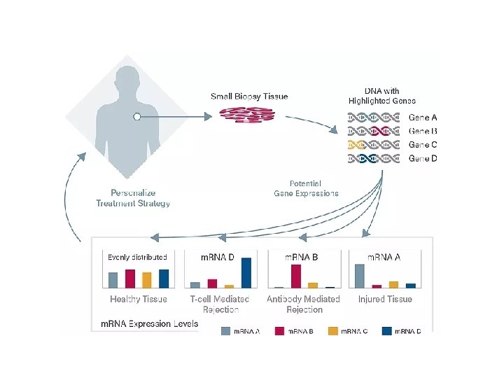
Molecular microscope diagnostic system: MMDx • • • Advanced tool Provide more precise information Smarter analysis Can detect injury at the molecular level Allow rapid target treatment
Molecular microscope RNA extracted from the biopsy: microarray analysis is performed to measure m. RNA levels Predefined computer algorithms used to generate a report Report= Molecular scores: reflect the probability that the biopsy sample has histologic features of TCMR, ABMR, rejection of any type, AKI , or IF/TA
The Banff classification of acute TCMR • Borderline – Mild interstitial inflammation (<25 percent of nonsclerotic cortical parenchyma; i 0 or i 1) plus any tubulitis (t 1, t 2, or t 3) or significant interstitial inflammation (>25 percent of nonsclerotic cortical parenchyma; i 2 or i 3) plus foci of mild tubulitis (t 1) • Type IA – Significant interstitial inflammation (>25 percent of nonsclerotic cortical parenchyma; i 2 or i 3) and foci of moderate tubulitis (t 2) moderate Type IB – Significant interstitial inflammation (>25 percent of nonsclerotic cortical parenchyma; i 2 or i 3) and foci of severe tubulitis (t 3) severe • • Type IIA – Mild-to-moderate intimal arteritis (v 1) with or without interstitial inflammation and tubulitis Type IIB – Severe intimal arteritis comprising >25 percent of the luminal area (v 2) with or without interstitial inflammation and tubulitis Type III – Transmural arteritis and/or arterial fibrinoid change and necrosis of medial smooth muscle cells with accompanying lymphocytic inflammation (v 3)
Treatment TCMR • Banff grade IA or IB - inpatient admission - pulse IV methylprednisolone at 3 to 5 mg/kg daily for 3 to 5 doses, maximum daily dose of 500 mg - oral glucocorticoids are tapered immediately to the maintenance dose of oral prednisone the patient had been taking prior to the episode • Banff grade II or III rejection - pulse IV methylprednisolone 3 to 5 mg/kg daily for three to five doses+ short tapered dose of oral prednisone. - Daily r. ATG-Thymoglobulin at 1. 5 to 3 mg/kg per dose for a total dose of 5 to 10 mg/kg. The total number of doses is determined by severity of Banff grading.
Treatment ABMR Primary aims : • Remove circulating donor-specific antibodies • Block their effects • Reduce their production No therapeutics have received FDA approval for the treatment Mainstays of contemporary therapy: - Plasma exchange - Intravenous immune globulin - Glucocorticoids Clin. Transplant 2015; 29: 118 -23.
Treatment ABMR • Anti-CD 20 monoclonal antibody: Rituximab • Proteasome inhibitor: Bortezomib Two randomized, controlled trials - RITUX ERAH (Sautenet B: Transplantation, 2016) - BORTEJECT (Eskandary F: J Am Soc Nephrol, 2018) neither trial showed clinical benefits!!!
Treatment ABMR • Anti-C 5 monoclonal antibody : Eculizumab reported to decrease the incidence of ABMR in HLAsensitized renal-transplant recipients. Stegal M: Am J Transplant, 2011 • Ig. G-degrading enzyme of Streptococcus pyogenes (Ide. S): Imlifidase Cleaves Human Ig. G Reduced/eliminated DSA before kidney transplantation HLA-incompatible Jordan SC: N Engl J Med, 2017
A Loupy: N Engl J Med, 2018
THANK YOU