Acute kidney injury Khozama jehad Defintion Acute kidney
- Slides: 38
Acute kidney injury Khozama jehad
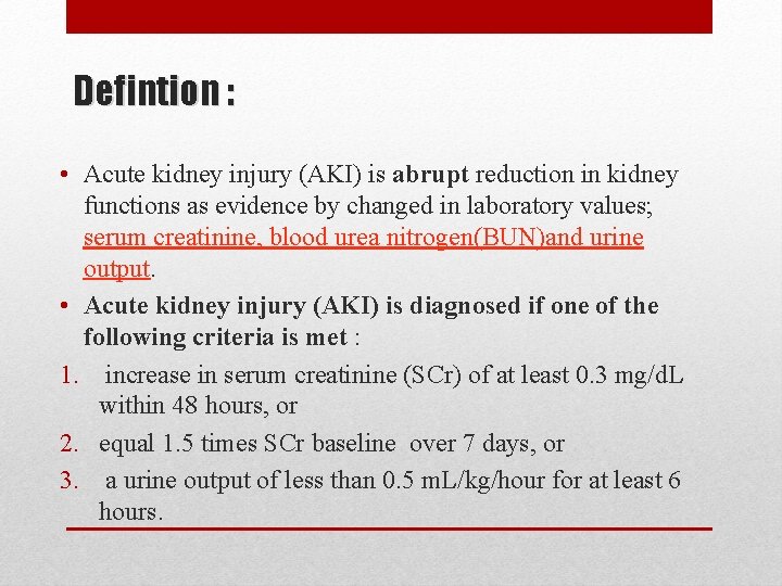
Defintion : • Acute kidney injury (AKI) is abrupt reduction in kidney functions as evidence by changed in laboratory values; serum creatinine, blood urea nitrogen(BUN)and urine output. • Acute kidney injury (AKI) is diagnosed if one of the following criteria is met : 1. increase in serum creatinine (SCr) of at least 0. 3 mg/d. L within 48 hours, or 2. equal 1. 5 times SCr baseline over 7 days, or 3. a urine output of less than 0. 5 m. L/kg/hour for at least 6 hours.
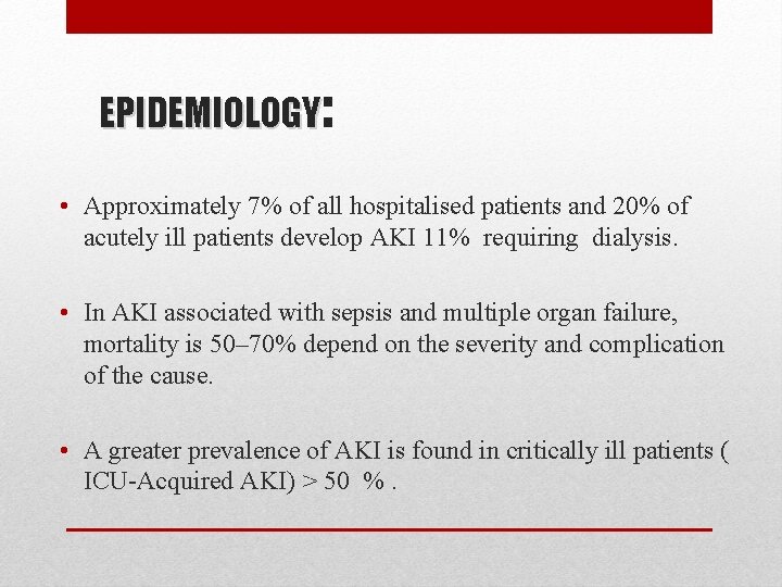
EPIDEMIOLOGY: • Approximately 7% of all hospitalised patients and 20% of acutely ill patients develop AKI 11% requiring dialysis. • In AKI associated with sepsis and multiple organ failure, mortality is 50– 70% depend on the severity and complication of the cause. • A greater prevalence of AKI is found in critically ill patients ( ICU-Acquired AKI) > 50 %.
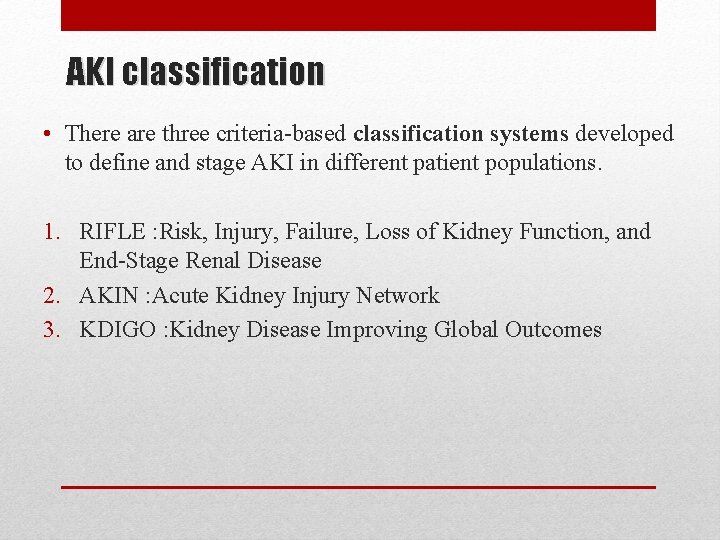
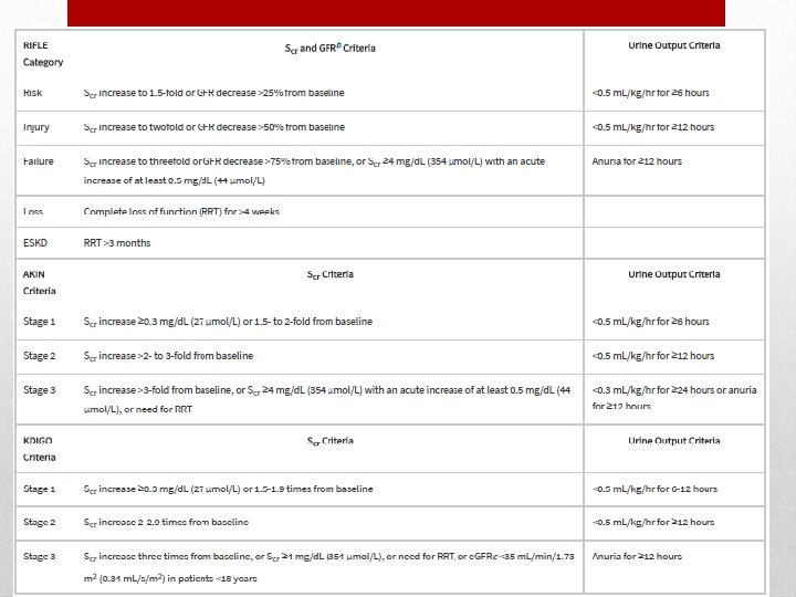
AKI classification • There are three criteria-based classification systems developed to define and stage AKI in different patient populations. 1. RIFLE : Risk, Injury, Failure, Loss of Kidney Function, and End-Stage Renal Disease 2. AKIN : Acute Kidney Injury Network 3. KDIGO : Kidney Disease Improving Global Outcomes
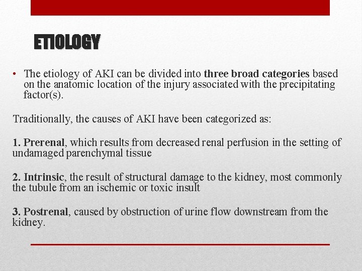
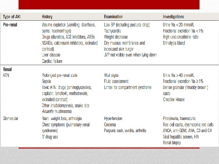
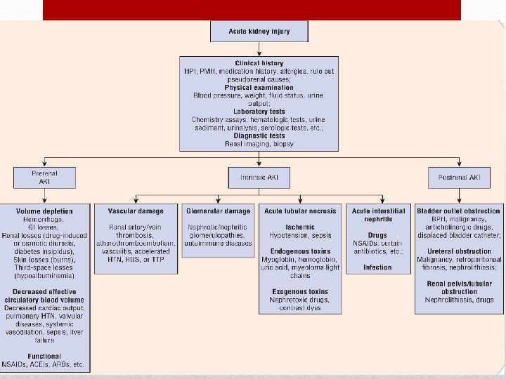
ETIOLOGY • The etiology of AKI can be divided into three broad categories based on the anatomic location of the injury associated with the precipitating factor(s). Traditionally, the causes of AKI have been categorized as: 1. Prerenal, which results from decreased renal perfusion in the setting of undamaged parenchymal tissue 2. Intrinsic, the result of structural damage to the kidney, most commonly the tubule from an ischemic or toxic insult 3. Postrenal, caused by obstruction of urine flow downstream from the kidney.
PRERENAL AKI Results from hypoperfusion of the renal parenchyma, with or without systemic arterial hypotension. Renal hypoperfusion associated with systemic arterial hypotension: • Intravascular volume depletion 1. Hemorrhage 2. excessive gastrointestinal (GI) losses (severe vomiting or diarrhea) 3. dehydration 4. extensive burns 5. diuretic therapy. • Decrease effective circulatory blood volume 1. Decrease cardiac output (CHF, MI, hypotension) 2. Pulmonary hypertension 3. Liver failure 4. Sepsis
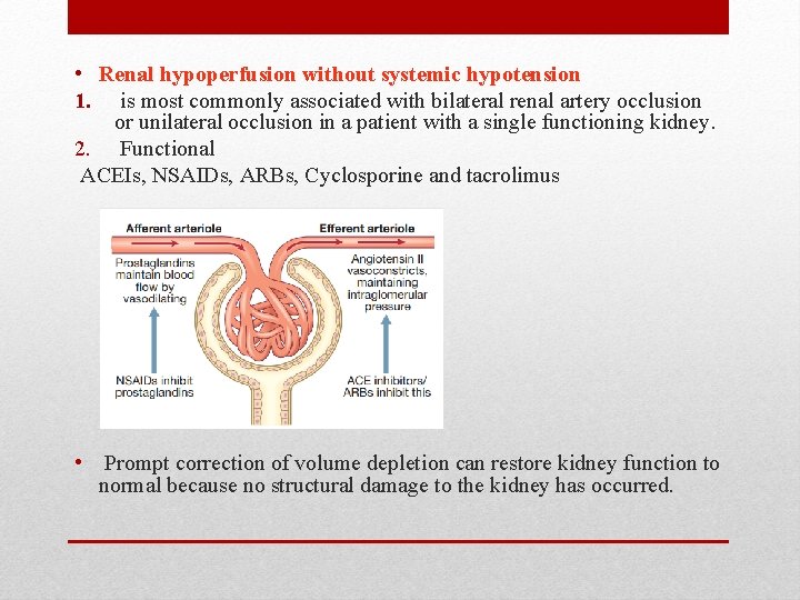
• Renal hypoperfusion without systemic hypotension 1. is most commonly associated with bilateral renal artery occlusion or unilateral occlusion in a patient with a single functioning kidney. 2. Functional ACEIs, NSAIDs, ARBs, Cyclosporine and tacrolimus • Prompt correction of volume depletion can restore kidney function to normal because no structural damage to the kidney has occurred.
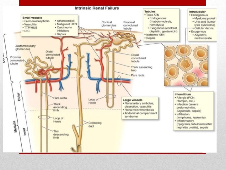
Intrinsic Acute Kidney Injury Renal Vasculature Damage: Acute vascular syndrome Micro vascular or macro vascular • Macrovascular : Sever abdominal aortic disease Major renal artery occlusion Renal vein thrombosis Can cause AKI and renal infarction ( abdominal pain , hematuria ) Polyarteritis nodosa causing microanueyrsm leading to acute kidney injury Atherosclerosis can affect the renal artery causing AKI Atheroembolic disease caused by procedure during catheterization .
• Microvascular : HUS “(hemolytic uremic syndrome ) Hellp syndrome ( hemolysis , elevated liver enzymes , low PLT ) Sclerodrema renal crisis Drug induced microangiopathy
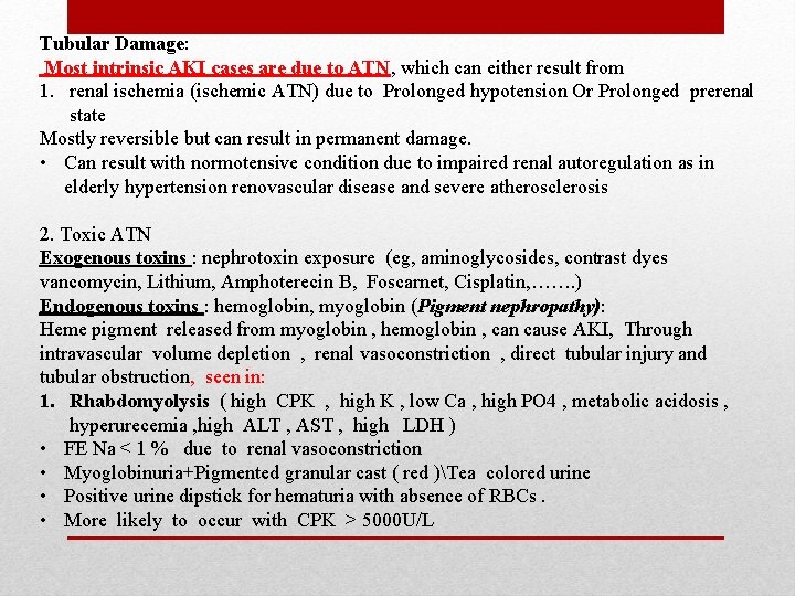
Tubular Damage: Most intrinsic AKI cases are due to ATN, which can either result from 1. renal ischemia (ischemic ATN) due to Prolonged hypotension Or Prolonged prerenal state Mostly reversible but can result in permanent damage. • Can result with normotensive condition due to impaired renal autoregulation as in elderly hypertension renovascular disease and severe atherosclerosis 2. Toxic ATN Exogenous toxins : nephrotoxin exposure (eg, aminoglycosides, contrast dyes vancomycin, Lithium, Amphoterecin B, Foscarnet, Cisplatin, ……. ) Endogenous toxins : hemoglobin, myoglobin (Pigment nephropathy): Heme pigment released from myoglobin , hemoglobin , can cause AKI, Through intravascular volume depletion , renal vasoconstriction , direct tubular injury and tubular obstruction, seen in: 1. Rhabdomyolysis ( high CPK , high K , low Ca , high PO 4 , metabolic acidosis , hyperurecemia , high ALT , AST , high LDH ) • FE Na < 1 % due to renal vasoconstriction • Myoglobinuria+Pigmented granular cast ( red )Tea colored urine • Positive urine dipstick for hematuria with absence of RBCs. • More likely to occur with CPK > 5000 U/L
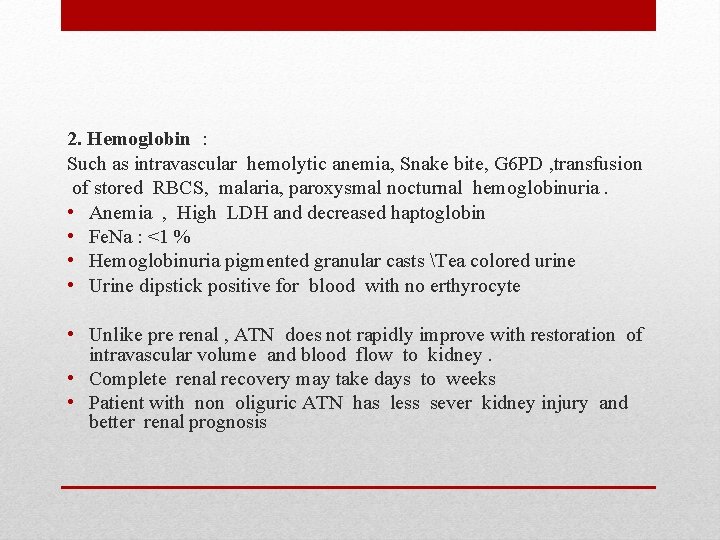
2. Hemoglobin : Such as intravascular hemolytic anemia, Snake bite, G 6 PD , transfusion of stored RBCS, malaria, paroxysmal nocturnal hemoglobinuria. • Anemia , High LDH and decreased haptoglobin • Fe. Na : <1 % • Hemoglobinuria pigmented granular casts Tea colored urine • Urine dipstick positive for blood with no erthyrocyte • Unlike pre renal , ATN does not rapidly improve with restoration of intravascular volume and blood flow to kidney. • Complete renal recovery may take days to weeks • Patient with non oliguric ATN has less sever kidney injury and better renal prognosis
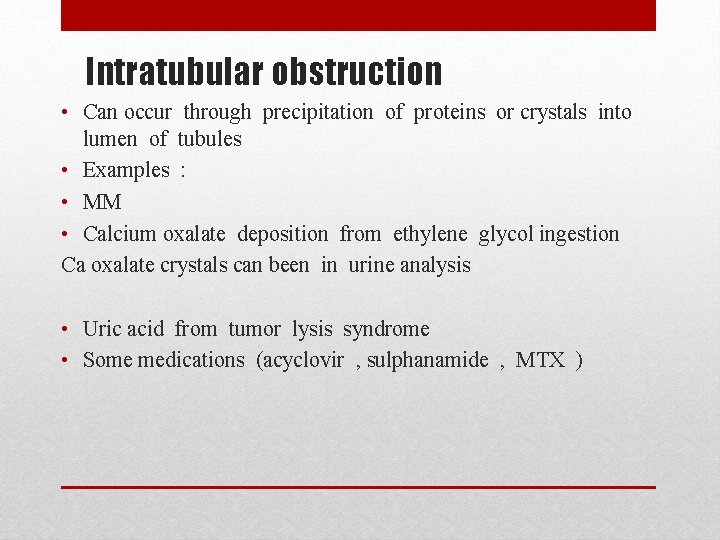
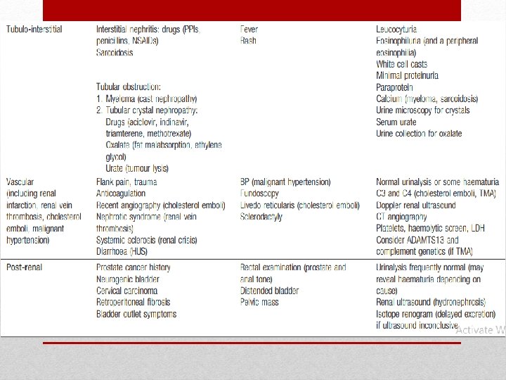
Intratubular obstruction • Can occur through precipitation of proteins or crystals into lumen of tubules • Examples : • MM • Calcium oxalate deposition from ethylene glycol ingestion Ca oxalate crystals can been in urine analysis • Uric acid from tumor lysis syndrome • Some medications (acyclovir , sulphanamide , MTX )
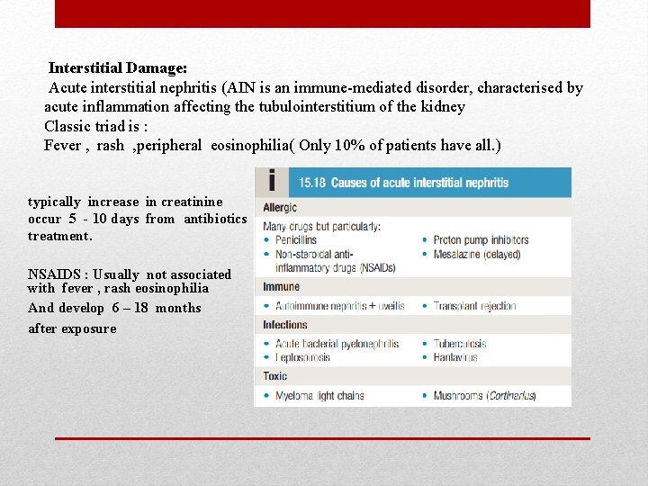
Interstitial Damage: Acute interstitial nephritis (AIN is an immune-mediated disorder, characterised by acute inflammation affecting the tubulointerstitium of the kidney Classic triad is : Fever , rash , peripheral eosinophilia( Only 10% of patients have all. ) typically increase in creatinine occur 5 - 10 days from antibiotics treatment. NSAIDS : Usually not associated with fever , rash eosinophilia And develop 6 – 18 months after exposure
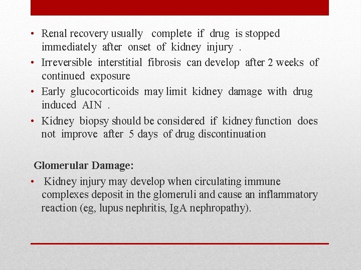
• Renal recovery usually complete if drug is stopped immediately after onset of kidney injury . • Irreversible interstitial fibrosis can develop after 2 weeks of continued exposure • Early glucocorticoids may limit kidney damage with drug induced AIN . • Kidney biopsy should be considered if kidney function does not improve after 5 days of drug discontinuation Glomerular Damage: • Kidney injury may develop when circulating immune complexes deposit in the glomeruli and cause an inflammatory reaction (eg, lupus nephritis, Ig. A nephropathy).
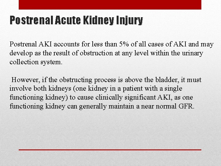
Postrenal Acute Kidney Injury Postrenal AKI accounts for less than 5% of all cases of AKI and may develop as the result of obstruction at any level within the urinary collection system. However, if the obstructing process is above the bladder, it must involve both kidneys (one kidney in a patient with a single functioning kidney) to cause clinically significant AKI, as one functioning kidney can generally maintain a near normal GFR.
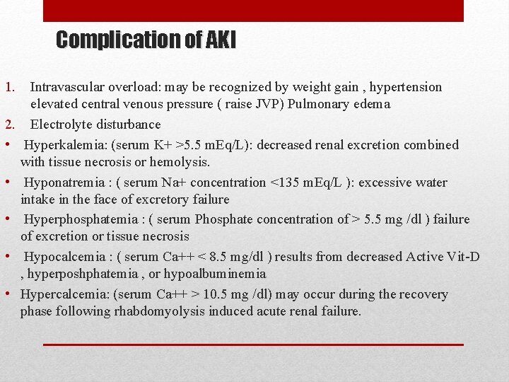
Complication of AKI 1. 2. • • • Intravascular overload: may be recognized by weight gain , hypertension elevated central venous pressure ( raise JVP) Pulmonary edema Electrolyte disturbance Hyperkalemia: (serum K+ >5. 5 m. Eq/L): decreased renal excretion combined with tissue necrosis or hemolysis. Hyponatremia : ( serum Na+ concentration <135 m. Eq/L ): excessive water intake in the face of excretory failure Hyperphosphatemia : ( serum Phosphate concentration of > 5. 5 mg /dl ) failure of excretion or tissue necrosis Hypocalcemia : ( serum Ca++ < 8. 5 mg/dl ) results from decreased Active Vit-D , hyperposhphatemia , or hypoalbuminemia Hypercalcemia: (serum Ca++ > 10. 5 mg /dl) may occur during the recovery phase following rhabdomyolysis induced acute renal failure.
• Metabolic acidosis : ( arterial blood PH < 7. 35 ) is associated with sepsis or severe heart failure • Hyperuricemia: due to decreased uric acid excretion • Bleeding tendency : may occur due to platelet dysfunction and coagulopathy associated with sepsis • Seizure: may occur related to uremia 3. Infection : a common and serious complication of aki (occur in 50% to 60% ) the cause is probably multifactorial, but uremia itself is thought to impair immune function. Example : pneumonia, UTI, wound infection, and sepsis.
Clinical manifestations • Patient presentation varies widely and depends on the underlying cause. • Outpatients often are not in acute distress; hospitalized patients may develop AKI after a catastrophic event (eg, shock). • Symptoms in the outpatient setting include acute change in urinary habits, sudden weight gain, or severe abdominal or flank pain. Can be asymptomatic • Signs include edema, colored or foamy urine, and, in volumedepleted patients postural hypotension. • AKI in hospitalized patients is often detected much earlier in its course due to frequent laboratory studies and daily patient assessment Oliguria : < 500 ml / d Anuria < 50 ml / d
• • Symptoms of uremia : Nausea seizure Vomiting mental status changes Anorexia pericarditis Wt loss pruritis Fatigue PLT dysfunction Muscle cramps anemia Restless leg
DIAGNOSIS • Thorough medical and medication histories, physical examination, assessment of laboratory values, and, if needed, imaging studies are important in the diagnosis of AKI. 1. labs : • Scr cannot be used alone to diagnose AKI because it is insensitive to rapid changes in glomerular filtration rate (GFR) lagging behind the GRF’s decline by 1 to 2 days leading to a potential delay in diagnosis. • The use of BUN in AKI is very limited because urea production and renal clearance are heavily influenced by extrarenal factors such as critical illness, volume status, protein intake, and medications. • Electolytes • CBC , LFTS • ABG (metabolic acidosis)
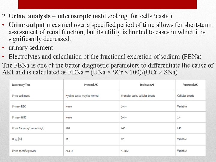
2. Urine analysis + microscopic test(Looking for cells casts ) • Urine output measured over a specified period of time allows for short-term assessment of renal function, but its utility is limited to cases in which it is significantly decreased. • urinary sediment • Electrolytes and calculation of the fractional excretion of sodium (FENa) The FENa is one of the better diagnostic parameters to differentiate the cause of AKI and is calculated as FENa = (UNa × SCr × 100)/(UCr × SNa)
3. U/S : Should be obtained from suspected obstruction Kidney size can help to distinguish between acute or chronic kidney injury Kidney size can be normal in Patient with CKD from infiltrative disorder such as DM , amyloidosis and MM 4. Biopsy : Obtained if no clear cause is obvious
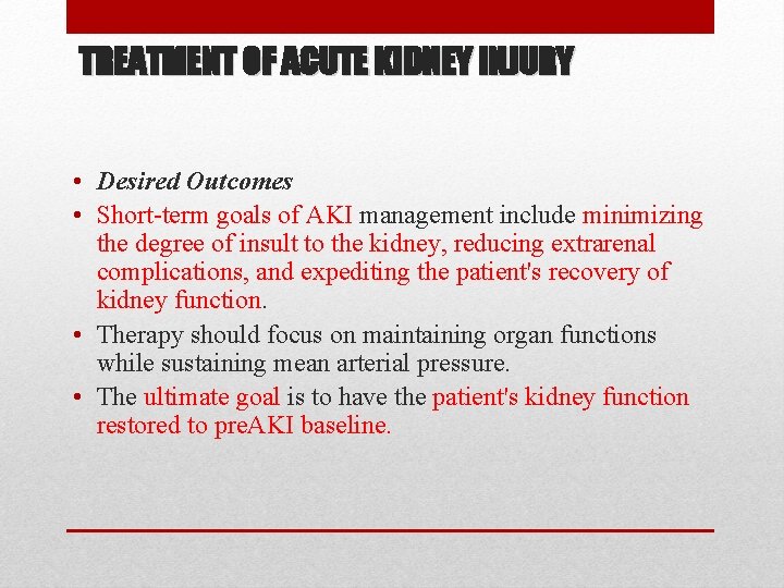
TREATMENT OF ACUTE KIDNEY INJURY • Desired Outcomes • Short-term goals of AKI management include minimizing the degree of insult to the kidney, reducing extrarenal complications, and expediting the patient's recovery of kidney function. • Therapy should focus on maintaining organ functions while sustaining mean arterial pressure. • The ultimate goal is to have the patient's kidney function restored to pre. AKI baseline.
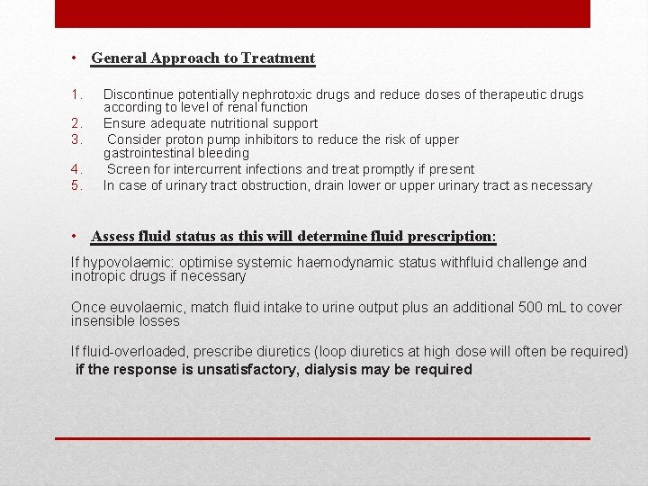
• General Approach to Treatment 1. 2. 3. 4. 5. Discontinue potentially nephrotoxic drugs and reduce doses of therapeutic drugs according to level of renal function Ensure adequate nutritional support Consider proton pump inhibitors to reduce the risk of upper gastrointestinal bleeding Screen for intercurrent infections and treat promptly if present In case of urinary tract obstruction, drain lower or upper urinary tract as necessary • Assess fluid status as this will determine fluid prescription: If hypovolaemic: optimise systemic haemodynamic status withfluid challenge and inotropic drugs if necessary Once euvolaemic, match fluid intake to urine output plus an additional 500 m. L to cover insensible losses If fluid-overloaded, prescribe diuretics (loop diuretics at high dose will often be required) if the response is unsatisfactory, dialysis may be required
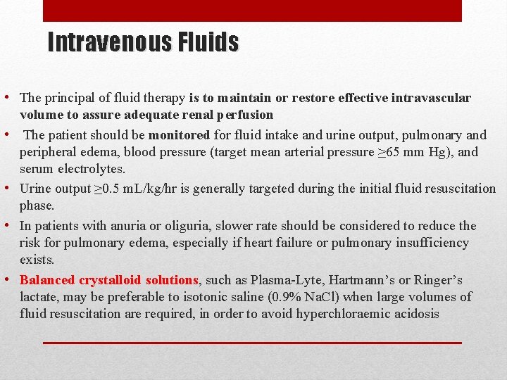
Intravenous Fluids • The principal of fluid therapy is to maintain or restore effective intravascular volume to assure adequate renal perfusion • The patient should be monitored for fluid intake and urine output, pulmonary and peripheral edema, blood pressure (target mean arterial pressure ≥ 65 mm Hg), and serum electrolytes. • Urine output ≥ 0. 5 m. L/kg/hr is generally targeted during the initial fluid resuscitation phase. • In patients with anuria or oliguria, slower rate should be considered to reduce the risk for pulmonary edema, especially if heart failure or pulmonary insufficiency exists. • Balanced crystalloid solutions, such as Plasma-Lyte, Hartmann’s or Ringer’s lactate, may be preferable to isotonic saline (0. 9% Na. Cl) when large volumes of fluid resuscitation are required, in order to avoid hyperchloraemic acidosis
• If AKI is a result of blood loss or is complicated by symptomatic anemia, red blood cell transfusion to a hemoglobin >7 g/d. L is the treatment of choice. • Once a hemoglobin of >7 g/d. L is reached, balanced solutions or normal saline can be used to restore intravascular volume. • Albumin is typically preferred in individuals with severe hypoalbuminemia secondary to cirrhosis or nephrotic syndrome. • In critically ill patients with vasodilatory shock, vasopressors such as norepinephrine, vasopressin, or dopamine may be used in conjunction with fluids in order to maintain adequate hemodynamics and renal perfusion
Diuretics • Loop diuretics are frequently prescribed for the management of fluid overload in patients with established kidney injury. • Loop diuretics have several theoretical advantages: 1. Increased urine output; 2. Decreased risk of ischemic injury by inhibiting the Na-K-Cl cotransporter and thus decreasing oxygen demand; and 3. Enhanced renal blood flow due to increased availability of renal prostaglandins • However, clinical studies have found that even though the loop diuretics increase urine output, they neither reduce the incidence of AKI nor improve patient outcomes for patients with established AKI • Therefore, the KDIGO guidelines recommend limiting the use of loop diuretics to the management of fluid overload and avoiding their use for the sole purpose of prevention or treatment of AKI. • Furosemide, bumetanide, torsemide and ethacrinic acid ) all have similar efficacy • Dose: Initial iv loading dose equivalent to (40 -60 mg furosemide ) + Continuous infusion equivalent to 10 -20 mg/h
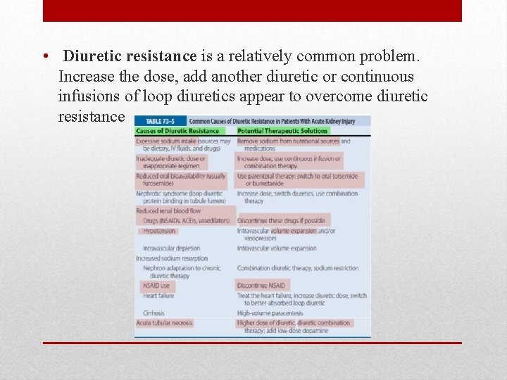
• Diuretic resistance is a relatively common problem. Increase the dose, add another diuretic or continuous infusions of loop diuretics appear to overcome diuretic resistance
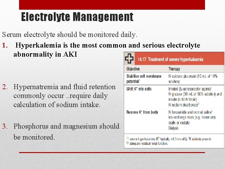
Electrolyte Management Serum electrolyte should be monitored daily. 1. Hyperkalemia is the most common and serious electrolyte abnormality in AKI 2. Hypernatremia and fluid retention commonly occur. . require daily calculation of sodium intake. 3. Phosphorus and magnesium should be monitored.
Nutritional Considerations in AKI • Nutritional management of critically ill patients with AKI can be extremely complex, as it needs to account for metabolic derangements resulting from both impaired kidney function and underlying disease processes
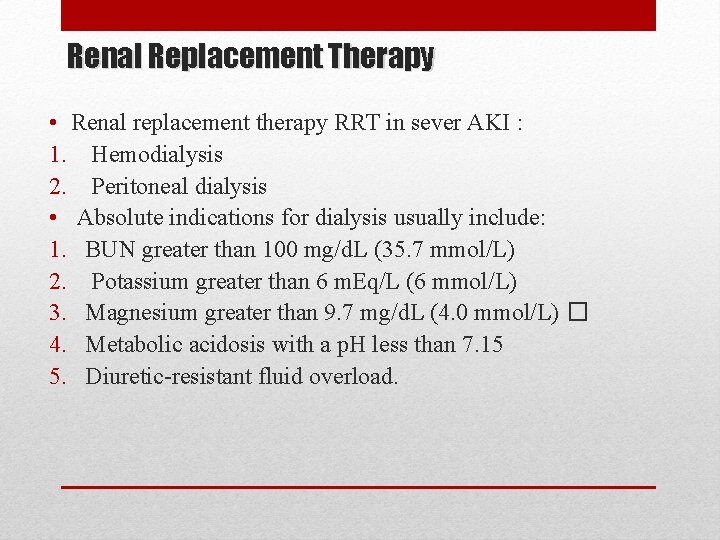
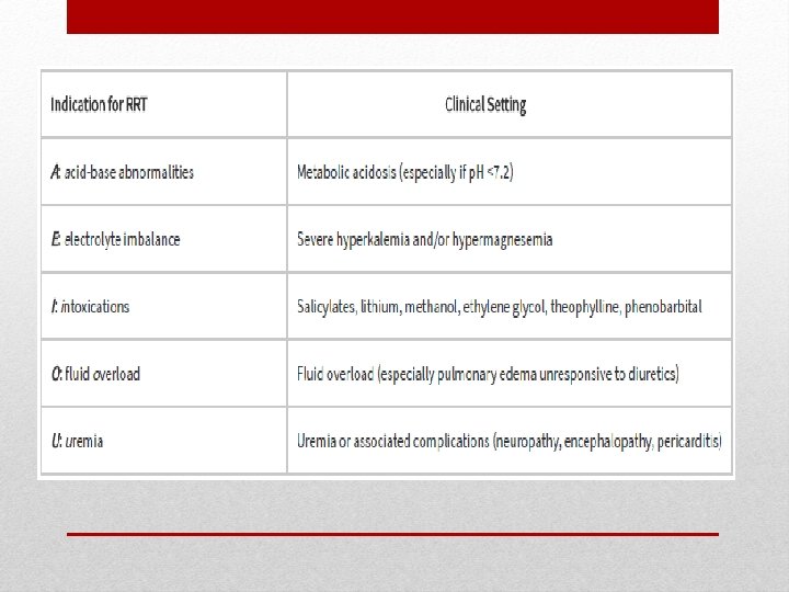
Renal Replacement Therapy • Renal replacement therapy RRT in sever AKI : 1. Hemodialysis 2. Peritoneal dialysis • Absolute indications for dialysis usually include: 1. BUN greater than 100 mg/d. L (35. 7 mmol/L) 2. Potassium greater than 6 m. Eq/L (6 mmol/L) 3. Magnesium greater than 9. 7 mg/d. L (4. 0 mmol/L) � 4. Metabolic acidosis with a p. H less than 7. 15 5. Diuretic-resistant fluid overload.
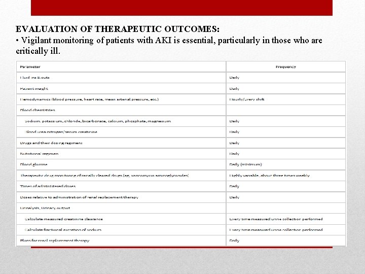
EVALUATION OF THERAPEUTIC OUTCOMES: • Vigilant monitoring of patients with AKI is essential, particularly in those who are critically ill.