Endothelial injury post implantation Implanted stent Plaque Stent


 Cell responds to growth factor stimulation Mitosis Cell")


 Paclitaxel-eluting non– polymer-based")


 Paclitaxel is a mitotic inhibitor used in cancer chemotherapy. It was")
 Σε εξέλιξη μελέτη από το Βέλγιο Αναμένονται τα αποτελέσματά")


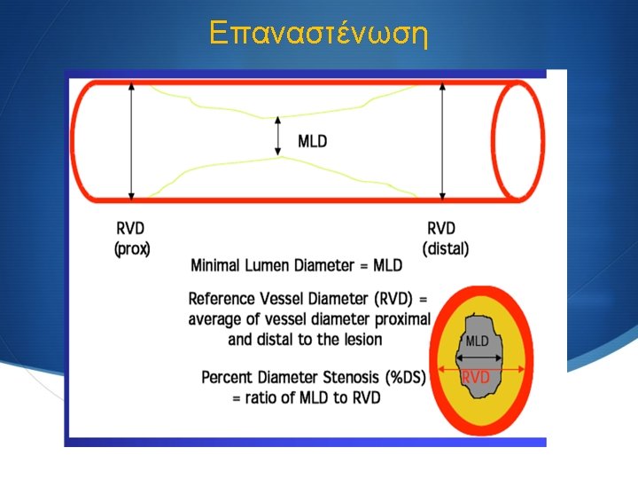
 balloon) Scheller B et al. Circulation. 2004;")
 trial")


 production S Normal vessels")

- Slides: 33
Endothelial injury post implantation Implanted stent Plaque Stent implantation causes arterial injury, which can initiate restenosis. The restenosis process includes inflammation, migration of smooth muscle cells, smooth muscle cell proliferation and extracellular matrix formation. 2
Platelet aggregation and activation Drug-eluting stent struts Platelets Red blood cells Inflammatory cells Platelet deposition and activation occur at the injury site, leading to the release of cell-signaling molecules. 3
Transmigration of inflammatory cells Endothelial cells Transmigration of inflammatory cells Smooth muscle cells Inflammatory cells secreting cell-signaling molecules Once activated, these inflammatory cells roll across the endothelial surface and transmigrate into the lesion. 4
Activation of smooth muscle cells Smooth muscle cell extracellular view Cell signaling molecules activate smooth muscle cells Smooth muscle cell surface receptor The activated inflammatory cells secrete molecules that bind to specific receptors 5 smooth muscle cells. on
Activation of smooth muscle cells Smooth muscle cell intracellular view Activated smooth muscle cell receptor m. TOR activates smooth muscle cells to enter cell cycle Bound smooth muscle cell receptors activate various intracellular smooth muscle cell 6 proteins. One such protein, m. TOR, plays a central regulatory role in the cell cycle.
Activation of smooth muscle cells (III) Cell responds to growth factor stimulation Mitosis Cell resting phase Restriction point Cell prepares for mitosis DNA synthesis Activated m. TOR stimulates smooth muscle cells to advance from the G 1 phase to the S phase where DNA replication occurs, causing the smooth muscle cells to undergo mitosis (ie, cell proliferation). 7
Fraction of Maximal Response Differential Events Leading to In-Stent Restenosis 1 0 Time Platelet Deposition Leukocyte recruitment VSMC migration / proliferation Matrix deposition
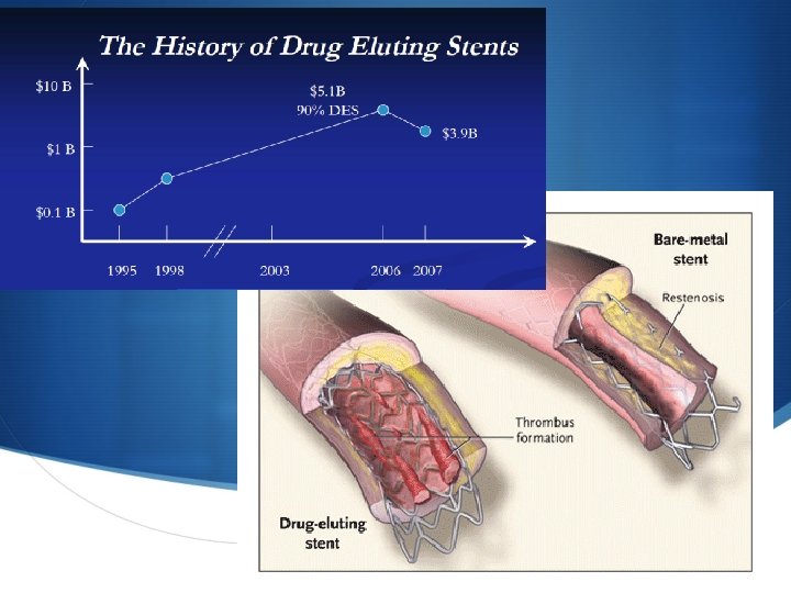
There are three major components to a drug-eluting stent: S Type of stent that carries the drug coating S Method by which the drug is delivered (eluted) by the coating to the arterial wall (polymeric or other) S The drug itself – how does it act in the body to prevent restenosis? S Cordis CYPHER™ sirolimus-eluting stent S Boston Scientific TAXUS™ paclitaxel-eluting stent system, S Medtronic's Endeavor stent which uses ABT-578 S XIENCE PRIME Everolimus Eluting Coronary Stent System
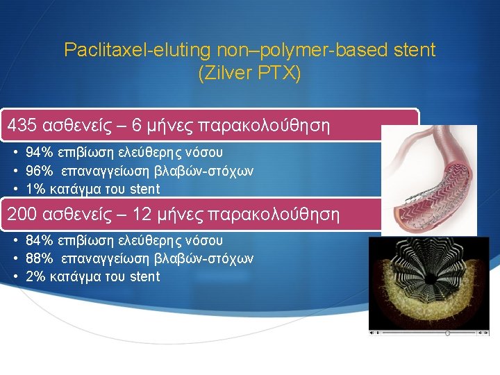
Τύποι drug-eluting stents με εφαρμογή στην αγγειοχειρουργική Sirolimus-eluting stents (SMART stents) Paclitaxel-eluting non– polymer-based stent (Zilver PTX) Self-expanding polymerbased everolimuseluting stent (Dynalink. E)
Rapamycin Analogs EVEROLIMUS O H H N O H 3 C O HO O H O O ABT-578 N NN N Chiral OH CH 3 O O H 3 C OH H 3 C SIROLIMUS CH 3 H 3 C O O CH 3 Chemical Formula C 53 H 83 NO 14 Molecular Wt: 958. 25 C 51 H 79 NO 13 Molecular Wt: 914. 2 C 52 H 79 NO 12 Molecular Wt: 966. 23 Intended Pharma Indications Chronic & Acute Rejection – Heart, Kidney, Lung Acute Rejection – Kidney, Liver None Approvals OUS US – H 2 04 (Est. ) OUS & US None
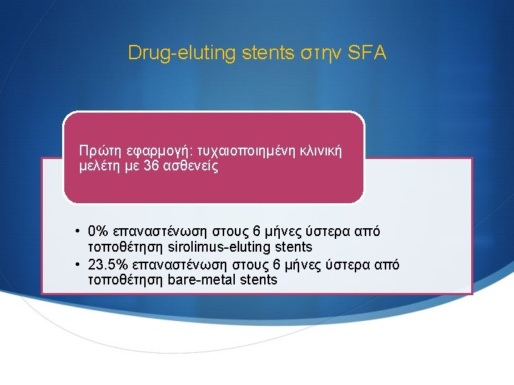
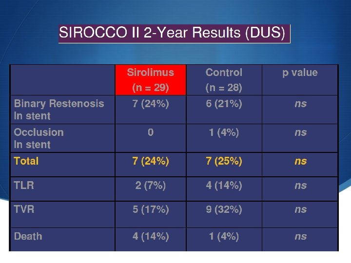
SMART stents στην SFA The only study which reported local drug delivery in the SFA was the Sirolimus-Coated Cordis Self-Expandable Stent (SIROCCO) trial, in which sirolimus-coated stents were not significantly superior to uncoated stents SIROCCO I & SIROCCO II trials μη στατιστικά σημαντική διαφορά μεταξύ των ασθενών που έφεραν sirolimus-eluting stents και αυτών που έφεραν baremetal stents στους 6 μήνες 0% Vs. 7. 7% επαναστένωση στην SFA δεν επιβεβαίωσαν την αποτελεσματικότητα του sirolimus Duda SH. Circulation 2002; 106: 1505– 1509. Duda SH. J Vasc Interv Radiol 2005; 16: 331– 338
SMART stents στην SFA Duda SH. J Vasc Interv Radiol 2005; 16: 331– 338
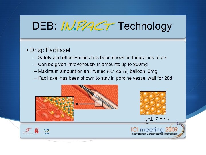
Zilver PTX (paclitaxel) Paclitaxel is a mitotic inhibitor used in cancer chemotherapy. It was discovered in a National Cancer Institute program at the Research Triangle Institute in 1967 when Monroe E. Wall and Mansukh C. Wani isolated it from the bark of the Pacific Yew tree, Taxus brevifolia and named it 'taxol' First, it allows targeted delivery of a drug (paclitaxel) proven to reduce the renarrowing (restenosis) of arteries opened using balloon angioplasty. Second, by eliminating the need for a polymer, Zilver PTX avoids the potential patient risks posed by leaving a permanent foreign, plastic substance in the body. Zilver PTX mechanisms of action: Hydrophobic—PTX won't wash off. It adheres to the stent without the need for a synthetic polymer Lipophilic—PTX seeks the lipids in the vessel wall and attaches Antiproliferative—once in the cell, PTX blocks cell division (proliferation) for the life of the cell
Self-expanding polymer-based everolimus-eluting stent (Dynalink-E) Σε εξέλιξη μελέτη από το Βέλγιο Αναμένονται τα αποτελέσματά της Bosiers M. Vasc Health Risk Manag. 2008; 4: 553– 559.
Drug eluting Ballons
Drug-eluting Ballons
Drug-coated balloons for femoropopliteal PTA: Paccocath (Cotavance) balloon) Scheller B et al. Circulation. 2004; 110: 810– 814. Scheller B et al. N Engl J Med. 2006; 355: 2113– 2124. Scheller B. Euro. Intervention. 2008; 4(suppl C): C 63–C 66. Scheller B et al. Heart. 2007; 93: 539– 541. standard angioplasty balloon catheter with a paclitaxel coating (a mixture of paclitaxel and contrast) 10% to 20% of the drug is taken up by the vessel wall short-term contact prolonged inhibition of neointimal proliferation
Local Taxane with Short Exposure for Reduction of Restenosis in Distal Arteries (THUNDER) trial S 154 patients (24% smokers, 49% diabetics) with femoropopliteal lesions S Paccocath (n=48 patients) S no adverse event S 6 months mean late lumen loss 0. 461. 2 mm vs. 1. 761. 8 mm for controls (p=0. 001) S 6 -month & 12 -month angiographic binary restenosis were 10% and 25% for the Paccocath patients vs. 41% and 59% for the control patients (p=0. 01) Currently, the use of antiproliferative agents, either exposed by stents or balloon catheters in preventing restenosis in infrainguinal arteries, is still investigational. Tepe G, et al. N Engl J Med. 2008; 358: 689– 99.
Ανεπιθύμητες ενέργειες § Vascular toxicity rather than cytotoxicity – – Late incomplete apposition Medial thinning Aneurysm/rupture Delayed re-endothelialization Vasculo-toxic effects in pig coronaries: 90 days High dose, fast release Low dose, slow release Rogers C et al. Circ. 2000.
Late incomplete apposition Potential for stent thrombosis Baseline Follow-up In a Taxus and Cypher study of patients with late incomplete apposition upon clopidogrel discontinuation: No remodeling Positive remodeling 20% had stent thrombosis*
Percent struts endothelialized Human analysis: DES vs BMS Percentage endothelialization 100 90 80 70 60 50 40 30 20 Taxus and Cypher BMS 10 0 1 30 2 3 4 5 6 7 8 9 11 15 16 17 20 > 40 Duration in months Conclusions: DES (solid line) consistently show less endothelialization compared with BMS (dashed line) regardless of time point, even beyond 40 months DES are not fully endothelialized, whereas BMS are completely covered by 6 to 7 months Joner, Virmani et al. Circulation. 2005; 112: 3210.
Exposed stent struts at 6 months > 80% Cypher struts exposed vs BMS struts Percent 100 Sirolimus-eluting stent 75 50 25 0 Incomplete coverage Percent 0 Grade 0 Complete coverage Grade 1 Grade 2 25 50 75 100 31 et al. JACC. 2006; 47: 2108 -2111. Kotani Bare-metal stent Grade 3
Endothelial dysfunction Reduction in e. NOS and nitric oxide (NO) production S Normal vessels dilate in response to exercise or acetylcholine (ACH) This response is dependent on endothelial production of NO S Atherosclerotic vessels are characterized by having endothelial dysfunction and constrict in response to exercise or ACH Cai H, Harrison DG. Circ Res. 2000; 8 This is explained by either a loss of endothelial cells or loss of e. NOS expression and NO production 7: 840 -844. 32 PO et al. ATVB. 2003; 23: 168 -175. Bonetti