
OSCE answers JCM 1 Feb 2017 POH Joseph

OSCE answers JCM 1 Feb 2017 POH Joseph Tse 1
Case 1 49/F Good past health. Not on medication C/O Epigastric pain for 2 hours BP 210/90 Sp. O 2 100% on room air Abdomen soft Name 3 life threatening ddx 2
Life threatening ddx – Acute Coronary syndrome – Acute Aortic syndrome – Pancreatitis – Diabetic Ketoacidosis – Mesenteric ischemia – Ruptured viscera eg PPU – Pregnancy complications: HELLP/ ectopic 3
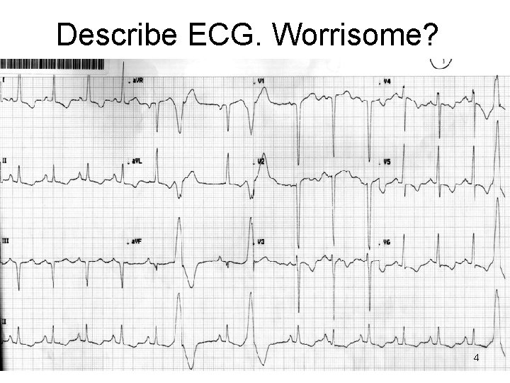
Describe ECG. Worrisome? 4
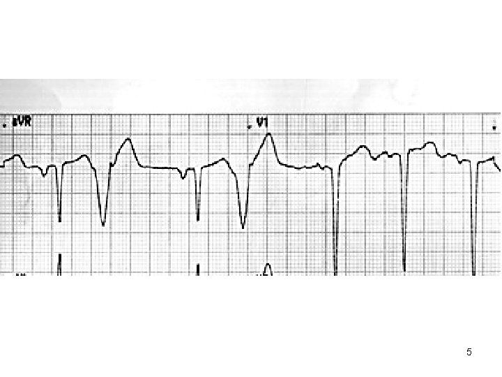
5
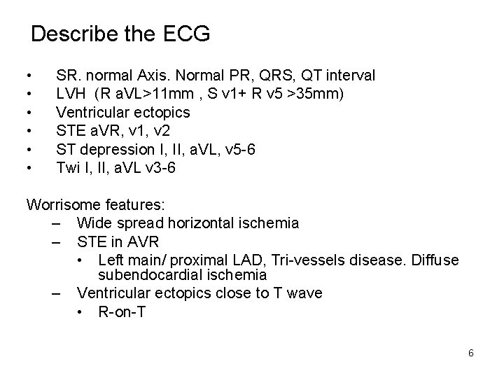
Describe the ECG • • • SR. normal Axis. Normal PR, QRS, QT interval LVH (R a. VL>11 mm , S v 1+ R v 5 >35 mm) Ventricular ectopics STE a. VR, v 1, v 2 ST depression I, II, a. VL, v 5 -6 Twi I, II, a. VL v 3 -6 Worrisome features: – Wide spread horizontal ischemia – STE in AVR • Left main/ proximal LAD, Tri-vessels disease. Diffuse subendocardial ischemia – Ventricular ectopics close to T wave • R-on-T 6
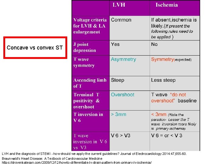
Concave vs convex ST LVH and the diagnosis of STEMI - how should we apply the current guidelines? Journal of Electrocardiology 2014: 47, 655 -60. Braunwald's Heart Disease: A Textbook of Cardiovascular Medicine https: //drsvenkatesan. com/2009/12/12/how-to-differentiate-lv-strain-pattern-from-primary-lv-ischemia/
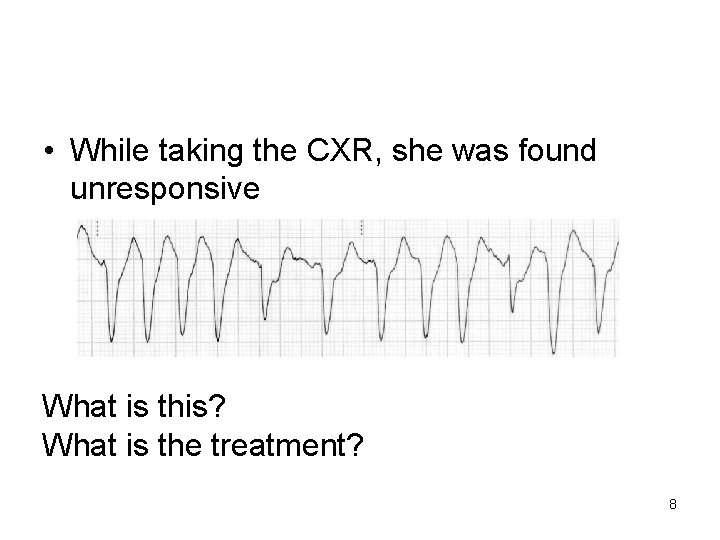
• While taking the CXR, she was found unresponsive What is this? What is the treatment? 8
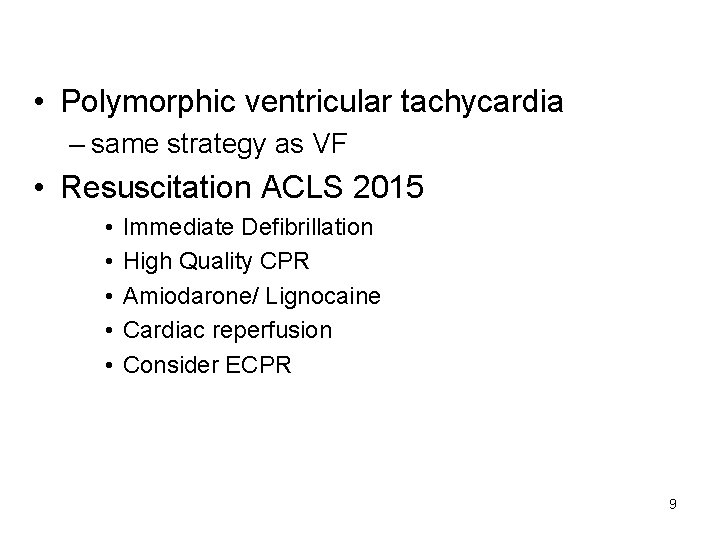
• Polymorphic ventricular tachycardia – same strategy as VF • Resuscitation ACLS 2015 • • • Immediate Defibrillation High Quality CPR Amiodarone/ Lignocaine Cardiac reperfusion Consider ECPR 9
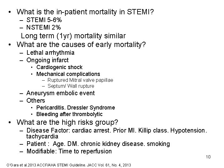
• What is the in-patient mortality in STEMI? – STEMI 5 -6% – NSTEMI 2% Long term (1 yr) mortality similar • What are the causes of early mortality? – Lethal arrhythmia – Ongoing infarct • Cardiogenic shock • Mechanical complications – Ruptured Mitral valve papillae – Septum/ Wall rupture – Aneurysm embolic event – Others • Pericarditis. Dressler Syndrome • Bleeding after thrombolytic • What are the high risks group? – Disease Factor: cardiac arrest. Prior MI. Killip class. Hypotension. tachycardia – Patient : Age. DM. chronic kidney disease. smoking – Modifiable: Time to reperfusion 10 O’Gara et al. 2013 ACCF/AHA STEMI Guideline. JACC Vol. 61, No. 4, 2013
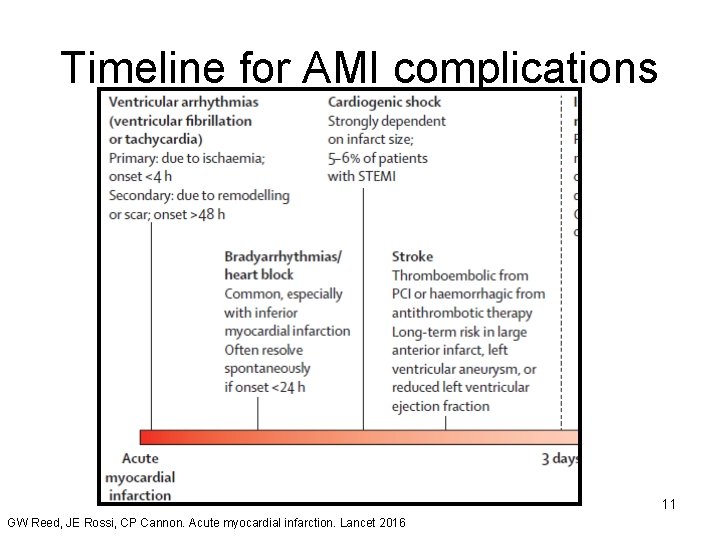
Timeline for AMI complications 11 GW Reed, JE Rossi, CP Cannon. Acute myocardial infarction. Lancet 2016
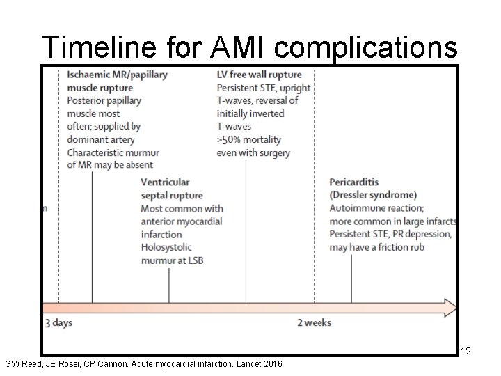
Timeline for AMI complications 12 GW Reed, JE Rossi, CP Cannon. Acute myocardial infarction. Lancet 2016
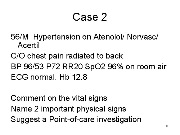
Case 2 56/M Hypertension on Atenolol/ Norvasc/ Acertil C/O chest pain radiated to back BP 96/53 P 72 RR 20 Sp. O 2 96% on room air ECG normal. Hb 12. 8 Comment on the vital signs Name 2 important physical signs Suggest a Point-of-care investigation 13
Comment on the vital signs – In Shock – Pulse rate not raised (B-blocker effect) – Tachypnea Name 2 important physical signs – Vascular insufficiency • Murmur (AR early diastole) • Pulse deficit (radio-radial, radiofemoral delay) – Tamponade • Beck’s Triad: distended neck veins. muffled heart sound • Pulsus paradoxus Suggest a Point-of-care investigation Ultrasound- cardiac probe – Parasternal long axis (Aortic root, effusion) – Suprasternal notch (Aortic arch) 14
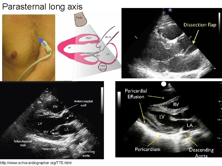
Parasternal long axis http: //www. echocardiographer. org/TTE. html
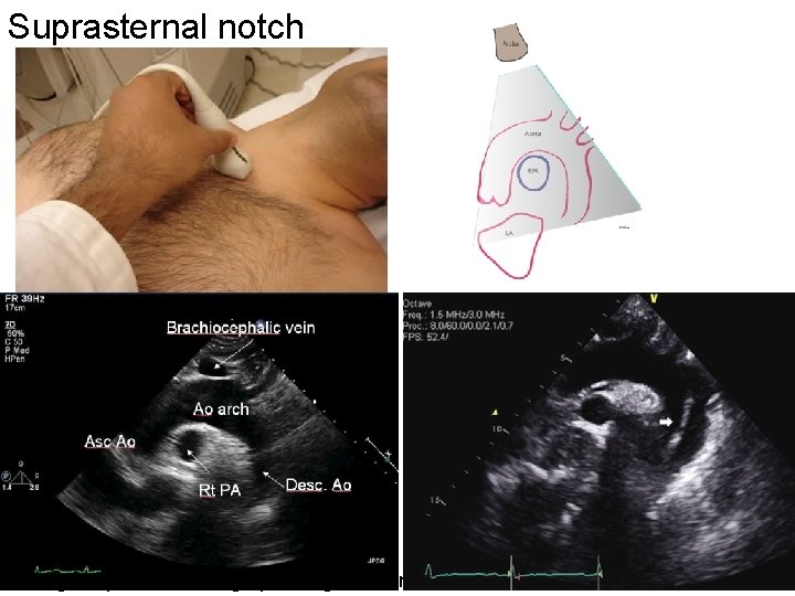
Suprasternal notch Image http: //echocardiographer. org/TTE. html
17
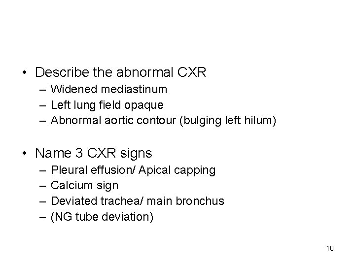
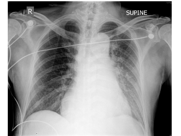
• Describe the abnormal CXR – Widened mediastinum – Left lung field opaque – Abnormal aortic contour (bulging left hilum) • Name 3 CXR signs – – Pleural effusion/ Apical capping Calcium sign Deviated trachea/ main bronchus (NG tube deviation) 18
19
20
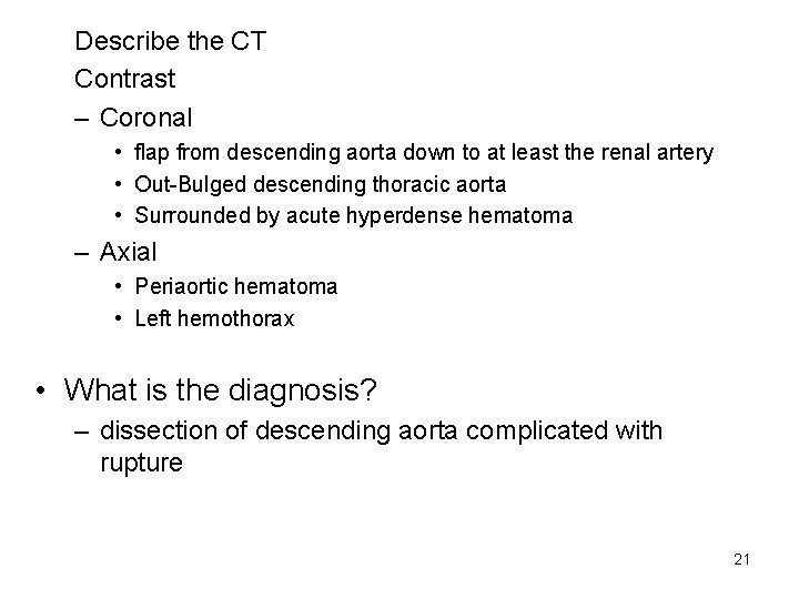
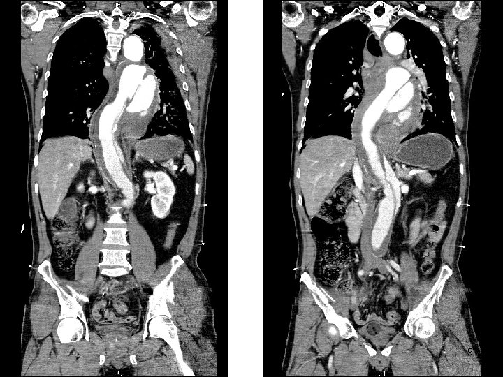
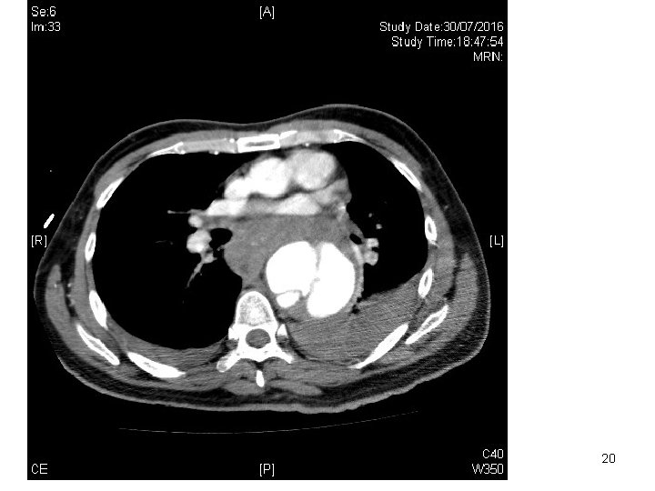
Describe the CT Contrast – Coronal • flap from descending aorta down to at least the renal artery • Out-Bulged descending thoracic aorta • Surrounded by acute hyperdense hematoma – Axial • Periaortic hematoma • Left hemothorax • What is the diagnosis? – dissection of descending aorta complicated with rupture 21
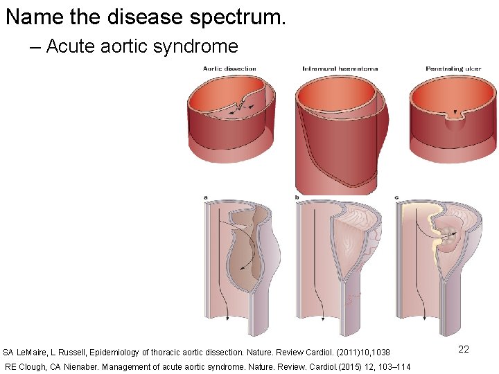
Name the disease spectrum. – Acute aortic syndrome SA Le. Maire, L Russell, Epidemiology of thoracic aortic dissection. Nature. Review Cardiol. (2011)10, 1038 RE Clough, CA Nienaber. Management of acute aortic syndrome. Nature. Review. Cardiol. (2015) 12, 103– 114 22
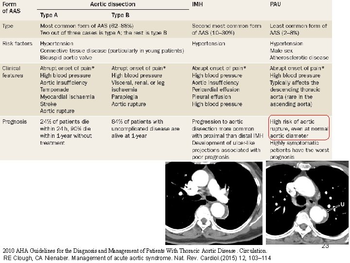
2010 AHA Guidelines for the Diagnosis and Management of Patients With Thoracic Aortic Disease. Circulation. RE Clough, CA Nienaber. Management of acute aortic syndrome. Nat. Rev. Cardiol. (2015) 12, 103– 114 23
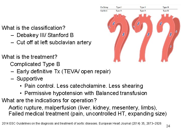
What is the classification? – Debakey III/ Stanford B – Cut off at left subclavian artery What is the treatment? Complicated Type B – Early definitive Tx (TEVA/ open repair) – Supportive • Pain control. Less catecholamine. Less shearing • Permissive hypotension with Balanced transfusion What are the indications for operation? Aortic rupture, malperfusion (liver, kidney, mesentery, limbs), Failed medical treatment (pain, uncontrolled HT, expanding size) 2014 ESC Guidelines on the diagnosis and treatment of aortic diseases. European Heart Journal (2014) 35, 2873– 2926 24
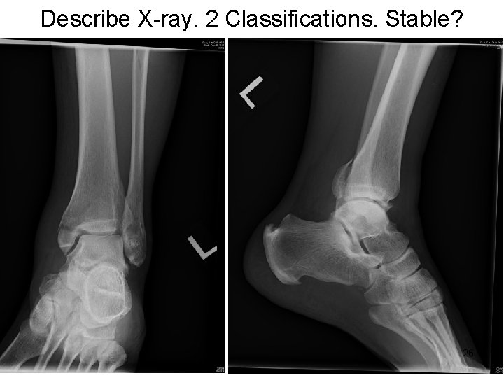
Case 3 • 46/M fell while climbing down from lorry driver seat • C/O left ankle pain • Difficult to walk 25
Describe X-ray. 2 Classifications. Stable? 26
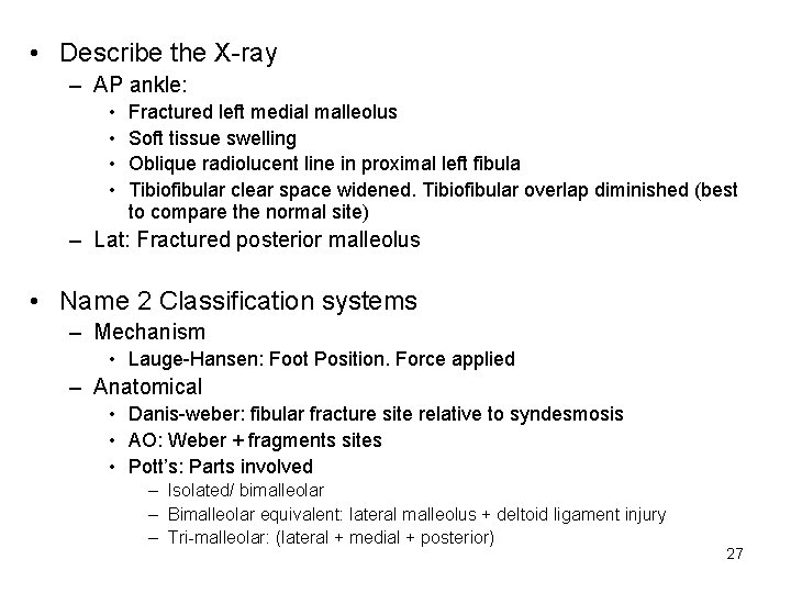
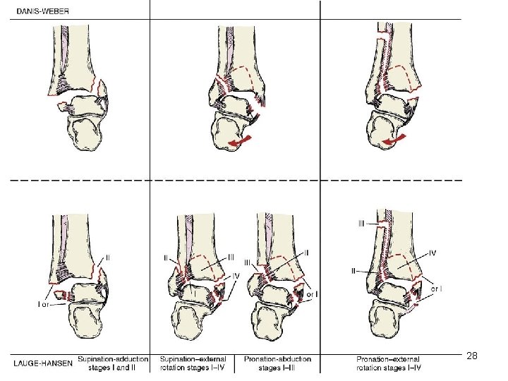
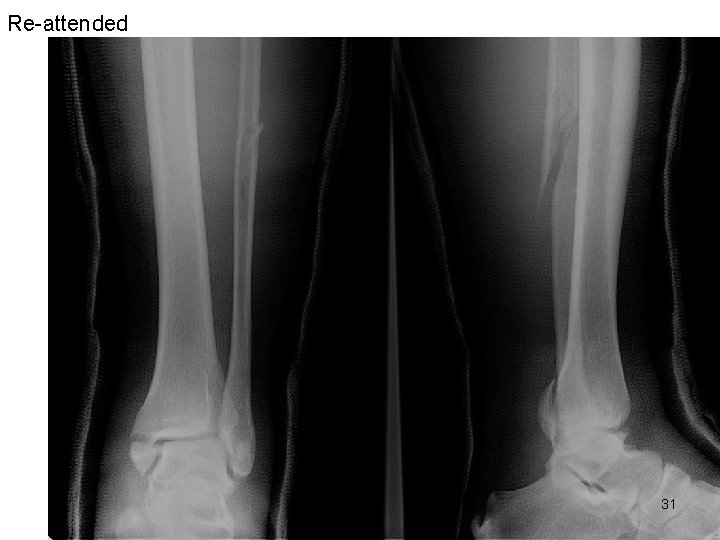
• Describe the X-ray – AP ankle: • • Fractured left medial malleolus Soft tissue swelling Oblique radiolucent line in proximal left fibula Tibiofibular clear space widened. Tibiofibular overlap diminished (best to compare the normal site) – Lat: Fractured posterior malleolus • Name 2 Classification systems – Mechanism • Lauge-Hansen: Foot Position. Force applied – Anatomical • Danis-weber: fibular fracture site relative to syndesmosis • AO: Weber + fragments sites • Pott’s: Parts involved – Isolated/ bimalleolar – Bimalleolar equivalent: lateral malleolus + deltoid ligament injury – Tri-malleolar: (lateral + medial + posterior) 27
28
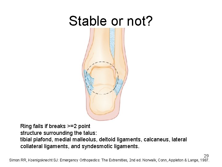
Stable or not? Ring fails if breaks >=2 point structure surrounding the talus: tibial plafond, medial malleolus, deltoid ligaments, calcaneus, lateral collateral ligaments, and syndesmotic ligaments. 29 Simon RR, Koenigsknecht SJ: Emergency Orthopedics: The Extremities, 2 nd ed. Norwalk, Conn, Appleton & Lange, 1987.
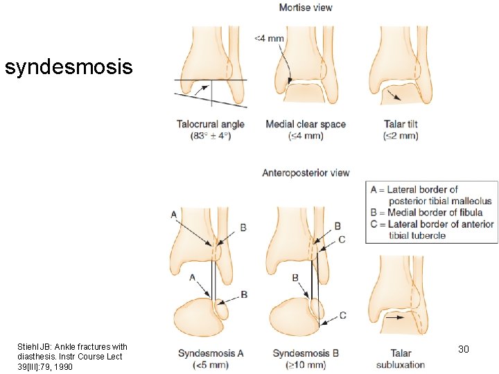
syndesmosis Stiehl JB: Ankle fractures with diasthesis. Instr Course Lect 39[III]: 79, 1990 30
Re-attended 31
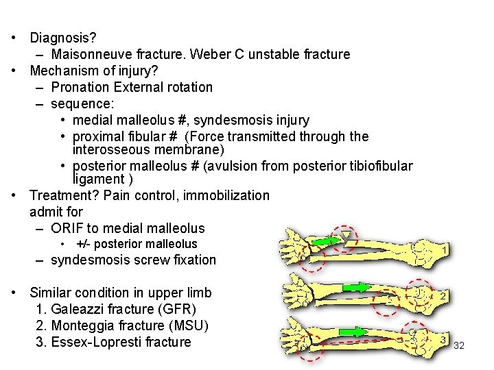
• Diagnosis? – Maisonneuve fracture. Weber C unstable fracture • Mechanism of injury? – Pronation External rotation – sequence: • medial malleolus #, syndesmosis injury • proximal fibular # (Force transmitted through the interosseous membrane) • posterior malleolus # (avulsion from posterior tibiofibular ligament ) • Treatment? Pain control, immobilization admit for – ORIF to medial malleolus • +/- posterior malleolus – syndesmosis screw fixation • Similar condition in upper limb 1. Galeazzi fracture (GFR) 2. Monteggia fracture (MSU) 3. Essex-Lopresti fracture 32
Case 4 M/51 Good past health. No recent travel Chills for 1 week Hiccup and abdominal bloating discomfort 2 days Temp 37 What is hiccup? 33
• What is hiccup? – Involuntary, spasmotic contraction of diaphragm and intercostal muscles – Results in sudden inspiration and ends in abruption closure of glottis • What is the reflex arc? – Afferent: phrenic, vagus nerve, sympathetic chain – Central mediator: cord c 3 -5, brainstem, hypothalamus – Efferent: phrenic nerve to glottis and intercostal muscle • Name 2 drugs for symptomatic relief of hiccup – Chlorpromazine (Largactil ) – Metoclopramide (Maxolon) – Baclofen/ nifedipine/ valporate/ gabapentin/ haloperidol 34 Tintinalli’s Emergency Medicine, 2016. Chapter 62 Respiratory distress p. 433
2 alarming features. 2 causes 35
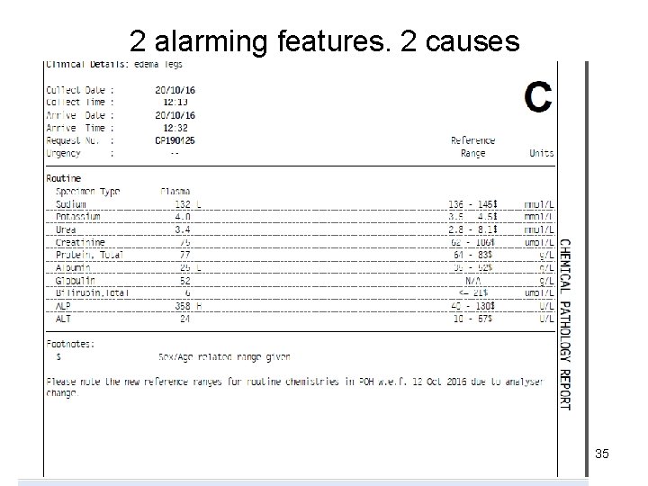
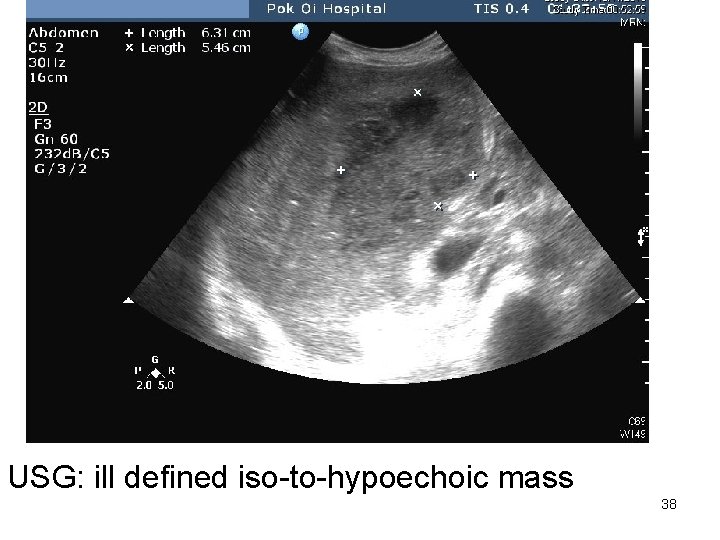
• What are the 2 alarming features? – Hypoalbuminemia – Raised ALP • What are the causes? Give 2 in each feature – Reversed AG ratio (normal 1. 2 -1. 7) • Low albumin – Production (malnutrition/ liver cirrhosis) – Loss (renal: nephrotic. GI: enteropathy) • Raised globulin: infection, paraproteinemia – ALP • Bone vs Liver origin » GGT » isoenzymes. (Bone Burns, Liver Lasts) – Liver: cholestasis/ infiltrative SOL » Intrahepatic: drugs, abscess » Extrahepatic: cholangio. CA/ pancreatic cancer 36 – Bone: Paget’s disease. Myeloma. Bone metastasis.
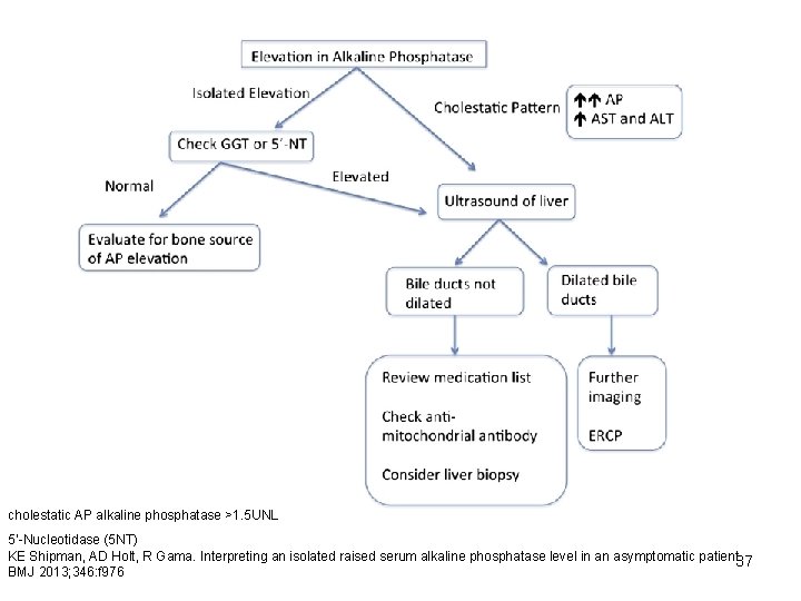
cholestatic AP alkaline phosphatase >1. 5 UNL 5’-Nucleotidase (5 NT) KE Shipman, AD Holt, R Gama. Interpreting an isolated raised serum alkaline phosphatase level in an asymptomatic patient. 37 BMJ 2013; 346: f 976
USG: ill defined iso-to-hypoechoic mass 38
39
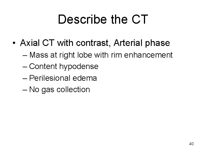
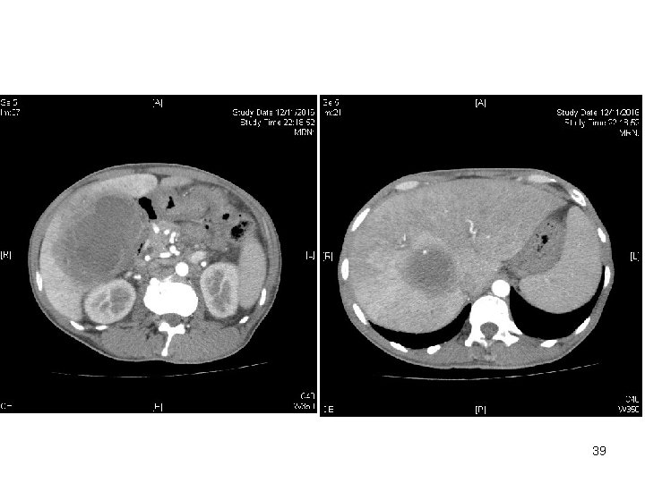
Describe the CT • Axial CT with contrast, Arterial phase – Mass at right lobe with rim enhancement – Content hypodense – Perilesional edema – No gas collection 40
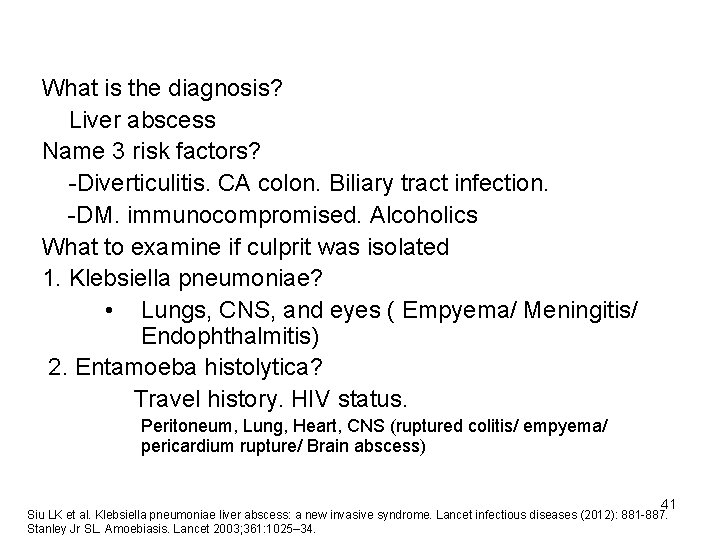
What is the diagnosis? Liver abscess Name 3 risk factors? -Diverticulitis. CA colon. Biliary tract infection. -DM. immunocompromised. Alcoholics What to examine if culprit was isolated 1. Klebsiella pneumoniae? • Lungs, CNS, and eyes ( Empyema/ Meningitis/ Endophthalmitis) 2. Entamoeba histolytica? Travel history. HIV status. Peritoneum, Lung, Heart, CNS (ruptured colitis/ empyema/ pericardium rupture/ Brain abscess) 41 Siu LK et al. Klebsiella pneumoniae liver abscess: a new invasive syndrome. Lancet infectious diseases (2012): 881 -887. Stanley Jr SL. Amoebiasis. Lancet 2003; 361: 1025– 34.
Case 5 • 65/F ADL-I • Known HT, poor memory await psy • Noted disoriented to time, place, person for 2 days Afebrile. BP 120/80 P 72 GCS 15. 4 limbs Power full Name 2 point-of-care investigations 42
Give 2 point-of-care investigations – Hemoglucostix – I-stat for sodium What is delirium? How to differentiate from dementia? Name 4 components of mental state exam Name 2 assessment tools Suggest 3 red-flag signs in a confused patient Name 3 types of dementia 43
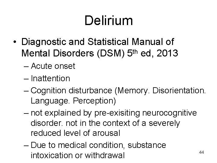
Delirium • Diagnostic and Statistical Manual of Mental Disorders (DSM) 5 th ed, 2013 – Acute onset – Inattention – Cognition disturbance (Memory. Disorientation. Language. Perception) – not explained by pre-exisiting neurocognitive disorder. not in the context of a severely reduced level of arousal – Due to medical condition, substance 44 intoxication or withdrawal
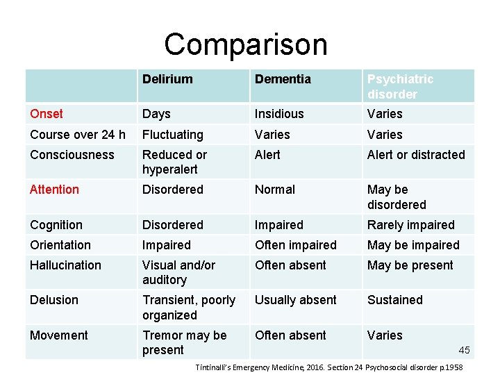
Comparison Delirium Dementia Psychiatric disorder Onset Days Insidious Varies Course over 24 h Fluctuating Varies Consciousness Reduced or hyperalert Alert or distracted Attention Disordered Normal May be disordered Cognition Disordered Impaired Rarely impaired Orientation Impaired Often impaired May be impaired Hallucination Visual and/or auditory Often absent May be present Delusion Transient, poorly organized Usually absent Sustained Movement Tremor may be present Often absent Varies 45 Tintinalli’s Emergency Medicine, 2016. Section 24 Psychosocial disorder p. 1958

Mental state exam • Objective – Appearance, behavior, speech, cognition • Subjective – Mood, thoughts, perception and insight 46
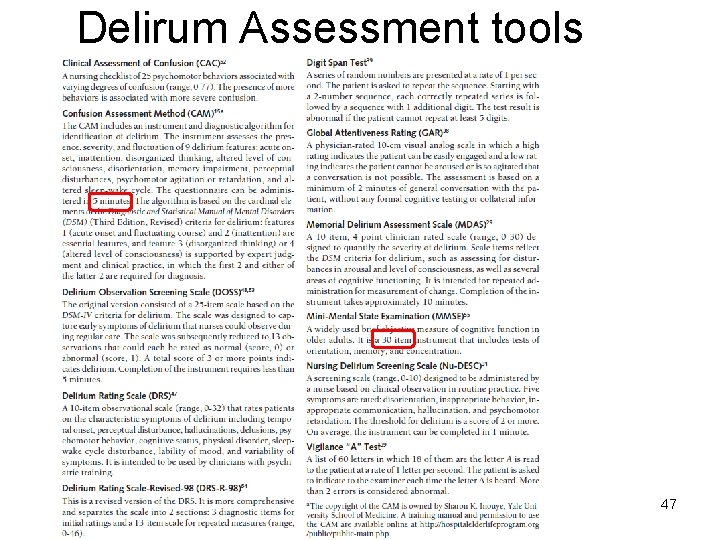
Delirum Assessment tools 47
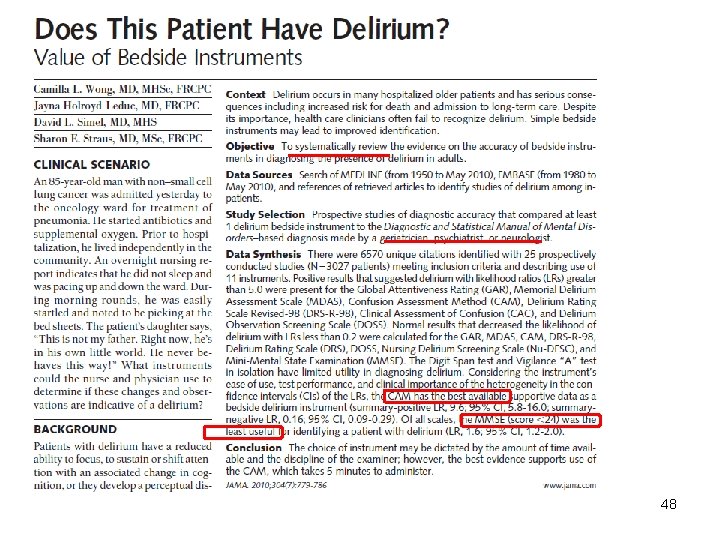
48
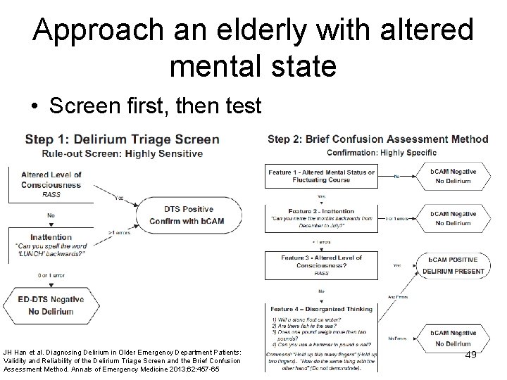
Approach an elderly with altered mental state • Screen first, then test JH Han et al. Diagnosing Delirium in Older Emergency Department Patients: Validity and Reliability of the Delirium Triage Screen and the Brief Confusion Assessment Method. Annals of Emergency Medicine 2013; 62: 457 -65 49
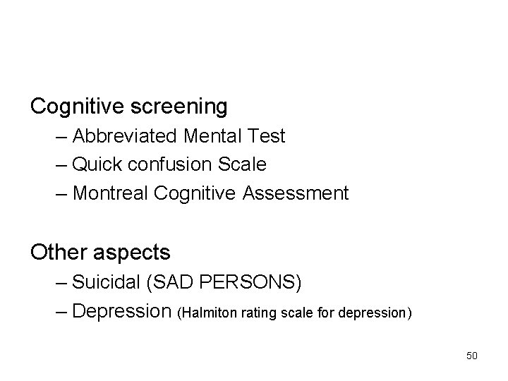
Cognitive screening – Abbreviated Mental Test – Quick confusion Scale – Montreal Cognitive Assessment Other aspects – Suicidal (SAD PERSONS) – Depression (Halmiton rating scale for depression) 50
• Red flags in confused patient – Injury. Morning headache. Vomiting. Personality change. Visual/ tactile hallucination. Delirium. Fever. Seizure • Name 3 common types of dementia? – Alzheimer’s disease – Vascular dementia – Lewy body Dementia • Parkinson’s disease dementia – Frontotemporal dementia • Name the type of dementia which had cognitive, psychomotor disturbance and hallucination – Lewy body dementia Dementia—not all about Alzheimer’s. Lancet 2015; 386: 1600 51
Reversible causes of DEMENTIA • Drugs – anticholinergics (Diphenhydramine, chlorpheniramine, amitriptyline) – Opioids – Sedatives (Benzodiazepines) • • Endocrine hypothyroidism hyperparathyroidism Metabolic uremia Emotion (depression) Nutritional (B 12 deficiency), Normal pressure hydrocephalus • Toxin heavy metals eg Lead • Infection Neurosyphilis HIV. Lyme disease • Alcohol
CT Brain 53
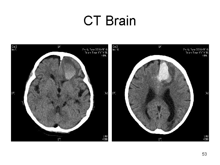
Describe the CT • Hyperdense lesion at left frontal lobe • Mass effect – Midline shift – Ventricle compression • Peri-lesional edema Radiological diagnosis: • Acute left frontal lobar hemorrhage with pressure effect Pathology: amyloid angiopathy 54
Give 3 common causes of non-traumatic Intracerebral hemorrhage • Primary: small vessels spontaneous rupture – Hypertension. Amyloid angiopathy • Secondary – AV malformation. Aneurysm. Tumor. Coagulopathy Give 3 common locations of bleeding in hemorrhagic stroke -Basal ganglia. Cerebellum. Pons AI Quershi et al. Spontaneous intracerebral hemorrhage. N Engl J Med, Vol. 344, 2001 55
- Slides: 55