Acute Kidney Injury Acute kidney injury AKI formerly


- Slides: 49

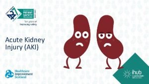
Acute Kidney Injury

• Acute kidney injury (AKI), formerly called acute renal failure, is a clinical syndrome in which a sudden deterioration in renal function results in the inability of the kidneys to maintain fluid and electrolyte homeostasis. • AKI occurs in 2 -3% of children admitted to pediatric tertiary care centers and in as many as 8% of infants in neonatal intensive care units.

• A classification system has been proposed to standardize the definition of AKI in adults. • These criteria of risk, injury, failure, loss, and end-stage renal disease were given the acronym of RIFLE. • A modified RIFLE criteria (p. RIFLE) was developed to characterize the pattern of AKI in critically ill children.

• Because RIFLE focuses on the glomerular filtration rate (GFR), a modification (Acute Kidney Injury Network) categorizes severity by rise in serum creatinine: stage 1 >150%, stage II >200%, stage III >300%.


PATHOGENESIS • AKI has been conventionally classified into 3 categories: prerenal, intrinsic renal, and postrenal.

Prerenal AKI • Prerenal AKI, also called prerenal azotemia, is characterized by diminished effective circulating arterial volume, which leads to inadequate renal perfusion and a decreased GFR. • Evidence of kidney damage is absent. • Common causes of prerenal AKI include dehydration, sepsis, hemorrhage, severe hypoalbuminemia, and cardiac failure. • If the underlying cause of the renal hypoperfusion is reversed promptly, renal function returns to normal. • If hypoperfusion is sustained, intrinsic renal parenchymal damage can develop.

Intrinsic renal AKI • Intrinsic renal AKI includes a variety of disorders characterized by renal parenchymal damage, including sustained hypoperfusion and ischemia. • Many forms of glomerulonephritis, including postinfectious glomerulonephritis, lupus nephritis, Henoch. Scnlein purpura nephritis, membranoproliferative glomerulonephritis, and anti– glomerular basement membrane nephritis, can cause AKI. • Ischemic/ hypoxic injury and nephrotoxic insults are the most common causes of intrinsic AKI in the United States, and are more common with an underlying comorbid condition; most are associated with cardiac, oncologic, urologic, renal, and genetic disorders or prematurity.

• Severe and prolonged ischemic/hypoxic injury and nephrotoxic insult lead to acute tubular necrosis (ATN), seen most often in critically ill infants and children. • Mechanisms leading to ischemic AKI include hypotension/intravascular volume depletion (hemorrhage, thirdspace fluid losses, diarrhea), decreased effective intravascular volume (heart failure, cirrhosis, hepatorenal syndrome, peritonitis, abdominal compartment syndrome), vasodilation/vasoconstriction (sepsis, hepatorenal syndrome), renal artery obstruction (thrombosis, embolization, stenosis), intrarenal artery disease (vasculitis, hemolytic-uremic syndrome, sickle cell anemia, transplant rejection), and impaired renal blood flow (cyclosporine, tacrolimus, angiotensinconverting enzyme [ACE] inhibitors, angiotensin-receptor blocking agents, radiocontrast agents).

Postrenal AKI • Postrenal AKI includes a variety of disorders characterized by obstruction of the urinary tract. • In neonates and infants, congenital conditions, such as posterior urethral valves and bilateral ureteropelvic junction obstruction, account for the majority of cases of AKI. • Other conditions, such as urolithiasis, tumor (intraabdominal lesion or within the urinary tract), hemorrhagic cystitis, and neurogenic bladder, can cause AKI in older children and adolescents. • In a patient with 2 functioning kidneys, obstruction must be bilateral to result in AKI. • Relief of the obstruction usually results in recovery of renal function, except in patients with associated renal dysplasia or prolonged urinary tract obstruction.


CLINICAL MANIFESTATIONS AND DIAGNOSIS • A carefully taken history is critical in defining the cause of AKI. • The physical examination must be thorough, with careful attention to volume status.

LABORATORY FINDINGS • Laboratory abnormalities can include anemia (the anemia is usually dilutional or hemolytic, as in SLE, renal vein thrombosis, HUS); leukopenia (SLE, sepsis); thrombocytopenia (SLE, renal vein thrombosis, sepsis, HUS); hyponatremia (dilutional); metabolic acidosis; elevated serum concentrations of blood urea nitrogen, creatinine, uric acid, potassium, and phosphate (diminished renal function); and hypocalcemia (hyperphosphatemia).

• The serum C 3 level may be depressed (postinfectious glomerulonephritis, SLE, or membranoproliferative glomerulonephritis), and antibodies may be detected in the serum to streptococcal (poststreptococcal glomerulonephritis), nuclear (SLE), neutrophil cytoplasmic (granulomatosis with polyangiitis, microscopic polyarteritis), or glomerular basement membrane (Goodpasture disease) antigens.

• The presence of hematuria, proteinuria, and red blood cell or granular urinary casts suggests intrinsic AKI, in particular glomerular disease and ATN. • The presence of white blood cells and white blood cell casts with low-grade hematuria and proteinuria suggests tubulointerstitial disease. • Urinary eosinophils may be present in children with drug-induced tubulointerstitial nephritis.

• Urinary indices may be useful in differentiating prerenal AKI from intrinsic AKI. • Patients whose urine shows an elevated specific gravity (>1. 020), elevated urine osmolality (UOsm > 500 m. Osm/kg), low urine sodium (UNa < 20 m. Eq/L), and fractional excretion of sodium <1% (<2. 5% in neonates) most likely have prerenal AKI. • Those with a specific gravity of <1. 010, low urine osmolality (UOsm < 350 m. Osm/kg), high urine sodium (UNa > 40 m. Eq/L), and fractional excretion of sodium >2% (>10% in neonates) most likely have intrinsic AKI.


• Chest radiography may reveal cardiomegaly, pulmonary congestion (fluid overload), or pleural effusions. • Renal ultrasonography can reveal hydronephrosis and/or hydroureter, which suggest urinary tract obstruction, or nephromegaly, consistent with intrinsic renal disease.

• Renal biopsy can ultimately be required to determine the precise cause of AKI in patients who do not have clearly defined prerenal or postrenal AKI.

• Although serum creatinine is used to measure kidney function, it is an insensitive and delayed measure of decreased kidney function following AKI. • Other biomarkers under investigation include changes in plasma neutrophil gelatinase– associated lipocalin and cystatin C levels and urinary changes in neutrophil gelatinaseassociated lipocalin, interleukin 18, and kidney injury molecule-1.

TREATMENT • Medical Management • Dialysis

Medical Management • In infants and children with urinary tract obstruction, such as in a newborn with suspected posterior ureteral valves, a bladder catheter should be placed immediately to ensure adequate drainage of the urinary tract. • The placement of a bladder catheter may also be considered in nonambulatory older children and adolescents to accurately monitor urine output during AKI; however, precautions to prevent iatrogenic infection should be taken.

• Determination of the volume status is of critical importance when initially evaluating a patient with AKI. • If there is no evidence of volume overload or cardiac failure, intravascular volume should be expanded by intravenous administration of isotonic saline, 20 m. L/kg over 30 min. • In the absence of blood loss or hypoproteinemia, colloid containing solutions are not required for volume expansion. • Severe hypovolemia may require additional fluid boluses. • Determination of the central venous pressure may be helpful if adequacy of the blood volume is difficult to determine. • After volume resuscitation, hypovolemic patients generally void within 2 hr; failure to do so suggests intrinsic or postrenal AKI. • Hypotension caused by sepsis requires vigorous fluid resuscitation followed by a continuous infusion of norepinephrine.

• Diuretic therapy should be considered only after the adequacy of the circulating blood volume has been established. • Furosemide (2 -4 mg/kg) and mannitol (0. 5 g/kg) may be administered as a single IV dose. • Bumetanide (0. 1 mg/kg) may be given as an alternative to furosemide. • If urine output is not improved, then a continuous diuretic infusion may be considered. • To increase renal cortical blood flow, many clinicians administer dopamine (2 -3 μg/kg/min) in conjunction with diuretic therapy, although no controlled data support this practice. • There is little evidence that diuretics or dopamine can prevent AKI or hasten recovery. • Mannitol may be effective in prevention of pigment (myoglobin, hemoglobin)-induced renal failure. • Atrial natriuretic peptide may be of value in preventing or treating AKI, although there is little pediatric evidence to support its use.

• If there is no response to a diuretic challenge, diuretics should be discontinued and fluid restriction is essential. • Patients with a relatively normal intravascular volume should initially be limited to 400 m. L/ m 2/24 hr (insensible losses) plus an amount of fluid equal to the urine output for that day. • Extrarenal (blood, gastrointestinal tract) fluid losses should be replaced, milliliter for milliliter, with appropriate fluids. • Markedly hypervolemic patients can require further fluid restriction, omitting the replacement of insensible fluid losses, urine output, and extrarenal losses to diminish the expanded intravascular volume. • Fluid intake, urine and stool output, body weight, and serum chemistries should be monitored on a daily basis.

• In AKI, rapid development of hyperkalemia (serum potassium level > 6 m. Eq/L) can lead to cardiac arrhythmia, cardiac arrest, and death. • The earliest electrocardiographic change seen in patients with developing hyperkalemia is the appearance of peaked T waves. • This may be followed by widening of the QRS intervals, ST segment depression, ventricular arrhythmias, and cardiac arrest.

• Procedures to deplete body potassium stores should be initiated when the serum potassium value rises to >6. 0 m. Eq/L. • Exogenous sources of potassium (dietary, intravenous fluids, total parenteral nutrition) should be eliminated. • Sodium polystyrene sulfonate resin (Kayexalate), 1 g/kg, should be given orally or by retention enema. • This resin exchanges sodium for potassium and can take several hr to take effect. • A single dose of 1 g/kg can be expected to lower the serum potassium level by about 1 m. Eq/L. • Resin therapy may be repeated every 2 hr, the frequency being limited primarily by the risk of sodium overload.

• More severe elevations in serum potassium (>7 m. Eq/L), especially if accompanied by electrocardiographic changes, require emergency measures in addition to Kayexalate. • The following agents should be administered: • • Calcium gluconate 10% solution, 1. 0 m. L/kg IV, over 3 -5 min • • Sodium bicarbonate, 1 -2 m. Eq/kg IV, over 5 -10 min • • Regular insulin, 0. 1 units/kg, with glucose 50% solution, 1 m. L/kg, over 1 hr

• Calcium gluconate counteracts the potassium-induced increase in myocardial irritability but does not lower the serum potassium level. • Administration of sodium bicarbonate, insulin, or glucose lowers the serum potassium level by shifting potassium from the extracellular to the intracellular compartment. • A similar effect has been reported with the acute administration of β-adrenergic agonists in adults, but there are no controlled data in pediatric patients. • Because the duration of action of these emergency measures is just a few hours, persistent hyperkalemia should be managed by dialysis.

• Mild metabolic acidosis is common in AKI because of retention of hydrogen ions, phosphate, and sulfate, but it rarely requires treatment. • If acidosis is severe (arterial p. H < 7. 15; serum bicarbonate < 8 m. Eq/L) or contributes to significant hyperkalemia, treatment is indicated. • The acidosis should be corrected partially by the intravenous route, generally giving enough bicarbonate to raise the arterial p. H to 7. 20 (which approximates a serum bicarbonate level of 12 m. Eq/L).

• The remainder of the correction may be accomplished by oral administration of sodium bicarbonate after normalization of the serum calcium and phosphorus levels. • Correction of metabolic acidosis with intravenous bicarbonate can precipitate tetany in patients with renal failure as rapid correction of acidosis reduces the ionized calcium concentration.

• Hypocalcemia is primarily treated by lowering the serum phosphorus level. • Calcium should not be given intravenously, except in cases of tetany, to avoid deposition of calcium salts into tissues. • Patients should be instructed to follow a low-phosphorus diet, and phosphate binders should be orally administered to bind any ingested phosphate and increase GI phosphate excretion. • Common agents include sevelamer (Renagel), calcium carbonate (Tums tablets or Titralac suspension), and calcium acetate (Phos. Lo). • Aluminum-based binders, commonly employed in the past, should be avoided because of the risk of aluminum toxicity.

• Hyponatremia is most commonly a dilutional disturbance that must be corrected by fluid restriction rather than sodium chloride administration. • Administration of hypertonic (3%) saline should be limited to patients with symptomatic hyponatremia (seizures, lethargy) or those with a serum sodium level <120 m. Eq/L. • Acute correction of the serum sodium to 125 m. Eq/L (mmol/L) should be accomplished using the following formula: • m. Eq sodium required = 0. 6 × weight in kg ×(125− serum sodium in m. Eq/L)

• AKI patients are predisposed to GI bleeding because of uremic platelet dysfunction, increased stress, and heparin exposure if treated with hemodialysis or continuous renal replacement therapy. • Oral or intravenous H 2 blockers such as ranitidine are commonly administered to prevent this complication.

• Hypertension can result from hyperreninemia associated with the primary disease process and/or expansion of the extracellular fluid volume and is most common in AKI patients with acute glomerulonephritis or HUS. • Salt and water restriction is critical, and diuretic administration may be useful. • Isradipine (0. 05 -0. 15 mg/kg/dose, maximum dose 5 mg qid) may be administered for relatively rapid reduction in blood pressure.

• Longer-acting agents such as calcium channel blockers (amlodipine, 0. 1 -0. 6 mg/ kg/24 hr qd or divided bid) or β blockers (propranolol, 0. 5 -8. 0 mg/ kg/24 hr divided bid or tid; labetalol, 4 -40 mg/kg/24 hr divided bid or tid) may be helpful in maintaining control of blood pressure. • Children with severe symptomatic hypertension (hypertensive urgency or emergency) should be treated with continuous infusions of nicardipine (0. 55. 0 μg/kg/min), sodium nitroprusside (0. 5 -10. 0 μg/kg/min), labetalol (0. 25 -3. 0 mg/kg/hr), or esmolol (150 -300 μg/kg/min) and converted to intermittently dosed antihypertensives when more stable.

• Neurologic symptoms in AKI can include headache, seizures, lethargy, and confusion (encephalopathy). • Potential etiologic factors include hypertensive encephalopathy, hyponatremia, hypocalcemia, cerebral hemorrhage, cerebral vasculitis, and the uremic state. • Benzodiazepams are the most effective agents in acutely controlling seizures, and subsequent therapy should be directed toward the precipitating cause.

• The anemia of AKI is generally mild (hemoglobin 9 -10 g/d. L) and primarily results from volume expansion (hemodilution). • Children with HUS, SLE, active bleeding, or prolonged AKI can require transfusion of packed red blood cells if their hemoglobin level falls below 7 g/d. L. • In hypervolemic patients, blood transfusion carries the risk of further volume expansion, which can precipitate hypertension, heart failure, and pulmonary edema. • Slow (4 -6 hr) transfusion with packed red blood cells (10 m. L/kg) diminishes the risk of hypervolemia.

• The use of fresh, washed red blood cells minimizes the acute risk of hyperkalemia, and the chronic risk of sensitization if the patient becomes a future candidate for renal replacement therapy. • In the presence of severe hypervolemia or hyperkalemia, blood transfusions are most safely administered during dialysis or ultrafiltration.

• Nutrition is of critical importance in children who develop AKI. • In most cases, sodium, potassium, and phosphorus should be restricted. • Protein intake should be moderately restricted while maximizing caloric intake to minimize the accumulation of nitrogenous wastes. • In critically ill patients with AKI, parenteral hyperalimentation with essential amino acids should be considered.

• Indications for dialysis in AKI include the following: • • Anuria/oliguria • • Volume overload with evidence of hypertension and/or pulmonary edema refractory to diuretic therapy • • Persistent hyperkalemia • • Severe metabolic acidosis unresponsive to medical management • • Uremia (encephalopathy, pericarditis, neuropathy) • • Blood urea nitrogen >100 -150 mg/d. L (or lower if rapidly rising) • • Calcium: phosphorus imbalance, with hypocalcemic tetany that cannot be controlled by other measures

• An additional indication for dialysis is the inability to provide adequate nutritional intake because of the need for severe fluid restriction. • In patients with AKI, dialysis support may be necessary for days or for up to 12 wk. • Many patients with AKI require dialysis support for 1 -3 wk.

• Intermittent hemodialysis is useful in patients with relatively stable hemodynamic status. • This highly efficient process accomplishes both fluid and electrolyte removal in 3 -4 hr sessions using a pump-driven extracorporeal circuit and large central venous catheter. • Intermittent hemodialysis may be performed 3 -7 times per week based on the patient’s fluid and electrolyte balance.

• Peritoneal dialysis is most commonly employed in neonates and infants with AKI, although this modality may be used in children and adolescents of all ages. • Hyperosmolar dialysate is infused into the peritoneal cavity via a surgically or percutaneously placed peritoneal dialysis catheter. • The fluid is allowed to dwell for 45 -60 min and is then drained from the patient by gravity (manually or with the use of machine-driven cycling), accomplishing fluid and electrolyte removal. • Cycles are repeated for 8 -24 hr/day based on the patient’s fluid and electrolyte balance. • Anticoagulation is not necessary. • Peritoneal dialysis is contraindicated in patients with significant abdominal pathology.

• Continuous renal replacement therapy (CRRT) is useful in patients with unstable hemodynamic status, concomitant sepsis, or multiorgan failure in the intensive care setting. • CRRT is an extracorporeal therapy in which fluid, electrolytes, and small- and medium-size solutes are continuously removed from the blood (24 hr/day) using a specialized pump-driven machine. • Usually, a double-lumen catheter is placed into the subclavian, internal jugular, or femoral vein. • The patient is then connected to the pump-driven CRRT circuit, which continuously passes the patient’s blood across a highly permeable filter.

• CRRT may be performed in 3 basic fashions. • In continuous venous hemofiltration, a large volume of fluid is driven by systemic or pump-assisted pressure across the filter, bringing with it by convection other molecules such as urea, creatinine, phosphorus, and uric acid. • The blood volume is reconstituted by IV infusion of a replacement fluid having a desirable electrolyte composition similar to that of blood. • Continuous venous hemofiltration dialysis uses the principle of diffusion by circulating dialysate in a countercurrent direction on the ultrafiltrate side of the membrane. • No replacement fluid is used. • Continuous hemodiafiltration employs both replacement fluid and dialysate, offering the most effective solute removal of all forms of CRRT


PROGNOSIS • The mortality rate in children with AKI is variable and depends entirely on the nature of the underlying disease process rather than on the renal failure itself. • Children with AKI caused by a renal-limited condition such as postinfectious glomerulonephritis have a very low mortality rate (<1%); those with AKI related to multiorgan failure have a very high mortality rate (>90%).

• The prognosis for recovery of renal function depends on the disorder that precipitated AKI. • Recovery of renal function is likely after AKI resulting from prerenal causes ATN, acute interstitial nephritis, or tumor lysis syndrome. • Recovery of renal function is unusual when AKI results from most types of rapidly progressive glomerulonephritis, bilateral renal vein thrombosis, or bilateral cortical necrosis. • Medical management may be necessary for a prolonged period to treat the sequelae of AKI, including chronic renal insufficiency, hypertension, renal tubular acidosis, and urinary concentrating defect.
 Aki aki aki
 Aki kifelé tekint álmodik aki befelé az ébred
 Formerly restricted data
 Absence management formerly aesop
 Bank alfalah formerly
 Salish design
 Intentional injury
 Frame semantics
 Nem azert szeretlek aki vagy hanem
 Likisi
 Manyetik akı formülü
 Aki sorsa
 Aki kdigo 2012
 Manyetik tork
 Manyetik akı
 Proyecto aki
 Aki security training
 Okhatározói mellékmondat
 Aki parviainen business finland
 Shews score
 Aki hietanen
 Metolasona
 Aki bosnian boy
 Jussi rasku
 Aki stages
 Kdigo aki classification
 Skt aki
 és aki száz meg százezret rabolt
 Normaalijakauma excel
 Gauss
Gauss A feladat a reformációhoz kapcsolódik
 Aki rasku
 Aki niemi
 Istennek fia aki született
 Menhir cihunjuran
 Kidney biopsy core curriculum
 Kidney
 Mesonephric duct
 Kidney infection
 Albumin kidney disease
 Kidney donor transplant
 Kidney space
 Regulation of kidney function class 11
 Order of urine flow through kidney
 Kidney epithelial tissue
 Horseshoe kidney
 Nih stroke scale
 Kidney
 Where is kidney pain felt diagram
 Hyperplastic arteriolitis
