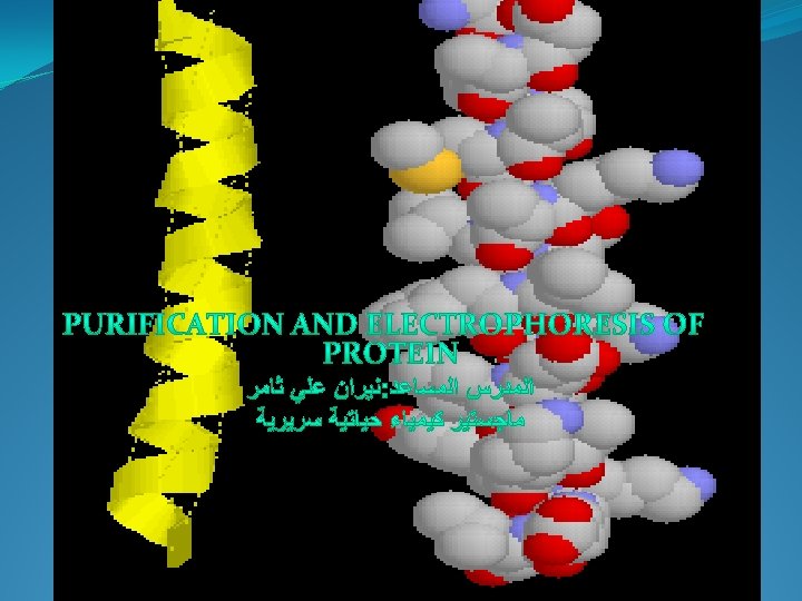
protein isolation purification Chromatography Electrophoresis I Protein Isolation


 be insoluble in the buffer. (2)")


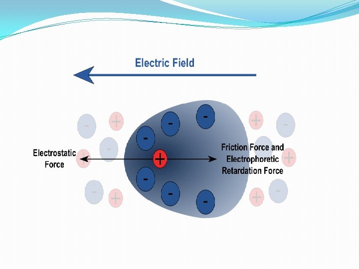
like sedimentation) F=q. E [q")


 is the most direct method for")

. The")

. dissolving 29. 2 gm of acrylamide and 0. 8 gm")


.")
. Freshly prepared on the analysis day")


- Slides: 61
protein; isolation ; purification Chromatography Electrophoresis
I. Protein Isolation A. Selection of a Protein Source-often can obtain the same or similar protein several different sources chose a source with 1. . which can be obtained in large amounts and has. . 2. high concentration of the protein 3. . molecular cloning techniques allow production and purification of proteins from E. coli, yeast or other cells. B. Method of solubilization 1. . serum proteins or secreted proteins are already soluble 2. . otherwise cells must be broken up • osmotic lysis; perhaps aided by enzymes or chemicals to weaken cell membranes (detergents solvents, or lysozyme for bacteria ) • mechanical disruption by grinding, blending, homogenizing or ultrasonic disruption 3. filter or centrifuge crude lysate to remove cell debris (membranes, cell walls etc, ) from
C. Stabilization of proteins--proteins are delicate 1. . may be denatured by high temperature • keep solution at appropriate temperature, usually fairly cold. • can use denaturation of some proteins to help purify a protein which is stable high temperature 2. Proteases are enzymes which break peptide bonds. • maintain conditions which inhibit proteases ==> low temperature or change in or addition of chemical inhibitors. • may use proteases to digest labile proteins if the protein of interest is stable to proteases. at p. H
D. Assay of Proteins-need some way of measuring the concentration of a specific protein, so we know when we're doing something right • • • enzymes can be measured by the reactions they catalyze--either measure products produced or reactants used up. other proteins may be measured by their biological effects: ability to bind specific molecules or the effect of a hormone on cells, tissue or organism. . Immunochemical techniques-can produce antibodies which bind specifically to particular proteins
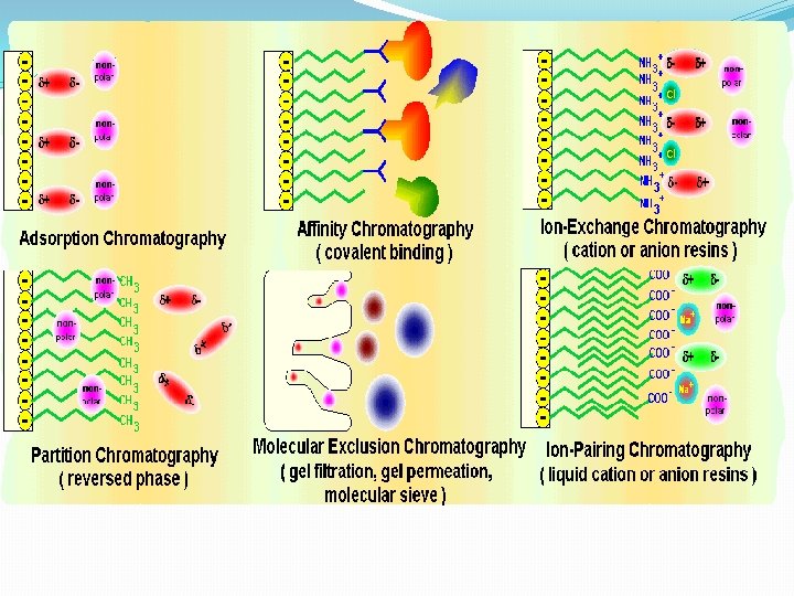
E. General Strategy of Protein Purificationfractionate based on different characteristic Proteins are purified by fractionation procedures in a series of steps. 1. • • • Charge ion exchange chromatography Electrophoresis Isoelectric focusing • 2. Polarity • adsorption chromatography • paper chromatography • reverse-phase chromatography • hydrophobic interaction chromatography
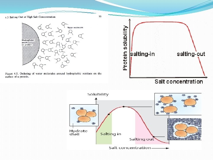
3. Size • gel electrophoresis • gel filtration chromatography • ultracentrifugation • dialysis and ultrafiltration II. Solubilities of Proteins A. Effects of Salt Concentration 1. salting in : at low concentrations of salt, solubility of the proteins usually increase. 2. salting out –at high concentrations of salt , solubility of the protein decreased sharply (precipitate), because the salt ions bind most of the water molecules Salt ions compete with protein globules for water and, eventually, at a sufficiently high concentration, strip the water of aqueous shell. Aqueous salt solution becomes a poor solvent for proteins, which precipitate out.
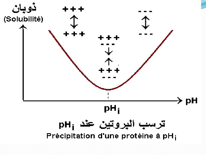
• The number and distribution of charges, • nonionic polar groups, • and hydrophobic residues on the surface of the protein • The size and shape of the protein. • determines the concentration of the salt needed to cause precipitation of the protein. B. Effects of p. H All proteins have an isoelectric point, p. I, a p. H at which they have no net charge, and they are least soluble at their p. I because there are no net electrostatic repulsions between protein molecules: . Different proteins have different p. I's, so one can manipulate the relative solubilities of a mixture of proteins by changing the p. H. C. Crystallization Once a protein is reasonably pure, one may try to crystallize it which is the ultimate criterion of purity.
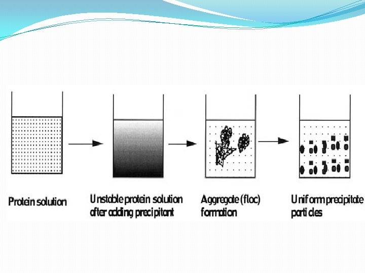
kinetic of protein precipitation involve the following. First, the protein environment is altered by the addition of a precipitating agent, causing the solution to become unstable disrupting bipolarity. Second, solid phase appears as small spherical ‘‘primary’’ particles of solid protein, which grow by diffusional transport of protein molecules to the solid surface. Third, primary particles aggregate as a result of convective transport and lead to floc formation. Finally, the aggregate (floc) size is limited by hydrodynamic disruption of the aggregates, generating smooth and uniform precipitate particles. maximizing the aggregate’s size and density.
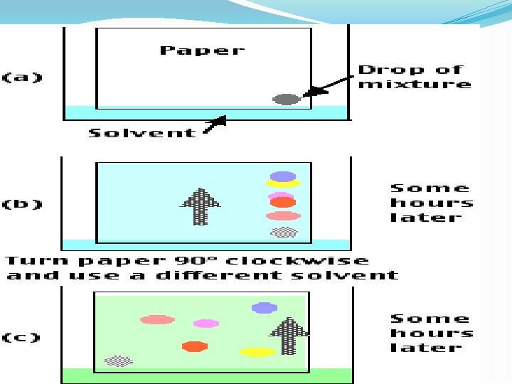
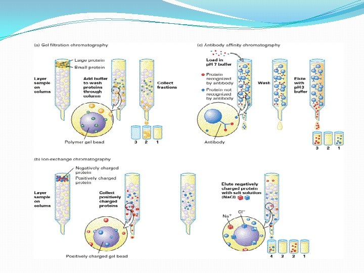
III. Chromatographic Separations One of the most power class of separation procedures is chromatography; this technique can take various forms based upon the physical apparatus column chromatography, paper chromatography, or thin layer chromatography –. All depend upon having a mobile phase, usually a liquid, and a stationary phase, usually a solid coated with a liquid. The sample is applied in the mobile phase which passes down the column (or paper or thin layer plate) and the different solutes move at different speeds depending upon their relative affinities for the mobile and stationary phases; we say that they partition between the mobile and stationary phases.
Paper chromotography
Column chromotography
The Chromatographic Matrix A chromatographic matrix should. (1) be insoluble in the buffer. (2) hydrophilic. (3) easily activated and coupled to a ligand (for affinity chromatography. (4) have large or small pores accessible to the protein (5) have a large surface area. (6) be physically and chemically stable to withstand the conditions during sterilization.
• A variety of materials have been used as matrices. These include --inorganic materials glass, silica, and hydroxyapatite; -synthetic organic polymers like polyacrylamide, . -agarose based (e. g. , Sepharose and Superose) - dextran based (e. g. , Sephadex) - cellulose based (e. g. , Sephacel).
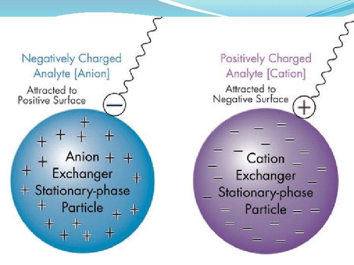
A. Ion Exchange Chromatography--adsorption chromatography 1. . Stationary Phase -- chemically bound charged groups with counter ions bound; these may be positively charged groups which bind anion(anion exchanger) or negatively charged groups which bind cations )cation exchanger) 2. Mobile Phase -- an aqueous buffer solution characterized by: p. H and ionic strength 3. Eluting Ion Exchange Columns -- molecules usually adsorb tightly in the buffer in which they're applied ==> must weaken this interaction. • Increase ionic strength (most common method): F = q 1 q 2 / D r 2 q 1 and q 2 are the charges on 2 groups, r is the distance between the groups, and D is the dielectric constant of the solvent which is increased with higher ionic strength thus weakening the force between the solute and the ion exchanger. Another way to look at this is that other ions in the buffer compete for the ion exchanger binding site. • change p. H -- changes the charges on the molecules being separated; also can change the charge of a weak ion exchanger • These changes can be made stepwise by changing the buffer reservoir (step gradient) or as gradient -- by mixing two buffers
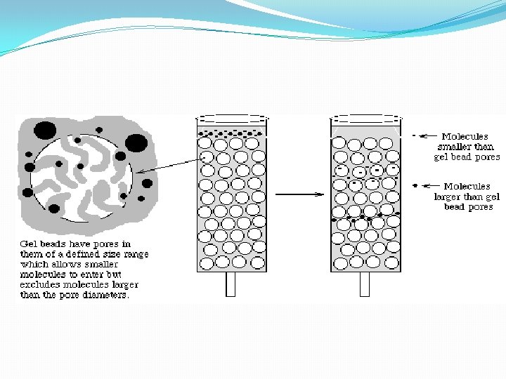
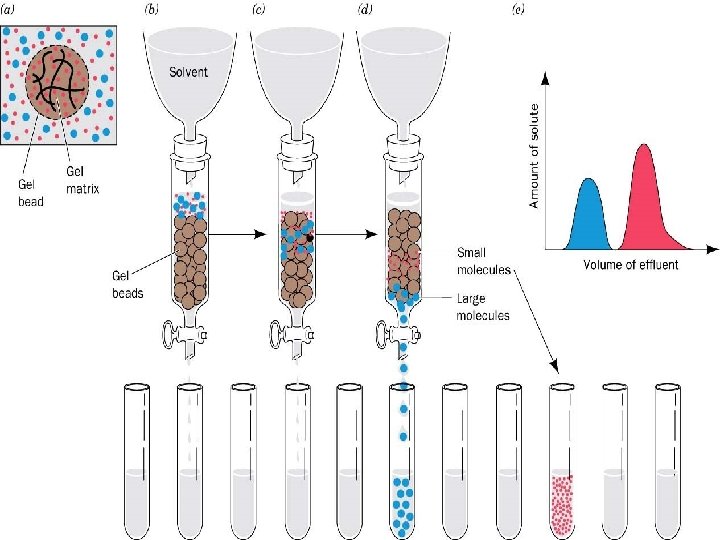
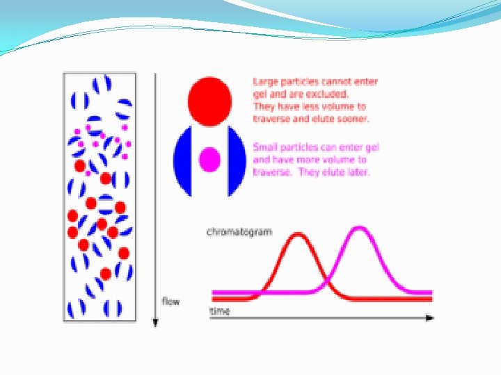
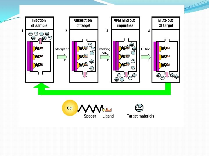
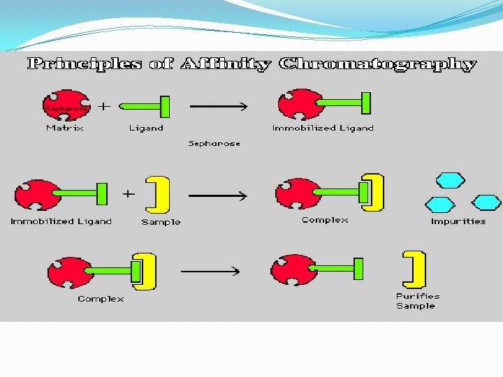
B. Gel Filtration Chromatography 1. Column Packing -- spherical porous beads of defined size crosslinked dextrans -Sephadex (Pharmacia) crosslinked polyacrylamide -- Bio-Gel P (Bio-Rad) crosslinked agarose -- Sepharose (Pharmacia) or Biogel A (Bio-Rad). Agarose beads have very large pores and are, therefore, good for separating very large molecules other materials developed by other companies 2. Gel beads are designed to have a distribution of pore sizes around a mean pore size. The mean pore size and the distribution determines the size range of molecules which can be separated. 3. Dialysis is a form of molecular Filtration. C. Affinity Chromatography based upon specific binding of the target protein to a particular ligand which is bound to an inert matrix.
D. Other Chromatographic Techniques 1. Reverse Phase Chromatography stationary phase is more hydrophobic liquid adsorbed to inert matrix mobile phase is more hydrophilic liquid 2. Hydrophobic Interaction Chromatography--similar to reverse phase chromtography but with less densely packed hdrophobic groups ==> less denaturing. 3. HPLC = High Performance Chromatography--a form of column chromatogrpahy with very small particles in the stationary phase to increase resolution; speed increased by using very high pressures. Commonly used in reverse phase mode but also with ion exchange, gel filtration, or hydrophobic interaction.
Gas chromotography
HPLC( High performance liquid chromotography
spleen cells cytoplasm was applied on top of sepharose 6 B gel filtration column. Flow rate was 30 ml /hrs, 5 ml fraction collected. 0, 045 0, 04 Absorbance 280 nm 0, 035 0, 03 0, 025 0, 02 0, 015 0, 01 0, 005 0 1 3 5 7 9 11 13 15 17 19 21 23 25 27 29 31 33 35 37 39 41 43 45 47 49 Fraction Numbre
Electrophoresis Protein Electrophoresis is used to separate mixtures of protein (e. g. , from cells, subcellular fractions, column fractions, or immunoprecipitates) inorder to investigate subunit compositions to verify homogeneity of protein samples to purify proteins for use in further application
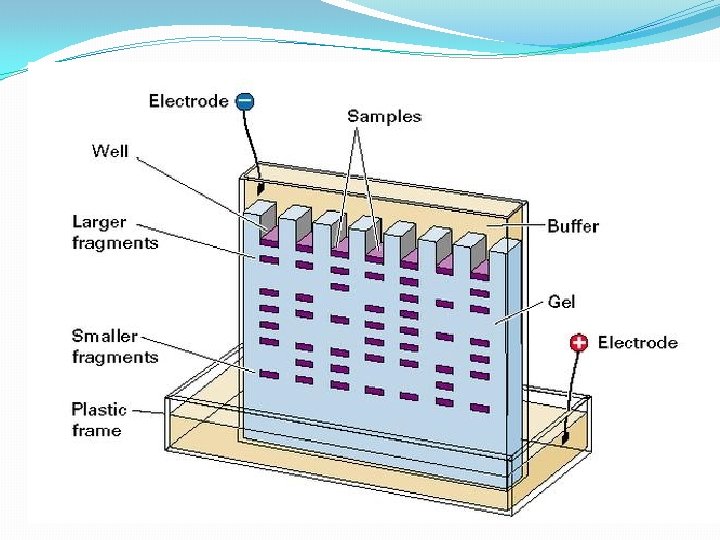
Electrophoresi. S A. Macromolecule is accelerated by a force )like sedimentation) F=q. E [q is the net charge on the molecule; E is the electric field strength experienced by the molecule] 2. This causes an acceleration until the velocity, v, causes a frictional force equal to but opposite in direction to the applied force F = q. E=fv 3. Zonal: most common mode used • sample is applied in a zone (small region: a spot on moistened paper or a band on a gel) and an applied field causes the molecules to separate into zones based upon different U's. • Like Zonal sedimentation, need some way of stabilizing the zones to prevent mechanical mixing (from vibrations) or convection mixing (from temperature differences -- a particularly severe problem with resistance heating caused by the electric field.
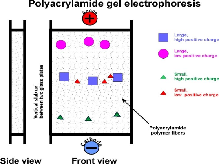
Gel electrophoresis is a method used in clinical chemistry to separate proteins by charge and or size (IEF agarose, essentially size independent) and in biochemistry and molecular biology to separate a mixed population of DNA and RNA fragments by length, to estimate the size of DNA and RNA fragments or to separate proteins by charge. Nucleic acid molecules are separated by applying an electric field to move the negatively charged molecules through an agarose matrix. Shorter molecules move faster and migrate farther than longer ones because shorter molecules migrate more easily through the pores of the gel. This phenomenon is called sieving. Proteins are separated by charge in agarose because the pores of the gel are too large to sieve proteins. Gel electrophoresis can also be used for separation of nanoparticles
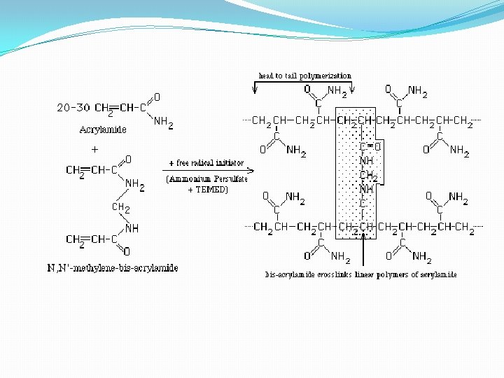
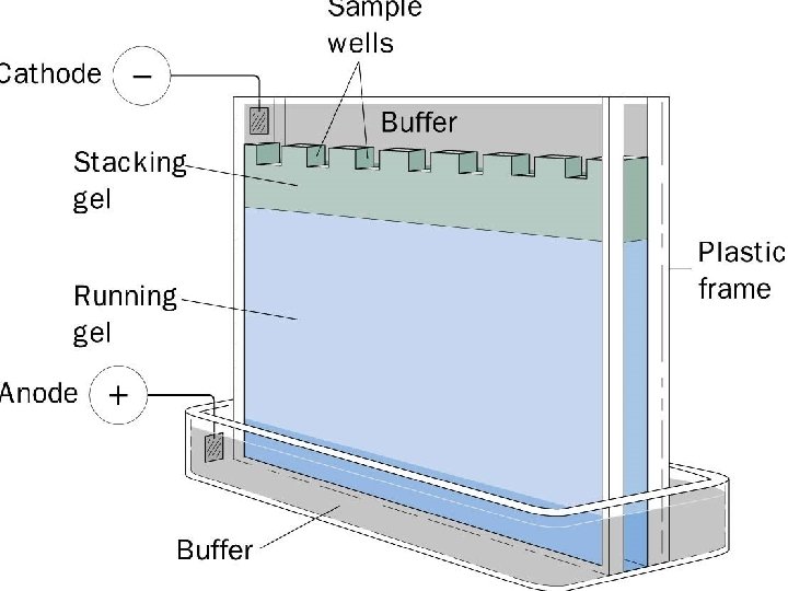
• 4 - Gel electrophoresis media ; • - Three types • Starch Gel -- swollen potato starch granules (little used now except for prep isoelectric focusing) • Agarose Gel -- purified large MW polysaccharide (from agar) ==> very open (large pore) gel used frequently for large DNA molecules • Polyacrylamide Gels -- most commonly used gel because they are very stable and can be made at a wide variety of concentrations or even with a gradient of concentrations ==> large variety of pore sizes • Acrylamide Concentrations -- typically 5 -20% by weight (5%, 7. 5%, 10%, 12. 5%, 15%, 20% are commonly used values) ==> gel is mostly water. • Acrylamide polymerizes in head-to-tail fashion to form long polymers which form a complex network held together by bis-acrylamide crosslinks. The criscrossing polymers create pores in the gel; the size of pores is determined by the acrylamide concentraion. • 5 -. Acrylamide can be polymerized into any desired shape -- two shapes used for electrophoresis • Tube Gels -- polymerize in glass tubing ==> cylindrical shape • Slab Gels -- polymerize between glass plates _--
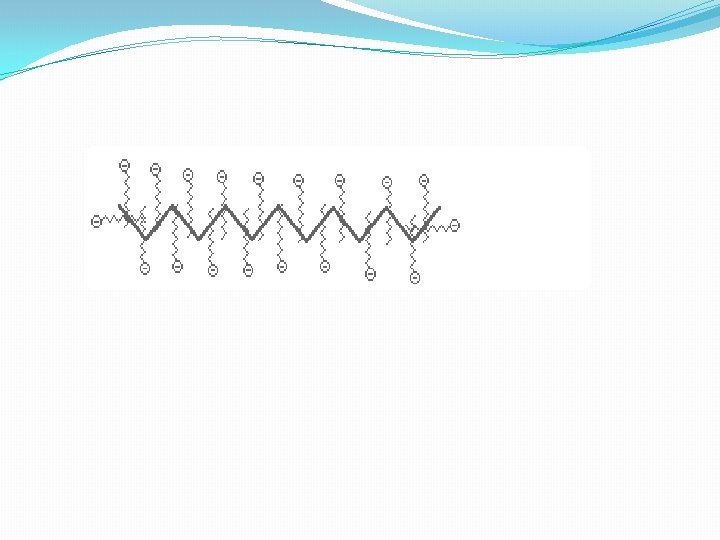
B. SDS Poly. Acrylamide Gel Electrophoresis -- SDS PAGE 1. Sodium Dodecyl Sulfate = Sodium Lauryl Sulfate: CH 3( CH 2) 11 SO Na+ This is a detergent because it contains a hydrophobic region, the CH 3( CH 2) 11 tail, attached to a hydrophilic group, SO 3 -Na+, making it amphipathic. It is a very strong detergent which denatures proteins by binding to the polypeptide backbone ((( -3 estimation of purity and molecular weight makes use of the detergent sodium dodecyl sulfate (SDS). Na+ - SO 4(CH 2)11 CH 3 SDS binds to most proteins in amounts proportional to the molecular weight of the protein, about one molecule of SDS for every two amino acid residues. SDS, net negative charge, rendering the intrinsic charge of the protein insignificant and conferring on each protein a similar charge-to-mass ratio. Electrophoresis in the presence of SDS therefore separates proteins almost exclusively on the basis of mass (molecular weight).
Sodium dodecyl sulfate – polyacrylamide gel electrophoresis (SDS-PAGE) is the most direct method for assessing in a fast and reproducible manner, the relative molecular weight (Mr) of denatured polypeptide chains and the purity of a protein preparation. In SDS-PAGE, the sample to be applied to the gel is first treated with the anionic detergent SDS which denatures the proteins in the sample and binds tightly to the protein molecules. The SDS molecules confer a relatively uniform negative charge to the polypeptide in proportion to its length. When an electric current is applied across the gel, all proteins will migrate through the gel matrix toward the anode. In this way, SDS-PAGE separates proteins according to size because the SDScoated proteins have a uniform charge: mass ratio. Proteins with less mass travel more quickly through the gel than those with larger mass because of the sieving effect of the gel matrix
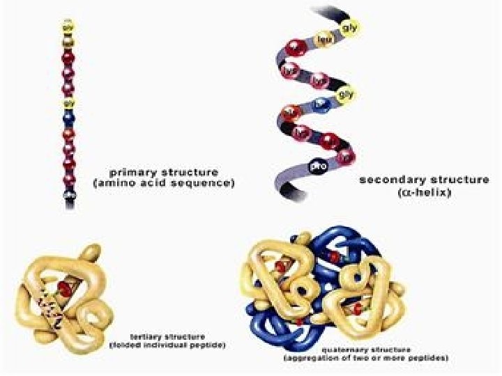
. C. Iso. Electric Focusing: All protein charges vary from a net positive charge at low p. H ( -COOH and –NH 3 +forms of acidic and basic functional groups) through 0 at some intermediate p. H to a net negative charge (-COO- and -NH 3 forms) at high p. H. 1. p. I - Iso. Electric Point: p. H at which a protein has a net 0 charge (positive and negative charges balance). Depends mostly on the amino acid composition and a little on the tertiary structure 2. Create a p. H gradient in a gel: Can be done on a slab (vertical or horizontal) or a tube. the final positions of each band depend only on an intrinsic property of the proteins, their p. I's, and not on where they started in the gel D. Two-Dimensional Electrophoresis: There are many variations; all basically combine 2 types of electrophoresis
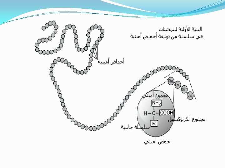
Proteins Electrophoresis carried out in gels made up of the cross-linked polymer (polyacrylamide). The polyacrylamide gel acts as a molecular sieve, slowing the migration of proteins approximately in proportion to their charge-to-mass ratio. µ=V/E=Z/f µ, The electrophoretic mobility of the molecule. V, the velocity of the particle molecule, E, moving force of the molecule(electrical potential) Z, the net charge of the molecule. f, frictional coefficient. ----- reflects in part a protein’s shape. Different PAA Concentrations for different purposes
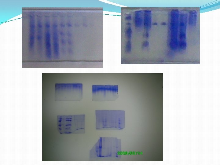
Non. SDS-PAGE , Denatured SDS-PAGE, Nondenatured The proteins are visualized by adding a dye such as Coomassie blue, which binds to proteins but not to the gel itself. In compares' with the positions to which proteins of known molecular weight migrate in the gel, the position of an unidentified protein can provide a measure of its molecular weight. If the protein has two or more different subunits, the subunits will generally be separated by the SDS treatment and a separate band will appear for each
Reagents and Buffers. Acrylamide-bisacrylamide(30%). dissolving 29. 2 gm of acrylamide and 0. 8 gm of bisacrylamide in 50 ml of distilled water. When the acrylamide was completely dissolved, the volume is completed to 100 ml with distilled water. The solution is stored in a refrigerator at 4°C in a dark bottle for no longer than one month. 2 -Concentrated Resolving Gel Buffer (1. 5 M Tris-HCl, p. H 8. 8). dissolving 18. 2 gm of Tris-HCl in 80 ml of distilled water. The p. H adjust to 8. 8 with 1 N HCl, and the volume completed to 100 ml with distilled water. It was stored at 4°C in a refrigerator.
Most modern commercial equipment is color-coded so that the red or positive terminal of the power supply is connected to the red lead of the gel apparatus. The red lead goes to the lower buffer chamber. The black lead is connected to the black or negative terminal and goes to the upper buffer chamber. In slab gel electrophoreses negatively charged proteins or nucleic acids move to the positive electrode in the lower buffer chamber (an anionic system). Proteins separated in cationic (+) systems. In these gels, the proteins are positively charged because of the very low p. H of the gel buffers (e. g. , acetic acid/urea gels for histone separations) or a cationic detergent (e. g. , CTAB). Proteins move toward the negative electrode (cathode) the polarity is reversed compared to SDS-PAGE: the red lead from the lower buffer chamber is attached to the black outlet of the power supply, and the black lead from the upper buffer chamber is attached to the red outlet of the power supply.
Power supplies usually have more than one pair of outlets. The pairs are connected in parallel with each other internally. gel is connected directly to the outlets of a power supply, then these gels are connected in parallel = voltage is the same across each gel. power supply reads 100 V, then each gel has 100 V across its electrodes. The total current, however, is the sum of the individual currents Therefore, under constant current it is necessary to increase the to the power supply. Two identical gels require double the current to achieve the same starting voltages and electrophoresis separation times.
3 - Concentrated Stacking Gel Buffer (0. 5 M Tris-HCl, p. H 6. 8). dissolving 6 gm Tris-HCl in 50 ml of distilled water. The p. H was adjusted to 6. 8 with 1 N HCl; volume was completed to 100 ml with distilled water. Solution was stored at 4°C in a refrigerator. 4 - Sodium Dodecyl Sulfate Solution 10% (SDS). Prepared by dissolving 10 gm SDS in 50 ml of distilled water and the volume was completed to 100 ml with distilled water. 5 - Stock Sample Buffer (0. 06 M Tris-HCl p. H 6. 8, 2% SDS, 10% glycerol, and 0. 025% bromophenol blue) mixing of the following; 0. 5 M Tris acid p. H 6. 8 ……………. . 1. 2 ml 10% SDS ………………………. . 2. 0 ml Glycerol ………………. . . 1. 0 ml 0. 5% bromophenol blue (w/v water). . . 0. 5 ml Sucrose ………………………. 0. 2 gm/ml Water ………………………… 4. 8 ml The mixture was stored at room temperature.
6 - Ammonium per Sulfate (10% as catalyst). Freshly prepared on the analysis day by dissolving 0. 1 gm of ammonium persulfate in 1 ml of distilled water. 7 - TEMED (N, N, N, N-tetramethylene diamine). prepared immediately before use by adding 0. 5 -1. 0 µl/ml of the final solution volume. 8 - Electrode Buffer p. H 8. 3 Tris-base. . . …. ……………. . 3 gm. Glycine …………………. …. . 14. 4 gm. SDS (10% w/v)…. ………. 10 ml. Distilled water ……. . …… 1 L. 9 - Stacking Gel Buffer Prepared by mixing the following ingredients: Tris-HCl buffer (0. 5 M p. H 6. 8) ………………. 20 ml SDS (10%)……………. . ………………. 1. 6 ml Distilled water …………… 18. 4 ml 10 - Resolving Gel Buffer Prepared by mixing the following ingredients: buffer( 1. 5 M p. H 8. 8) ……………. 20 ml SDS (10%)…………………. 1. 6 ml Distilled water …………………. . 18. 4 ml 11: Fixing Solution Prepared by dissolving 100 gm of tricholoracetic acid in 500 ml of distilled water, 400 ml of absolute methanol was added, the volume brought to 1 L with distilled water.
12 - Staining Solution. mix methanol, glacial acetic acid, and coomassie brilliant blue G-250 in a final concentration of 45% with 9% and 0. 05% respectivly. 13 -Destaining Solution. mix methanol, acetic acid, and distilled water in final volumes of 1: 4: 5 respectivly. 1 - Resolving Gel Preparation 10% mix the following volumes of the previously prepared stock solutions: Acrylamidebisacrylamide (30%) ……. . 5. 0 ml SDS (10%)……………………. . 0. 1 ml Resolving gel buffer …………… 3. 75 ml D. W. . …………………. 6. 25 ml This mixture must be degassed under vacuum. Ammonium persulphate and TEMED must be added at the ratios of 50µl / ml and 5µl / ml of the final volume, respectively.
2 - Stacking Gel Preparation prepare at concentration of 5% by mixing the following: acrylamide-bisacrylamide (30%)…. ……. . . 1. 3 ml SDS (10%) ……………………. . 0. 1 ml Stacking gel buffer …………. . . ………………. … 2. 5 ml D. W……………………. 6. 1 ml The mixture shoud be degassed under vacuum. 50 µl of 10% of ammonium persulphate and 10 µl of TEMED were added to each 10 ml of degassed monomer solution immediately before casting. 3 - Resolving Gel Casting. The 10 % resolving gel was applied to the electrophoresis unit. Its surface was immediately overlaid with water saturated butanol and lefted for 45 minutes to 1 hrs at room temperature to polymerize. butanol was removed after gel polymerization, the gel surface was rinsed with D. W. The stacking gel was added at the same way and lifted to polymeriz. The cast comb was then inserted in between the glasses and removed after hardening of the stacking gel.
Phosphorylase 94 k. Da Albumin 67 k. Da Ovalbumin 43 k. Da Carbonic anhydrase 30 k Da Trypsin inhibitor 20 k. Da a- Lactalbumin 14. 4 k. Da
Relative mobility = distance of the protein band / total distance 5, 2 phosphorelase 5 albumin ovalbumin Log MW 4, 8 carbonic anhydrase 4, 6 trypsin inhibitor 4, 4 α-lactalbumin 4, 2 4 3, 8 3, 6 0, 45 0, 55 0, 65 0, 75 0, 8 Relative mobility (Rm) 10%SDS- PAGE 0, 85 0, 95 1
THANK YOU