SODIUM DODECYL SULFATE POLYACRYLAMIDE GEL ELECTROPHORESIS Electrophoresis Electrophoresis


- Slides: 10

SODIUM DODECYL SULFATE POLYACRYLAMIDE GEL ELECTROPHORESIS

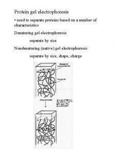
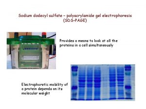
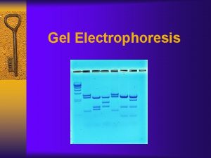
Electrophoresis - Electrophoresis is a method used to separate charged particles from one to another based on differences in their migration speed. - Electrophoresis is a technique widely used in biochemistry, forensic chemistry, genetics molecular biology and biotechnology to separate biological macromolecules, usually proteins or nucleic acids according to their electrophoretic mobility. - Applying the electric field to a solution (including protein molecule) - In electrophoresis, moving of protein molecules depending on their net charge, size and shape. • The development of sodium dodecyl sulfate-polyacrylamide gel electrophoresis (SDSPAGE) was breakthrough in routine protein analysis. • In SDS-PAGE, proteins are separated in a polyacrylamide gel based on their molecular weight. • In the SDS-PAGE method, polyacrylamide gel with a large number of cross-links is used as the medium in which the proteins move.

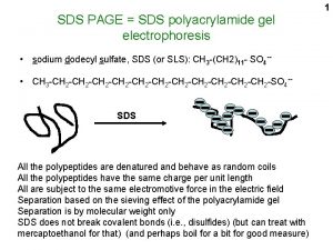
Sodium Dodecyl Sulfate (SDS) • • Strong anionic detergent Negatively charged Denature and linearize proteins Coats the proteins with negatively charged • Proteins are amphoteric molecules, i. e. they have both positive and negative charges. To make them move in a single direction, a uniform negative charge is created on them. When the proteins are mixed with SDS, they acquire a net negative charge. Thus, in SDS-PAGE, the separation is directly related to the molecular weights independent of their charge. • Also, reducing agents (Bromphenol blue/DTT) break the disulfide bonds

SDS PAGE includes two types of gel systems • The upper (stacking) gel, includes sample wells. The sample to be analyzed is applied to the wells of the gel with a tracer dye and electric current is passed through the system. • The lower (seperating) gel is responsible for actually seperating polypeptides by size.

Preparation of Acrylamide Gels v The gel formation occured by the polymerization of acrylamide and acrylamide derivative NN'-methylene bisacrylamide and the samples are run on this gel. v. For polymerization, the acrylamide molecules bind side by side and form straight chains. v Bisacrylamide molecules form cross-linkings between two acrylamide chains v Thus, a networked structure occurs v. The pore size depends on the acrylamide concentration v. The Ammonium persulfat (APS) is reaction initiator which causes free radical formation for polymerization v. N, N, N’ -tetramethyl-ethylenediamin (TEMED) acts as catalyst.

The ratio of bisacrylamide to acrylamide • The ratio of bisacrylamide to acrylamide can be different depending on protein target • Lower percentage gels are better for separating high molecular weight molecules. • Higher acrylamide percentages are needed for separating smaller proteins.

Loading samples into SDS-PAGE gel • A comb placed on top of the gel during polymerization allows the formation of small wells in the gel. • The comb is removed after polymerization. • The procedure is performed by placing the gels in an electrophoresis device containing an electrophoresis buffer. • Negatively charged proteins are loaded into the wells of the gel and electrical current is passed. • Electrophoresis buffer provides electric current in the medium and set and maintain the proper p. H during electrophoresis. • In electrophoresis, electric current must run from cathode to anode. • The upper side is negatively charged, the bottom side is positively charged.

Preparation of Sample Buffer: • Used for the preparation and loading of protein samples onto a gel for SDS-PAGE analysis. The contents of sample buffer • SDS: • denatures proteins and makes them negatively charged. • β-mercaptoethanol/DTT: • is used to break disulphide bonds • Glycerol: • increases the density of the sample relative to the surrounding running buffer making it easier to load in the well • Bromophenol blue • is used to follow the run of protein sample on the gel (Tracking dye)

Coomassie Brilliant Blue Anionic dye - Nonspesifically binds to proteins - After the run, PAGE gel is placed in a Coomassive Brilliant Blue dye solution for staining for a few hours and is de-stained to visualize the separated protein molecules as bands. - The proteins are detected as blue bands on a clear background -

References Ankara University Faculty of Pharmacy Biochemistry Practice book-2004 Practical Biochemistry (2015). Aljebory, A. , And Alsalman, A. A Laboratory Text Book of Biochemistry, Molecular Biology and Microbiology (2014) Lehninger Principles of Biochemistry- 5 th Edition (2008) 7 th edition Biochemistry Jeremy M. Berg John L. Tymoczko Lubert Stryer, Gregory J. Gatto, Jr. W. H. Freeman and Company. • Mahin Basha, Analytical Techniques in Biochemistry , 2019, Springer Protocol Handbooks • Gallagher, S. R. (2012). One-Dimensional SDS Gel Electrophoresis of Proteins. Current Protocols in Molecular Biology, 97(1), 10. 2 A. 1– 10. 2 A. 44. • One-dimensional SDS-polyacrylamide gel electrophoresis (1 D SDS-PAGE). Brunelle JL. , Green R. Methods Enzymol. 2014; 541: 151 -9. • • •
 Sdspage gel
 Polyacrylamide gel electrophoresis (page)
 Agarose gel
 Agarose gel vs polyacrylamide gel
 Spectator ions equation example
 Calcium carbonate nitric acid reaction
 Use of micropipette in agarose gel electrophoresis
 Translate image
 Gel electrophoresis definition
 Paper and gel electrophoresis
 Ap bio electrophoresis lab
