Overview of the Components of the MSS Lecture


 that moves around")


 • The structural classification of joints • Fibrous joints (bones held")
 • Lack a synovial cavity • The articulating bones are held")
 • Sutures • Occur only between bones of the skull •")
 • Lacks a synovial cavity • Allows little or no movement")
 • Synchondroses • Connecting tissue is hyaline cartilage • Epiphyseal (growth)")
 • Synovial cavity allows a joint to be freely movable •")
")
 • Accessory Ligaments and Articular Discs • Collateral ligaments of the")
 • Nerve and Blood Supply • Nerve endings convey information about")
 • Bursae and Tendon Sheaths • Bursae • Sac-like structures containing")


- Slides: 49
Overview of the Components of the MSS
Lecture Objectives • Discuss the components and functions of the MSS. • Describe the relation between bones and skeletal muscles in producing body movements. • Identify the major regions and compartments of upper and lower limbs. • Contrast the structural and functional classification of joints and identify factors that determine the degree of movement at a joint. • Describe the components of the synovial joints and give examples from the upper and lower limbs.
Upper Limb
Upper Limb Regions, Bones, & Joints
Upper Limb Fascia
Upper Limb Fascia
Arm Compartments
Forearm Compartments
Forearm Compartments
Upper Limb Arteries
Upper Limb Deep Veins
Upper Limb Superficial Veins
Upper Limb Lymphatics
Upper Limb Lymphatics
Upper Limb Lymphatics
Upper Limb Innervation
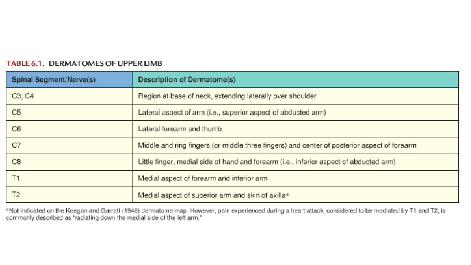
Upper Limb Dermatomes
Lower Limb
Lower Limb Regions, Bones, and Joints
Lower Limb Fascia and Compartments • Superficial facia • Deep facia • Fascia lata (in thigh) • Saphenous opening • Iliotibial tract • Crural fascia (in leg)
Thigh Compartments
Leg Compartments
Leg Compartments
Lower Limb Arteries
Lower Limb Deep Veins
Lower Limp Superficial Veins
Lower Limb Superficial Veins
Lower Limb Lymphatics
Lower Limb Innervation
Lower Limb Dermatomes
Lower Limb Dermatomes • Thigh • Anterior, lateral, and medial • Lumber plexus • Superioposterior • Posterior rami L 1 -S 3 • Inferioposterior • Sacral plexus • Leg • Anteromedial and posteromedial • Lumber plexus • Remaining • Sacral plexus
Muscle Attachment Sites: Origin and Insertion • Skeletal muscles shorten & pull on the bones they are attached to • Origin is the bone that does not move when muscle shortens (normally proximal) • Insertion is the movable bone (some 2 joint muscles) • Fleshy portion of the muscle in between attachment sites = belly
Lever Systems and Leverage • Muscle acts on rigid rod (bone) that moves around a fixed point called a fulcrum • Resistance is weight of body part & perhaps an object • Effort or load is work done by muscle contraction • Mechanical advantage • the muscle whose attachment is farther from the joint will produce the most force • the muscle attaching closer to the joint has the greater range of motion and the faster the speed it can produce
Fascicle Arrangements • A contracting muscle shortens to about 70% of its length • Fascicular arrangement represents a compromise between force of contraction (power) and range of motion • muscles with longer fibers have a greater range of motion • a short fiber can contract as forcefully as a long one.
Coordination Within Muscle Groups • Most movement is the result of several muscle working at the same time • Most muscles are arranged in opposing pairs at joints • prime mover or agonist contracts to cause the desired action • antagonist stretches and yields to prime mover • synergists contract to stabilize nearby joints • fixators stabilize the origin of the prime mover • scapula held steady so deltoid can raise arm
Joints • • Joint Classifications Fibrous Joints Cartilaginous Joints Synovial Joints Types of Movements at Synovial Joints Types of Synovial Joints Factors Affecting Contact and Range of Motion at Synovial Joints Selected Joints of the Body
Joints (Joint Classification) • The structural classification of joints • Fibrous joints (bones held together by dense collagen fibers) • Cartilaginous joints (bones held together by cartilage) • Synovial joints (bones held together by ligaments) • The functional classification of joints • Synarthrosis (an immovable joint) • Amphiarthrosis (a slightly movable joint) • Diarthrosis (a freely movable joint)
Joints (Fibrous Joints) • Lack a synovial cavity • The articulating bones are held very closely together by dense irregular connective tissue • Fibrous joints permit little or no movement • Three types of fibrous joints • Sutures • Syndesmoses • Gomphoses
Joints (Fibrous Joints) • Sutures • Occur only between bones of the skull • Syndesmoses • Permits slight movement • Interosseous membrane • Between the tibia and fibula in the leg • Gomphoses • Immovable joint • Joint in which a cone-shaped peg fits into a socket • Articulations of the teeth with the sockets of the maxillae and mandible
Joints (Cartilaginous Joints) • Lacks a synovial cavity • Allows little or no movement • Joint is tightly connected by either cartilage • Two types of cartilaginous joints • Synchondroses • Symphyses
Joints (Cartilaginous Joints) • Synchondroses • Connecting tissue is hyaline cartilage • Epiphyseal (growth) plate • Symphyses • Slightly movable joint • Ends of the articulating bones are covered with hyaline cartilage, but a disc of fibrocartilage connects the bones • Pubic symphysis • Between the anterior surfaces of the hip bones • Intervertebral joints between the vertebrae
Joints (Synovial Joints) • Synovial cavity allows a joint to be freely movable • Ligaments hold bones together in a synovial joint • Articular Capsule • A sleeve-like capsule encloses the synovial cavity • The articular capsule is composed of two layers • an outer fibrous capsule • an inner synovial membrane • Synovial Fluid • The synovial membrane secretes synovial fluid • Functions to reduce friction by: • • lubricating the joint absorbing shocks supplying oxygen and nutrients to the cartilage removing carbon dioxide and metabolic wastes from the cartilage
Joints (Synovial Joints)
Joints (Synovial Joints) • Accessory Ligaments and Articular Discs • Collateral ligaments of the knee joint • Anterior and posterior cruciate ligaments of the knee joint • Menisci • Pads of cartilage lie between the articular surfaces of the bones • Allow bones of different shapes to fit together more tightly
Joints (Synovial Joints) • Nerve and Blood Supply • Nerve endings convey information about pain from the joint to the spinal cord and brain • Nerve endings respond to the degree of movement and stretch at a joint • Arterial branches from several different arteries merge around a joint before penetrating the articular capsule
Joints (Synovial Joints) • Bursae and Tendon Sheaths • Bursae • Sac-like structures containing fluid similar to synovial fluid • Located between tendons, ligaments and bones • Cushion the movement of these body parts • Tendon sheaths • Wrap around tendons • Reduce friction at joints
Factors Affecting Contact and Range for Motion at Synovial Joints • Range of motion (ROM) • Refers to the range, measured in degrees of a circle, through which the bones of a joint can be moved • Factors contribute to keeping the articular surfaces in contact and affect range of motion: • Structure or shape of the articulating bones • Shape of bones determines how closely they fit together • Strength and tension of the joint ligaments • Ligaments are tense when the joint is in certain positions • Tense ligaments restrict the range of motion
Factors Affecting Contact and Range for Motion at Synovial Joints • Arrangement and tension of the muscles • Muscle tension reinforces the restraint placed on a joint by its ligaments , and thus restricts movement • Contact of soft parts • The point at which one body surface contacts another may limit mobility • Movement be restricted by the presence of adipose tissue • Hormones • Flexibility may also be affected by hormones • Relaxin increases the flexibility of the pubic symphysis and loosens the ligaments between the sacrum and hip bone toward the end of pregnancy • Disuse • Movement may be restricted if a joint has not been used for an extended period