NONINVASIVE VENTILATION IN ACUTE RESPIRATORY FAILURE Virginia Chung


 Non-cardiogenic Pulmonary Edema (ARDS,")


 Ø Dyspnea (moderate")


 group 31")


- Slides: 60
NON-INVASIVE VENTILATION IN ACUTE RESPIRATORY FAILURE Virginia Chung, MD Chief, Pulmonary & Critical Care Medicine Jacobi Medical Center January 30, 2013
OUTLINE Ø Acute respiratory failure Ø Definitions, Pathophysiology Ø NIPPV / NIV / BPAP / Bi. PAP vs CPAP Ø Indications / Contraindications Ø Use of NIV in: Ø COPD, Severe Asthma, CAP, ARDS, APE/CHF, DNI/DNR Ø Summary of Recommendations
BACKGROUND Ø Respiratory failure is a syndrome where the respiratory system fails in one or both of its gas exchange functions: oxygen uptake and carbon dioxide elimination. Ø Respiratory failure may be acute or chronic. Ø While acute respiratory failure (ARF) is characterized by life -threatening derangements in ABGs and acid-base status, manifestations of chronic respiratory failure are less dramatic and may not be as readily apparent.
CLASSIFICATION Ø Respiratory failure can be classified as HYPOXEMIC or HYPERCAPNIC and may be ACUTE or CHRONIC. Ø TYPE I : Hypoxemic Respiratory Failure is characterized by a Pa. O 2 < 60 mm. Hg with a normal or low Pa. CO 2. Most common form of respiratory failure Can be associated with virtually all acute diseases of the lung Examples: pulmonary edema, pneumonia, ARDS, PE
CLASSIFICATION Ø TYPE II : Hypercapnic respiratory failure is characterized by a Pa. CO 2 of > 50 mm. Hg. Hypoxemia is common in patients with Type II failure who are breathing room air. p. H depends on the serum bicarbonate level, which, in turn, is dependent on the duration of the hypercapnia Examples: opiate overdose, neuromuscular disease, status asthmaticus, severe COPD.
Acute vs. Chronic Hypercapnic Respiratory Failure Ø Acute hypercapnic respiratory failure develops over minutes to hours; therefore, p. H < 7. 3. Ø Chronic hypercapnic respiratory failure develops over several days or longer, allowing time for renal compensation and an increase in serum bicarbonate concentration; p. H is only slightly decreased.
PATHOPHYSIOLOGY Ø Hypoxemic Respiratory Failure Ø Hypoxemia can be caused by any one of these four mechanisms: Ventilation-Perfusion (V/Q) mismatch, Shunt, Diffusion Impairment, and Hypoventilation. Ø V/Q mismatch is the most important and common mechanism. Areas of low ventilation relative to perfusion (low V/Q units) lead to hypoxemia. Ø Shunts can be intracardiac or intrapulmonary.
Causes of Hypoxemic Respiratory Failure Pneumonia Cardiogenic Pulmonary Edema (CHF) Non-cardiogenic Pulmonary Edema (ARDS, seizure) Pulmonary Fibrosis (IPF, sarcoidosis) COPD / Asthma Pneumothorax Pulmonary Embolism Pulmonary Arterial Hypertension (Primary, Scleroderma) Pneumoconiosis (Coal-workers)
Causes of Hypoxemic Respiratory Failure Hypersensitivity Pneumonitis Congenital Heart Disease Bronchiectasis Fat Embolism Syndrome Kyphoscoliosis Obesity Massive Pleural Effusions Pulmonary Hemorrhage
Causes of Hypercapnic Respiratory Failure Tetanus Myxedema Severe ARDS Severe Pulmonary Edema Obesity Hypoventilation Syndrome Primary Alveolar Hypoventilation COPD Status Asthmaticus Drug Overdose Poisonings Myasthenia gravis Guillain-Barre Head and Cervical Cord Injury Poliomyelitis Polyneuropathy
SUMMARY Ø Two types of acute respiratory failure: Ø Type I : Hypoxemic , where Pa. O 2 < 60 mm. Hg Ø Type II : Hypercapnic , where Pa. CO 2 > 50 mm. Hg NB* : for status asthmaticus, Pa. CO 2 > 40 mm. Hg signifies hypercapnic respiratory failure. Ø V/Q mismatch is the most common mechanism for both types of respiratory failure. Ø Many conditions can cause both hypoxemia and hypercapnia : e. g. , COPD, Obesity, ARDS, severe pulmonary edema, neuromuscular disorders. Ø Avoid worsening hypercapnia by judiciously giving the patient supplemental oxygen. Ø Some patients may require NIPPV or mechanical ventilation.
NIPPV / NIV / BPAP/ Bi. PAP
Bi. PAP Graphics
BENEFITS OF NIV u Facilitate sleep u Avoid complications of ETI u VAP u Sepsis/shock u Tracheostomy u GI bleed u DVT u Correct mental status u Decrease mortality associated u Symptomatic relief of dyspnea u Correction of gas exchange u Improve lung mechanics u Pre-oxygenate for intubation u Prevent ETI with respiratory failure u Use NIV in the place of IMV u Assist DNI patients with respiratory failure
PHYSIOLOGIC MECHANSIMS u Unload respiratory muscles inspiratory cycle: hyperinflation >> respiratory muscle shortening/disadvantage Decreased compliance of respiratory system u NIPPV = augments respiratory effort, Increases Vt, decreases RR u Overcome intrinsic peep>> difficulty in generating pressure gradient for flow u CPAP u Stent open lower airway expiratory cycle u CPAP to reduce obstruction u Stent open upper airway u CPAP
PHYSIOLOGIC MECHANSIMS u Reduce CO 2 production u NIPPV u Improve gas exchange by decreasing atelectasis u CPAP/NIP u Reduce negative intra-thoracic pressure swings u CPAP u Redistribute pulmonary edema u CPAP/NIPPV u Increase CO by decreasing effective LV afterload u CPAP
Contraindications for NIV Absolute contraindications: Ø Coma Ø Cardiac arrest Ø Respiratory arrest Ø Any condition requiring immediate intubation Other contraindications (rare exceptions) Ø Cardiac instability (shock+need for vasopressors, ventricular dysrhythmias, complicated AMI) Ø GI bleeding – intractable emesis, uncontrolled bleeding
Contraindications for NIV Ø Inability to protect airway Ø impaired cough or swallowing Ø poor clearance of secretions Ø depressed sensorium and lethargy Ø Status epilepticus Ø Potential for upper airway obstruction Ø Extensive head / neck tumors Ø Any other tumor with extrinsic airway compromise Ø Angioedema or anaphylaxis causing airway compromise
Candidates for NIV Ø Patient cooperative (excludes agitated, belligerent, comatose patients) Ø Dyspnea (moderate to severe, short of respiratory failure / agonal breathing) Ø Tachypnea (rr> 24 /min) Ø Increased work of breathing (+accessory muscle use, pursed lip breathing) Ø Hypercapnic respiratory acidosis (p. H range 7. 10 – 7. 35) Ø Hypoxemia (Pa. O 2/Fi. O 2 < 200 mm Hg, best in rapidly reversible causes for hypoxemia)
Suitable Clinical Conditions for NIV Most patients with : Ø COPD Ø Cardiogenic pulmonary edema Selected patients with : Ø CAP + COPD Ø Asthma / CF Ø Decompensated OSA/OHS, cor pulmonale Ø ARDS Ø Immunocompromised state / mild PCP Ø Neuromuscular respiratory failure Ø DNI +/- DNR status Ø Post extubation COPD / post –op respiratory failure
NIV: utilization classification • mandatory ventilation • Alternative to intubation • severe ARF, meet criteria for IMV • Failed medical treatment • Trials: NIV vs IMV after failed MT • Primary outcome: mortality • supportive ventilation • Prevent intubation • mild-to-moderate ARF/does not meet criteria for IMV • Trials: NIV+MT vs MT • Primary outcome: intubation
NIV: utilization classification • prophylactic ventilation • To prevent ARF in patients • no substantial impairment of gas exchange • Trials: NIV+MT vs MT • Primary outcome: Blood gas values, FEV 1, etc • other purpose ventilation • bronchodilation • Pre-oxygenation • Facilitate sleep
NON-INVASIVE VENTILATION FOR ACUTE EXACERBATIONS OF COPD BROCHARD, MANCEBO, WYSOCKI: NEJM, 1995 Ø INCLUSION CRITERIA SUPPORTIVE VENTILATION RCT COPD with exacerbation of dyspnea > two days and at least two of the following: RR>30 Pa. O 2 < 45 mm Hg p. H < 7. 35 after > 10 min on RA Ø EXCLUSION CRITERIA RR< 12 breaths, sedative drugs within the previous 12 hours CNS disorder unrelated to hypercapnic encephalopathy or hypoxemia Cardiac arrest (within the previous five days) Cardiogenic pulmonary edema Asthma
NON-INVASIVE VENTILATION FOR ACUTE EXACERBATIONS OF COPD BROCHARD, MANCEBO, WYSOCKI: NEJM, 1995 SUPPORTIVE VENTILATION RCT kyphoscoliosis as the cause of chronic respiratory failure neuromuscular disorder as the cause of chronic respiratory failure Upper airway obstruction, facial deformity, tracheotomy need for immediate intubation = a clear cause of decompensation requiring specific treatment (e. g. , peritonitis, septic shock, AMI) pulmonary thromboembolism pneumothorax, hemoptysis severe pneumonia recent surgery or trauma
Primary outcome: need for intubation Secondary outcomes: LOS hosp, complications, length of MV, in hosp mortality Standard treatment arm `O 2 via NC up to 5 liters for target sat > 90% Medications: SQH, antibiotics, bronchodilators, IV corticosteroids or aminophylline NIPPV treatment arm: same as above and BIPAP at least 6 hours/day, NC for at least 2 hours/day IP=20, EP=0, flow cycled, PAC if patient is apneic
Primary outcome: need for intubation Secondary outcomes: LOS hosp, complications, length of MV, in hosp mortality Major Criteria for intubation: respiratory arrest, pauses with LOC, gasping, requiring sedation, HR<50 with lethargy, SPB<70 Minor Criteria for intubation: RR> 35 and > on admission, p. H < 7. 3 and < admission, Pa. O 2<45 despite O 2, worsening MS One Major Criteria or 2 Minor Criteria after one hour of RX would be indication for intubation. In the NIPPV group if 2 minor criteria met off NIV, they can be placed back on it. But if problem persisted then intubation performed
NIV for acute exacerbations COPD Brochard, NEJM, 1995
Primary outcome: need for intubation 85 patients total 42 standard rx (ST) group 31 intubated (74%) 43 NIPPV rx group 11 intubated (26%) ARR = 48%, NNT= 2 Major criteria for intubation met by 10/31 (ST) and 8/11 (NIPPV) At 1 hour: NIPPV group: improved encephalopathy, rr, Pa. O 2, p. H Standard group: worsening enceph, Pa. CO 2, p. H
Encephalopathy score 1= mild asterixis, 2= marked asterixis, mild confusion, sleepy during the day 3= major confusion with daytime sleepiness or agitation
Primary outcome: need for intubation Need for intubation was associated with: Higher SAP scores Higher encephalopathy scores on admission. v On admission prior to randomization: v ST 1. 6 v NIPPV 1. 8 v At one hour: v the scores worsened in ST 1. 9 v improved in NIPPV 1. 5 (and 0. 8 at 12 h) v Results: v ST group no ETI = 0. 7; +ETI = 1. 9 v NIPPV group no ETI = 1. 6; +ETI = 2. 5
NON-INVASIVE VENTILATION FOR ACUTE EXACERBATIONS OF COPD BROCHARD, MANCEBO, WYSOCKI: NEJM, 1995 SUPPORTIVE VENTILATION RCT Success probably related to rapid improvement in encephalopathy Mortality: ST 29% (32% intubated) NIPPV: 9% (25% intubated) Complications in ST 48%, NIPPV 16% NIPPV group: ST group: LOS: average NIPPV = 4 days; average MV = 25 days average MV =17 d ST 35 days, NIPPV 23 days
Noninvasive positive pressure ventilation in acute respiratory failure due to COPD vs other causes: Ritesh Agarwal, Rajesh Gupta, Ashutosh N Aggarwal, Dheeraj Gupta SUPPORTIVE VENTILATION: Both hypoxic and hypercapnic patients responded to NIV: COPD patients improved their PCO 2 and p. H PNA/ARDS patients improved their PAO 2 Avoided ETI in 87% of COPD patients and 61% all other etiologies Mortality: 12% in COPD, 18% other etiologies
Non-invasive positive pressure ventilation in acute respiratory failure due to COPD vs other causes: R Agarwal, R Gupta, A N Aggarwal, D Gupta: MIXED POPULATION STUDY Primary outcome: NIPPV failure defined as inability to stabilize or improve in 60 min gas exchange dyspnea mental status Supportive ventilation
Noninvasive positive pressure ventilation in acute respiratory failure due to COPD vs other causes: Ritesh Agarwal, Rajesh Gupta, Ashutosh N Aggarwal, Dheeraj Gupta Etiology is the only independent predictor of outcome: STUDIES WITH MIXED POPULATIONS ARE VIRTUALLY MEANINGLESS NIPPV failure rate is very high in Pneumonia, ARDS: Ø transient improvement in RR, HR and blood gas parameter does occur Ø the underlying process such as sepsis or pneumonia is not affected by NIPPV Ø improvement with antibiotics and other supportive measures takes at least 24 - 48 hours which can cause late NIPPV failure despite an improvement in the first few hours
RECOMMENDED ALGORITHM Noninvasive ventilation in acute exacerbations of COPD M. W. Elliott, Eur Respir Rev 2005
Factors for NIV Failure NIPPV failure: likely to need intubation v APACHE 2 score higher than 29 v Higher Pa. CO 2 on admission (>85) v Lower p. H( 7. 2 or less) leads to higher intubation rates but not worse outcomes v v Failure to reduce Pa. CO 2 in 1 -2 hours often related to air leak/poor interface Hypercapnic encephalopathy Asynchrony, copious secretions Despite higher ETI in the likely to fail group this did not lead to higher mortality from trial of NIV
SEVERE ACUTE ASTHMA Increased WOB secondary to inspiratory cycle: hyperinflation expiratory cycle: airway obstruction Increased CO 2 production secondary to increased WOB Decreased CO 2 elimination Mucus plugging resulting in atelectasis and hypoxemia Rational for BPAP/CPAP: unload respiratory muscles during inspiration and reduce obstruction with CPAP: airway stenting Improve gas exchange by eliminating atelectasis, distribute BD’s
A Pilot Prospective, Randomized, Placebo-Controlled Trial of Bilevel Positive Airway Pressure in Acute Asthmatic Attack, Arie Soroksky, MD, Chest 2003 PROPHYLACTIC Ventilation Patients in ED Nasal BPAP at EPAP 5, IPAP 8 -15 p. H both groups 7. 4, PCO 2= 34 FEV 1 37% 57% pred in NIV group 34% 44% pred in control Also significant improvement in ED d/c rates, RR
A Prospective RCT on the Efficacy of Noninvasive Ventilation in Severe Acute Asthma: Dheeraj Gupta MD DM, 2010 SUPPORTIVE Ventilation Clearly not the most severe status asthmaticus group but initial FEV 1= 23% pred and RR 37, P/F ratio < 300 and normocapnea 25 pt in each arm treated in a respiratory ICU Does not show significant statistical differences in improvement of FEV 1, RR, or P/F ratio between the two groups + trend toward a quicker reversal of bronchial obstruction= 50% improvement in (FEV 1) at 4 hours of treatment (64% vs 86%)
A Prospective RCT on the Efficacy of Noninvasive Ventilation in Severe Acute Asthma: Dheeraj Gupta MD DM, 2010 SUPPORTIVE Ventilation Shorter ICU stay (median 10 h vs 24 h) and hospital stay (median 38 h vs 54 h) Lower doses of BD were used in NIV group 4 pts in med arm had treatment failure but improved with NIV (masking potential benefit of NIV arm or need for intubation) (no one in the ST group was intubated) 2 patients on NIV required IMV for respiratory fatigue, hypoxia, and agitation There was no mortality in either group
Noninvasive Positive Pressure Ventilation in Status Asthmaticus, Meduri, G: Chest 1996 • MANDATORY VENTILATION • 17 patients with severe asthma exacerbation, not improved with medical management, and not immediately intubated in ED. • Average p. H 7. 25, PCO 2 67 • 2 required intubation due to rising PCO 2 • There were no controls
Non-invasive mechanical ventilation during status asthmaticus: M. M. Fernandez 2001 MANDATORY VENTILATION • Retrospective Observational Cohort Study • Status defined as: • hr > 140/min, +dyspnea, +accessory muscle use, • rr >35/min, pulsus paradoxus >18 mm. Hg, PEF <100 l/min, • hypercapnia • 14 medically managed patients improved and did not need MV or NIMV • 5/11 MV patients intubated in ED • NIMV not started until patients arrived in ICU • 22 pts were started on NIMV (CPAP 7 and BIPAP 10/5) because their PCO 2 was rising (53 63) • 3 were later intubated, 1/3 died of VAP, no other complications were noted
Non-invasive mechanical ventilation during Status Asthmaticus: M. M. Fernandez RR declined more slowly than in the MV both PCO 2 and RR did not improve at tx to ICU but improved rapidly after NIV initiation All blood gases eventually normalized P/F ratio: MV 212 improved to 285 NIV 261 improved to 292 Medical group 314 improved to 324 Overall: some improved with med therapy severe cases required intubation moderate cases were not harmed by NIV
SUMMARY of RESULTS: NIV for ASTHMA Some patients need to be intubated immediately: NIV is Contraindicated: CAC hemodynamic or electrical instability life threatening hypoxemia AMS Severe respiratory acidosis is a relative contraindication “Mandatory Ventilation” Has no RCT associated with it. Meduri and Fernandez retrospective studies show that a trial of NIV can correct impaired gas exchange (p. H 7. 2, 7. 25) without increasing risk to patient.
SUMMARY of RESULTS: NIV for ASTHMA “Supportive Ventilation” one RCT Did not show significant differences in improvement of FEV 1, RR, or P/F ratio Did show decreased ICU and hospital los, Intubation rates ? increased “Prophylactic Ventilation” one RCT Significant differences in improvement of FEV 1 and rr “Inhaler ventilation/ bronchodilator delivery” Some significant improvement in FEV 1 with or without BD’s
Non-invasive pressure support ventilation in severe CAP, Jolliet, Intensive care medicine, 2001, Observational study: SUPPORTIVE VENTILATION Oxygenation and RR improved in all Drager: PS 15/PEEP 5 Only 5 pts wore NIV continuously Effects of NIV dissipated 30 m post d/c Likely effect of NIV: recruitment, reduction in dyspnea, RR, WOB, oxygen consumption, improved gas mixing on inspiration. 16/24 were intubated Mortality IMV= 8/16, NIV only 0/8 Difference on admission between groups only in average ETI 55, NIV only 37 COPD, APE, restrictive lung dz patients were excluded.
NIV for PNA SUMMARY of FINDINGS 4 trials: observational, supportive RCT x 2, mandatory RCT x 1 Supportive ventilation 1 RCT Decreased mortality and intubation rates for PNA + COPD Increased mortality for non- COPD patients Supportive ventilation 2 RCT Decreased mortality and intubation rates Decreased HAP, septic shock Supportive ventilation 3 observational Decreased mortality in patients not requiring intubation 0/8 vs 8/16 ETI patients 16/24 were older Mandatory Ventilation 8/8 patients in the NIV arm were intubated Mortality trended toward better in NIV group
Observational case-control study of non-invasive ventilation in patients with ARDS, Domenighetti, G Mandatory Ventilation Ø 24 patients with ARDS: matched for age SAP score, P/F and p. H Ø 12 placed on NIV, Ø 12 immediately ETI NIV failed in 4/12 patients secondary to distant organ failures. NIV success patients had: Ø reduced cumulative time on ventilation ; reduced los in ICU After the first 60 h of ventilation: Ø Pa. O 2: NIV= 146 +/- 52 mm. Hg vs ETI= 109 +/- 34 mm. Hg; p = 0. 05 ICU mortality rate did not differ significantly between the groups but tended to be higher in the NIV group.
NIV for ARDS/ALI No RCT dedicated to ARDS/ALI Other trials: Ø Ferrer: intubation rates NIV 6/7, control 8/8 Ø mortality rates NIV 71%, control 88% Ø Ø Antonelli: Multicenter Survey: Ø Ø SAPS > 34 and P/F < 175 after 1 hour NIV associated with need for ETI Sameer Rana: ALI: cohort study: predictors of failure Shock but not sepsis, lactic acidosis Ø Severe hypoxemia Pa. O 2/Fi. O 2 < 147 Ø Higher Vt, minute ventilation causing lung injury Ø Patients who failed had a higher mortality than predicted by APACHE score Ø
Cardiogenic pulmonary edema The Rational: effects of CPAP/PS augmentation of cardiac output and oxygen delivery improved functional residual capacity improved respiratory mechanics decreased left ventricular afterload
Redistribution of H 2 O Application of CPAP/PEEP to the edematous lung decreases intra-alveolar fluid volume moves of water from interstitial spaces where gas exchange occurs (between the alveolar epithelium and pulmonary capillary endothelium) to the more compliant interstitial spaces (peribronchial and hilar regions) Redistribution of interstitial water improves oxygenation, lung compliance and V/Q matching.
Increasing FRC CPAP/PEEP results in an increased FRC by two distinct mechanisms: Ø 10 cm H 2 O or less increases the volume of patent alveoli Ø 10 cm H 2 O or more is generally responsible for alveolar recruitment
Effects of Nasal CPAP on Cardiac Output D M Baratz Responders vs non responders Mean PCWP 26 vs 27 HR 92 vs 109, EF 30 vs 23% Non responders c/w responders had higher HR, lower EF. were more preload dependent
Ventilatory and hemodynamic effects of CPAP in left heart failure. Lenique F, Habis M, Lafosa F, et Nine patients with acute heart failure PCWP >18, CI < 2. 8 CPAP pressures 5, 10 Results: no change in SV or CO lung compliance from 60 to 87 WOB 18 j/min to 12 j/min + reduction in LVEDP no change in CO noted al.
CPAP vs. BIPAP There appears to be trend in mortality benefit in BIPAP vs. CPAP No difference measured in avoidance of IMV Increased incidence of ACS may be attributable to: Lower PEEP levels used for BIPAP vs. CPAP ability to reduce Pa. CO 2 and vasoconstrict more readily with BIPAP than CPAP Asynchrony of patient with Bi. PAP
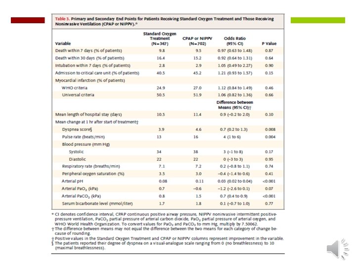
Gray, NEJM, 2008
Clinical practice guidelines for the use of noninvasive positive-pressure ventilation and noninvasive continuous positive airway pressure in the acute care setting Sean P. Keenan , MD, CMAJ, 2011 Pooled treatment failure: NIPPV RR 0. 36, 95% CI 0. 25– 0. 51 CPAP RR 0. 23, 95% CI 0. 17– 0. 32 Trend toward lower hospital mortality NIPPV RR 0. 84, 95% CI 0. 63– 1. 13 CPAP RR 0. 73, 95% CI 0. 51– 1. 05
Treatment of patients with DNI status Two basic uses For prolonged survival: Very effective in COPD and CPE Hospital survival rates > 50% High failure rates in hypoxemic respiratory failure, post-op and end stage cancer. For palliation of dyspnea or delay of death for arrival of family member Can be applied to any underlying diagnosis Reassess that palliation has actually occurred.
Evidence for efficacy and strength of recommendation: Noninvasive ventilation in acute respiratory failure Nicholas S. Hill, MD; John Brennan, MD; Erik Garpestad, MD; Stefano Nava, MD 2007 Level of evidence A: multiple randomized controlled trials and meta-analyses B: more than one randomized, controlled trial, case control series, or cohort studies C: case series or conflicting data Strength of Recommendation Recommended: Guideline: can be Option: suitable first choice for ventilatory support in selected patients used in appropriate patients but careful monitoring advised for a very carefully selected and monitored minority of patients.