Respiratory Physiology Acute Respiratory Failure Dr Priyanka Gupta


 Trachea- • • conduit for ventilation Clearance of tracheal &bronchial secretions")


: • Ease with which the lungs can expand. • Change")


 inspiratory reserve (~3100 ml)")


 • In a fetus: • After birth, vascular resistance decreases: •")
. • Alveoli at the")

 • I neurons project to, and stimulate spinal motor neurons that innervate respiratory")
: • Regulate activity")


 • Methemoglobin: • Has iron in the oxidized form (Fe 3+). • Lacks")
![Hemoglobin (continued) • Oxygen-carrying capacity of blood determined by its [hemoglobin]. • [Hemoglobin] below](https://slidetodoc.com/presentation_image/50e4f0ce12e8524cf02500c9d02a41a6/image-55.jpg "Hemoglobin (continued) • Oxygen-carrying capacity of blood determined by its [hemoglobin]. • [Hemoglobin] below")


![• Anemia: • RBCs total blood [hemoglobin] falls, each RBC produces greater amount](https://slidetodoc.com/presentation_image/50e4f0ce12e8524cf02500c9d02a41a6/image-58.jpg "• Anemia: • RBCs total blood [hemoglobin] falls, each RBC produces greater amount")
. • Dissolved C 02 (10%). •")
![Chloride Shift at Systemic Capillaries • Increased [HC 03 -] produced in RBC: •](https://slidetodoc.com/presentation_image/50e4f0ce12e8524cf02500c9d02a41a6/image-60.jpg "Chloride Shift at Systemic Capillaries • Increased [HC 03 -] produced in RBC: •")


 Intra-pulmonary • Small airways occluded ( e. g asthma, chronic")


 • • • Hypoxemia • Dyspnea,")


 • Mild to moderate RF •")


- Slides: 100
Respiratory Physiology &Acute Respiratory Failure Dr. Priyanka Gupta Dept of Anaesthesia AIIMS, Rishikesh
• Respiratory physiology is central to the practice of Anaesthesia
The most commonly used anaesthetics- the inhalational agents- depend on the lungs for uptake and elimination. The most important side effects of both inhalational and intravenously administered anaesthetics are primarily respiratory.
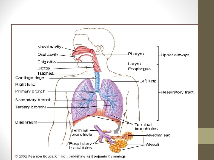
Functions of the Respiratory System • Gas Exchange • O 2, CO 2 • Acid-base balance • CO 2 +H 2 O←→ H 2 CO 3 ←→ H+ + HCO 3 • Phonation • Pulmonary defense • Pulmonary metabolism and handling of bioactive materials 6
• The term respiration includes 3 separate functions: • Ventilation: • Breathing. • Gas exchange: • Between air and capillaries in the lungs. • Between systemic capillaries and tissues of the body. • 02 utilization: • Cellular respiration. www. freelivedoctor. com Respiration
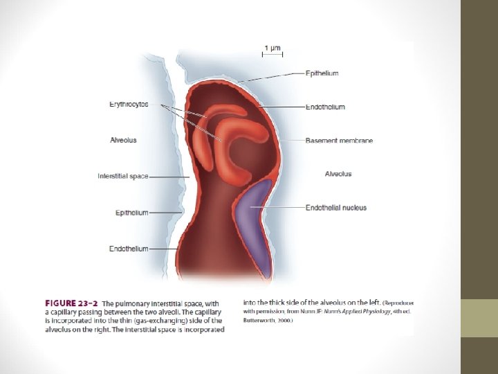
Ventilation • Diffusion is rapid because of the large surface area and the small diffusion distance. Insert 16. 1 www. freelivedoctor. com • Mechanical process that moves air in and out of the lungs. • [O 2] of air is higher in the lungs than in the blood, O 2 diffuses from air to the blood. • C 02 moves from the blood to the air by diffusing down its concentration gradient. • Gas exchange occurs entirely by diffusion:
• Region of gas exchange between air and blood. • Includes respiratory bronchioles and alveolar sacs. • Must contain alveoli. www. freelivedoctor. com Respiratory Zone
• Polyhedral in shape and clustered like units of honeycomb. • ~ 300 million air sacs (alveoli). • Large surface area (60– 80 m 2). • Each alveolus is 1 cell layer thick. • Total air barrier is 2 cells across (2 mm). • 2 types of cells: • Alveolar type I: • Structural cells. • Alveolar type II: • Secrete surfactant. www. freelivedoctor. com Alveoli
Conducting Zone • Mucus secreted to trap particles in the inspired air. • Mucus moved by cilia to be expectorated. Insert fig. 16. 5 www. freelivedoctor. com • All the structures air passes through before reaching the respiratory zone. • Warms and humidifies inspired air. • Filters and cleans:
Tracheobronchial Tree: a) Trachea- • • conduit for ventilation Clearance of tracheal &bronchial secretions Begins at the lower border of the cricoid cartilage and extends to the level of the carina Average length of 10– 13 cm The external diameters of the trachea is approximately 2. 3 cm coronally and 1. 8 cm sagitally in men and 2. 0 cm & 1. 4 cm, respectively, in women
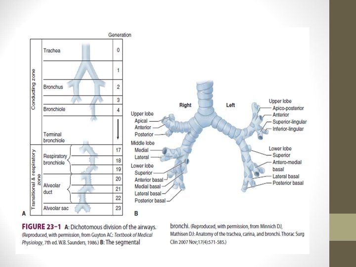
• The trachea bifurcates at the carina into the right and left main stem bronchi • Dichotomous division, starting with the trachea and ending in alveolar sacs, is estimated to involve 23 divisions. • An estimated 300 million alveoli provide an enormous membrane (50– 100 m 2 ) for gas exchange in the average adult • Gas exchange can occur only across the flat epithelium, which begins to appear on respiratory bronchioles
Pulmonary Circulation & Lymphatics • The lungs are supplied by two circulations, pulmonary and bronchial • The bronchial circulation arises from lt heart • Along their courses, the bronchial vessels anastomose with the pulmonary arterial circulation and continue as far as the alveolar duct. • The pulmonary circulation normally receives the total output of the right heart via the pulmonary artery, which divides into rt and lt branches to supply each lung
• Deoxygenated blood passes through the pulmonary capillaries, where O 2 is taken up and CO 2 is eliminated • The oxygenated blood is then returned to the lt heart by 4 main pulmonary veins (two from each lung)
Innervation • The diaphragm is innervated by the phrenic nerves, which arise from the C 3–C 5 nerve roots. • U/L phrenic nerve palsy only modestly reduces most indices of pulmonary function (~ 25%). • B/L phrenic nerve palsies produce more severe impairment • The vagus nerves provide sensory innervation to the tracheobronchial tree
Thoracic Cavity • Diaphragm: • Above diaphragm: thoracic cavity: www. freelivedoctor. com • Sheets of striated muscle divides anterior body cavity into 2 parts. • Contains heart, large blood vessels, trachea, esophagus, thymus, and lungs. • Below diaphragm: abdominopelvic cavity: • Contains liver, pancreas, GI tract, spleen, and genitourinary tract. • Intrapleural space: • Space between visceral and parietal pleurae.
• Visceral and parietal pleurae are flush against each other. • The intrapleural space contains only a film of fluid secreted by the membranes. • Lungs normally remain in contact with the chest walls. • Lungs expand contract along with the thoracic cavity. • Intrapulmonary pressure: • Intra-alveolar pressure (pressure in the alveoli). • Intrapleural pressure: • Pressure in the intrapleural space. • Pressure is negative, due to lack of air in the intrapleural space. www. freelivedoctor. com Intrapulmonary and Intrapleural Pressures
• Pressure difference across the wall of the lung. • Intrapulmonary pressure – intrapleural pressure. • Keeps the lungs against the chest wall. www. freelivedoctor. com Transpulmonary Pressure
Intrapulmonary and Intrapleural Pressures • During inspiration: • Atmospheric pressure is > intrapulmonary pressure (-3 mm Hg). • During expiration: • Intrapulmonary pressure (+3 mm Hg) is > atmospheric pressure. www. freelivedoctor. com (continued)
Boyle’s Law • Changes in intrapulmonary pressure occur as a result of changes in lung volume. • Increase in lung volume decreases intrapulmonary pressure. • Air goes in. • Decrease in lung volume, raises intrapulmonary pressure above atmosphere. • Air goes out. www. freelivedoctor. com • Pressure of gas is inversely proportional to its volume.
• Ventilation occurs as a result of pressure differences induced by changes in lung volume. • Physical properties that affect lung function: • Compliance. • Elasticity. • Surface tension. www. freelivedoctor. com Physical Properties of the Lungs
• Distensibility (stretchability): • Ease with which the lungs can expand. • Change in lung volume per change in transpulmonary pressure. DV/DP • 100 x more distensible than a balloon. • Compliance is reduced by factors that produce resistance to distension. www. freelivedoctor. com Compliance
• Tendency to return to initial size after distension. • High content of elastin proteins. • Very elastic and resist distension. • Recoil ability. • Elastic tension increases during inspiration and is reduced by recoil during expiration. www. freelivedoctor. com Elasticity
Surface Tension • Force exerted by fluid in alveoli to resist distension. • Lungs secrete and absorb fluid, leaving a very thin film of fluid. • H 20 molecules at the surface are attracted to other H 20 molecules by attractive forces. • Force is directed inward, raising pressure in alveoli. www. freelivedoctor. com • This film of fluid causes surface tension. • Fluid absorption is driven (osmosis) by Na+ active transport. • Fluid secretion is driven by the active transport of Cl- out of the alveolar epithelial cells.
Surfactant • Reduces attractive forces of hydrogen bonding by becoming interspersed between H 20 molecules. • Surface tension in alveoli is reduced. • As alveoli radius decreases, surfactant’s ability to lower surface tension increases. • Disorders: • RDS. • ARDS. Insert fig. 16. 12 www. freelivedoctor. com • Phospholipid produced by alveolar type II cells. • Lowers surface tension.
Quiet Inspiration • Active process: • Parasternal and external intercostals contract, raising the ribs; increasing thoracic volume laterally. • Pressure changes: • Alveolar changes from 0 to – 3 mm Hg. • Intrapleural changes from – 4 to – 6 mm Hg. • Transpulmonary pressure = +3 mm Hg. www. freelivedoctor. com • Contraction of diaphragm, increases thoracic volume vertically.
Expiration • Quiet expiration is a passive process. • Pressure changes: • Intrapulmonary pressure changes from – 3 to +3 mm Hg. • Intrapleural pressure changes from – 6 to – 3 mm Hg. • Transpulmonary pressure = +6 mm Hg. www. freelivedoctor. com • After being stretched by contractions of the diaphragm and thoracic muscles; the diaphragm, thoracic muscles, thorax, and lungs recoil. • Decrease in lung volume raises the pressure within alveoli above atmosphere, and pushes air out.
Pulmonary Ventilation www. freelivedoctor. com Insert fig. 16. 15
Lung volumes and capacities 4 lung volumes: tidal (~500 ml) inspiratory reserve (~3100 ml) expiratory reserve (~1200 ml) residual (~1200 ml) 4 lung capacities inspiratory (~3600 ml) functional residual (~2400 ml) vital (~4800 ml) total lung (~6000 ml)
Terms Used to Describe Lung Volumes and Capacities www. freelivedoctor. com
Anatomical Dead Space www. freelivedoctor. com • Not all of the inspired air reached the alveoli. • As fresh air is inhaled it is mixed with air in anatomical dead space. • Conducting zone and alveoli where [02] is lower than normal and [C 02] is higher than normal. • Alveolar ventilation = F x (TV- DS). • F = frequency (breaths/min. ). • TV = tidal volume. • DS = dead space.
Restrictive and Obstructive Disorders • Restrictive disorder: • Obstructive disorder: • Diagnosed by tests that measure the rate of expiration. • VC is normal. • FEV 1 is < 80%. Insert fig. 16. 17 www. freelivedoctor. com • Vital capacity is reduced. • FVC is normal.
Gas Exchange in the Lungs • Dalton’s Law: • Partial pressure: • The pressure that an particular gas exerts independently. • PATM = PN 2 + P 02 + PC 02 + PH 20= 760 mm Hg. • 02 is humidified = 105 mm Hg. • H 20 contributes to partial pressure (47 mm Hg). • P 02 (sea level) = 150 mm Hg. • PC 0 = 40 mm Hg. 2 www. freelivedoctor. com • Total pressure of a gas mixture is = to the sum of the pressures that each gas in the mixture would exert independently.
Partial Pressures of Gases in Inspired Air and Alveolar Air www. freelivedoctor. com Insert fig. 16. 20
Partial Pressures of Gases in Blood • The amount of gas dissolved in fluid reaches a maximum value (Henry’s Law). • Depends upon: • Solubility of gas in the fluid. • Temperature of the fluid. • Partial pressure of the gas. • [Gas] dissolved in a fluid depends directly on its partial pressure in the gas mixture. www. freelivedoctor. com • When a liquid or gas (blood and alveolar air) are at equilibrium:
Significance of Blood P 0 and PC 0 Measurements 2 • At normal P 0 arterial blood is about 100 mm Hg. • P 0 level in the systemic veins is about 40 mm Hg. 2 2 P is 46 mm Hg in the systemic veins. n. Provides a good index of lung function. n C 02 www. freelivedoctor. com 2
Pulmonary Circulation • Driving pressure is about 10 mm Hg. • Pulmonary vascular resistance is low. • Low pressure pathway produces less net filtration than produced in the systemic capillaries. • Avoids pulmonary edema. • Autoregulation: • Pulmonary arterioles constrict when alveolar P 0 decreases. • Matches ventilation/perfusion ratio. 2 www. freelivedoctor. com • Rate of blood flow through the pulmonary circulation is = flow rate through the systemic circulation.
Pulmonary Circulation (continued) • In a fetus: • After birth, vascular resistance decreases: • Opening the vessels as a result of subatmospheric intrapulmonary pressure. • Physical stretching of the lungs. • Dilation of pulmonary arterioles in response to increased alveolar P 0. 2 www. freelivedoctor. com • Pulmonary circulation has a higher vascular resistance, because the lungs are partially collapsed.
Lung Ventilation/Perfusion Ratios • Alveoli at apex are underperfused (overventilated). • Alveoli at the base are underventilated (overperfused). Insert fig. 16. 24 www. freelivedoctor. com • Functionally:
Brain Stem Respiratory Centers • Controls automatic breathing. • Consists of interacting neurons that fire either during inspiration (I neurons) or expiration (E neurons). Insert fig. 16. 25 www. freelivedoctor. com • Neurons in the reticular formation of the medulla oblongata form the rhythmicity center:
(continued) • I neurons project to, and stimulate spinal motor neurons that innervate respiratory muscles. • Expiration is a passive process that occurs when the I neurons are inhibited. • Activity varies in a reciprocal way. www. freelivedoctor. com Brain Stem Respiratory Centers
• I neurons located primarily in dorsal respiratory group (DRG): • Regulate activity of phrenic nerve. • Project to and stimulate spinal interneurons that innervate respiratory muscles. • E neurons located in ventral respiratory group (VRG): • Passive process. • Controls motor neurons to the internal intercostal muscles. • Activity of E neurons inhibit I neurons. • Rhythmicity of I and E neurons may be due to pacemaker neurons. www. freelivedoctor. com Rhythmicity Center
• Activities of medullary rhythmicity center is influenced by pons. • Apneustic center: • Promotes inspiration by stimulating the I neurons in the medulla. • Pneumotaxic center: • Antagonizes the apneustic center. • Inhibits inspiration. www. freelivedoctor. com Pons Respiratory Centers
Chemoreceptors 2 2 • Medulla. • Peripheral: • Carotid and aortic bodies. • Control breathing indirectly via sensory nerve fibers to the medulla (X, IX). Insert fig. 16. 27 www. freelivedoctor. com • 2 groups of chemoreceptors that monitor changes in blood PC 0 , P 0 , and p. H. • Central:
Effects of Blood PC 0 and p. H on Ventilation 2 • Oxygen content of blood decreases more slowly because of the large “reservoir” of oxygen attached to hemoglobin. • Chemoreceptors are more sensitive to changes in PC 0. H 20 + C 02 H 2 C 03 H+ + HC 03 • Rate and depth of ventilation adjusted to maintain arterial PC 0 of 40 mm Hg. 2 2 www. freelivedoctor. com • Chemoreceptor input modifies the rate and depth of breathing.
Chemoreceptor Control • Central chemoreceptors: H 20 + CO 2 H 2 C 03 2 H+ • H+ cannot cross the blood brain barrier. • C 02 can cross the blood brain barrier and will form H 2 C 03. • Lowers p. H of CSF. • Directly stimulates central chemoreceptors. www. freelivedoctor. com • More sensitive to changes in arterial PC 0.
www. freelivedoctor. com Chemoreceptor Control of Breathing
Effects of Blood P 0 on Ventilation • Blood PO 2 affected by breathing indirectly. • Influences chemoreceptor sensitivity to changes in PC 02. • Hypoxic drive: • Emphysema blunts the chemoreceptor response to PC 02. • Choroid plexus secrete more HC 03 - into CSF, buffering the fall in CSF p. H. • Abnormally high PC 02 enhances sensitivity of carotid bodies to fall in P 02. www. freelivedoctor. com 2
Effects of Pulmonary Receptors on Ventilation • Lungs contain receptors that influence the brain stem respiratory control centers via sensory fibers in vagus. • Capsaicin: • Produces apnea followed by rapid, shallow breathing. • Histamine and bradykinin: • Released in response to noxious agents. • Irritant receptors are rapidly adaptive receptors. • Hering-Breuer reflex: • Pulmonary stretch receptors activated during inspiration. • Inhibits respiratory centers to prevent undue tension on lungs. www. freelivedoctor. com • Unmyelinated C fibers can be stimulated by:
Hemoglobin and 02 Transport Insert fig. 16. 32 www. freelivedoctor. com • 280 million hemoglobin/RBC. • Each hemoglobin has 4 polypeptide chains and 4 hemes. • In the center of each heme group is 1 atom of iron that can combine with 1 molecule 02.
Hemoglobin • Oxyhemoglobin: • Deoxyhemoglobin: • When oxyhemoglobin dissociates to release oxygen, the heme iron is still in the reduced form. • Hemoglobin does not lose an electron when it combines with 02. www. freelivedoctor. com • Normal heme contains iron in the reduced form (Fe 2+). • Fe 2+ shares electrons and bonds with oxygen.
(continued) • Methemoglobin: • Has iron in the oxidized form (Fe 3+). • Lacks electrons and cannot bind with 02. • Blood normally contains a small amount. • Carboxyhemoglobin: • The reduced heme is combined with carbon monoxide. • The bond with carbon monoxide is 210 times stronger than the bond with oxygen. • Transport of 02 to tissues is impaired. www. freelivedoctor. com Hemoglobin
Hemoglobin (continued) • Oxygen-carrying capacity of blood determined by its [hemoglobin]. • [Hemoglobin] below normal. • Polycythemia: • [Hemoglobin] above normal. • Hemoglobin production controlled by erythropoietin. • Production stimulated by PC 0 delivery to kidneys. 2 • Loading/unloading depends: • P 0 of environment. • Affinity between hemoglobin and 02. 2 www. freelivedoctor. com • Anemia:
Insert fig. 16. 34 www. freelivedoctor. com Oxyhemoglobin Dissociation Curve
Effects of p. H and Temperature • Shift the curve to the right. Insert fig. 16. 35 www. freelivedoctor. com • The loading and unloading of O 2 influenced by the affinity of hemoglobin for 02. • Affinity is decreased when p. H is decreased. • Increased temperature and 2, 3 -DPG:
• Anemia: • RBCs total blood [hemoglobin] falls, each RBC produces greater amount of 2, 3 DPG. • Since RBCs lack both nuclei and mitochondria, produce ATP through anaerobic metabolism. • Fetal hemoglobin (hemoglobin f): • Has 2 g-chains in place of the b-chains. • Hemoglobin f cannot bind to 2, 3 DPG. • Has a higher affinity for 02. www. freelivedoctor. com Effect of 2, 3 DPG on 02 Transport
C 02 Transport • HC 03 - (70%). • Dissolved C 02 (10%). • Carbaminohemoglobin (20%). H 20 + C 02 ca H 2 C 03 High PC 0 2 www. freelivedoctor. com • C 02 transported in the blood:
Chloride Shift at Systemic Capillaries • Increased [HC 03 -] produced in RBC: • HC 03 - diffuses into the blood. • RBC becomes more +. • Cl- attracted in (Cl- shift). • H+ released buffered by combining with deoxyhemoglobin. • Hb. C 02 formed. • Unloading of 02. www. freelivedoctor. com • H 20 + C 02 H 2 C 03 H+ + HC 03 • At the tissues, C 02 diffuses into the RBC; shifts the reaction to the right.
Carbon Dioxide Transport and Chloride Shift www. freelivedoctor. com Insert fig. 16. 38
At Pulmonary Capillaries • H 20 + C 02 H 2 C 03 H+ + HC 03 • At the alveoli, C 02 diffuses into the alveoli; reaction shifts to the left. • Decreased [HC 03 -] in RBC, HC 03 - diffuses into the RBC. • RBC becomes more -. • Cl- diffuses out (reverse Cl- shift). • Deoxyhemoglobin converted to oxyhemoglobin. • Has weak affinity for H+. • Gives off Hb. C 02. www. freelivedoctor. com
Reverse Chloride Shift in Lungs www. freelivedoctor. com Insert fig. 16. 39
Respiratory Acidosis • PCO 2 increases. • p. H decreases. • Plasma HCO 3 - increases. www. freelivedoctor. com • Hypoventilation. • Accumulation of CO 2 in the tissues.
Respiratory Alkalosis • PCO 2 decreases. • p. H increases. • Plasma HCO 3 - decreases. www. freelivedoctor. com • Hyperventilation. • Excessive loss of CO 2.
Ventilation During Exercise • During exercise, breathing becomes deeper and more rapid. • Produce > total minute volume. • Neurogenic mechanism: • Sensory nerve activity from exercising muscles stimulates the respiratory muscles. • Cerebral cortex input may stimulate brain stem centers. • Humoral mechanism: • PC 0 and p. H may be different at chemoreceptors. • Cyclic variations in the values that cannot be detected by blood samples. 2 www. freelivedoctor. com Insert fig. 16. 41
Acute Respiratory Failure • Results from inadequate gas exchange • Insufficient O 2 transferred to the blood • Hypoxemia • Inadequate CO 2 removal • Hypercapnia
Classification of RF • Type 1 • • Hypoxemic RF • Pa. O 2 < 60 mm. Hg with • normal or ↓ Pa. CO 2 • q. Associated with acute • diseases of the lung q. Pulmonary edema (Cardiogenic, noncardiogenic (ARDS), pneumonia, pulmonary hemorrhage, and collapse • Type 2 Hypercapnic RF Pa. CO 2 > 50 mm. Hg Hypoxemia is common Drug overdose, neuromuscular disease, chest wall deformity, COPD, and Bronchial asthma
Pathophysiologic causes of Acute RF ●Hypoventilation ●V/P mismatch ●Shunt ●Diffusion abnormality
Classification of Respiratory Failure Fig. 68 -2
Mechanisms of hypoxemia • Alveolar hypoventilation • V/Q mismatch • Shunt • Diffusion limitation
Alveolar Hypoventilation Restrictive lung disease CNS disease Chest wall dysfunction Neuromuscular disease
Perfusion without ventilation (shunting) Intra-pulmonary • Small airways occluded ( e. g asthma, chronic bronchitis) • Alveoli are filled with fluid ( e. g pulm edema, pneumonia) • Alveolar collapse ( e. g atelectasis)
Dead space ventilation • DSV increase: • Alveolar-capillary interface destroyed e. g emphysema • Blood flow is reduced e. g CHF, PE • Overdistended alveoli e. g positivepressure ventilation
Diffusion limitation ØSevere emphysema ØRecurrent pulmonary emboli ØPulmonary fibrosis ØHypoxemia present during exercise
Diffusion Limitation Fig. 68 -5
Hypercarbia • Hypercarbia is always a reflection of inadequate ventilation • Pa. CO 2 is • directly related to CO 2 production • Inversely related to alveolar ventilation Pa. CO 2 = k x VCO 2 VA
Hypercarbia • When CO 2 production increases, ventilation increases rapidly to maintain normal Pa. CO 2 • Alveolar ventilation is only a fraction of total ventilation VA = V E – V D • Increased deadspace or low V/Q areas may adversely effect CO 2 removal • Normal response is to increase total ventilation to maintain appropriate alveolar ventilation
Hypercapnic Respiratory Failure • Imbalance between ventilatory supply and demand
Etiology and Pathophysiology • Airways and alveoli • Asthma • Emphysema • Chronic bronchitis • Cystic fibrosis. S
Etiology and Pathophysiology • Central nervous system • Drug overdose • Brainstem infarction • Spinal chord injuries
Etiology and Pathophysiology • Chest wall • Flail chest • Fractures • Mechanical restriction • Muscle spasm
Etiology and Pathophysiology • Neuromuscular conditions • Muscular dystrophy • Multiple sclerosis
Diagnosis of RF 1 – Clinical (symptoms, signs) • • • Hypoxemia • Dyspnea, Cyanosis Confusion, somnolence, fits • Tachycardia, arrhythmia • Tachypnea (good sign) • Use of accessory ms • Nasal flaring Recession of intercostal ms • Polycythemia Pulmonary HTN, • Corpulmonale, Rt. HF Hypercapnia ↑Cerebral blood flow, and CSF Pressure Headache Asterixis Papilloedema Warm extremities, collapsing pulse Acidosis (respiratory, and metabolic) ↓p. H, ↑ lactic acid
Respiratory Failure Symptoms • CNS: • • Headache Visual Disturbances Anxiety Confusion Memory Loss Weakness Decreased Functional Performance
Respiratory Failure Symptoms Cardiac: Orthopnea Peripheral edema Chest pain Other: Fever, Abdominal pain, Anemia, Bleeding
Clinical • Respiratory compensation • Sympathetic stimulation • Tissue hypoxia • Haemoglobin desaturation
Clinical • Respiratory compensation • Tachypnoea RR > 35 Breath /min • Accessory muscles • Recesssion • Nasal flaring • Sympathetic stimulation • Tissue hypoxia • Haemoglobin desaturation
Clinical • Respiratory compensation • Sympathetic stimulation • HR • BP • Sweating Tissue hypoxia • Altered mental state • HR and BP (late) • Haemoglobin desaturation cyanosis
Respiratory Failure Laboratory Testing Arterial blood gas Pa. O 2 Pa. CO 2 PH Chest imaging Chest x-ray CT sacn Ultrasound Ventilation–perfusion scan
Management of ARF • ICU admission • 1 -Airway management • Endotracheal intubation: • Indications • Severe Hypoxemia • Altered mental status • Importance • precise O 2 delivery to the lungs • remove secretion • ensures adequate ventilation
Management of ARF • Correction of hypoxemia • O 2 administration via nasal prongs, face mask, intubation and Mechanical ventilation • Goal: Adequate O 2 delivery to tissues • Pa. O 2 = > 60 mm. Hg • Arterial O 2 saturation >90%
Indications for intubation and mechanical ventilation • Innability to protect the airway • Respiratory acidosis (p. H<7. 2) • Refractory hypoxemia • Fatigue/increased metabolic demands • impending respiratory arrest • Pulmonary toilet
Management of ARF • Mechanical ventilation • Increase Pa. O 2 • Lower Pa. CO 2 • Rest respiratory ms (respiratory ms fatigue) • Ventilator • Assists or controls the patient breathing • The lowest FIO 2 that produces Sa. O 2 >90% and PO 2 >60 mm. Hg should be given to avoid O 2 toxicity
Management of ARF • PEEP (positive End. Expiratory pressure • Used with mechanical ventilation • Increase intrathoracic pressure • Keeps the alveoli open • Decrease shunting • Improve gas exchange • Hypoxemic RF (type 1) • ARDS • Pneumonias
Management of ARF • Noninvasive Ventilatory support (IPPV) • Mild to moderate RF • Patient should have • Intact airway, • Alert, normal airway protective reflexes • Nasal or full face mask • Improve oxygenation, • Reduce work of breathing • Increase cardiac output • AECOPD, asthma, CHF
Management of ARF • Treatment of the underlying causes • After correction of hypoxemia, hemodynamic stability • Antibiotics • Pneumonia • Infection • Bronchodilators (COPD, BA) • Salbutamol • reduce bronchospasm • airway resistance
Management of ARF • Treatment of the underlying causes • Physiotherapy • Chest percussion to loosen secretion • Suction of airways • Help to drain secretion • Maintain alveolar inflation • Prevent atelectasis, help lung expansion
Management of ARF • Weaning from mechanical ventilation • • • Stable underlying respiratory status Adequate oxygenation Intact respiratory drive Stable cardiovascular status Patient is a wake, has good nutrition, able to cough and breath deeply
THANK YOU