Management of Nystagmus the Ophthalmologists perspective Dr R

- Slides: 55
Management of Nystagmus – the Ophthalmologist’s perspective Dr. R. R. Battu Consultant Pediatric Ophthalmologist Narayana Nethralaya Bangalore
Historically What is the presenting feature? – Nystagmus - “Wobbly eyes” – Anomalous Head Posture – Poor vision – Photophobia – – – Informant: : : Night blindness Oscillopsia Vertigo Diplopia Head nodding Many times a combination of the above !!
Historically • Family history – Poor vision – Nystagmus – Neurological disease
Historically • When did this start? – At birth or shortly thereafter [ “Congenital” or infantile nystagmus ] • Congenital sensory or motor nystagmus • Congenital neurological nystagmus • Rare variants – PAN – Spasmus nutans
Historically • Medication – – – Anticonvulsants Sedatives “Psychiatric medications” • Occupation [ - and hobbies? ] • Epilepsy • Head Trauma • Neurological abnormalities……. . • Craniofacial anomalies
• Is there a visual defect? – If so, qualify and quantify • Is this likely to be an “ Ocular nystagmus” – Sensory defect nystagmus [ SDN ] – Latent nystagmus [ LN/ MLN ]
Observe • One time observation • Multiple session observation – Usually required in children – Tired adults
What to Observe • The eye • The alignment • The nystagmus • Anomalous Head position
The Eye • Evaluate refractive error • Evaluate the anterior segment • Evaluate the posterior segment
Visual Acuity • Behaviour – Eye poking • Pre verbal child or infant – Fix and follow – Other techniques
• Special problems with Latent nystagmus - Infantile Esotropia – Fogging – Polarised glasses – Vectograph – Neutral density filter – Remote occlusion – The Spielman Occluder
The Eye • Microphthalmos • Obvious malformations • AFFERENT PUPILLARY DEFECT
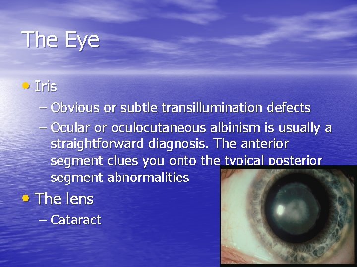
The Eye • Iris – Obvious or subtle transillumination defects – Ocular or oculocutaneous albinism is usually a straightforward diagnosis. The anterior segment clues you onto the typical posterior segment abnormalities • The lens – Cataract
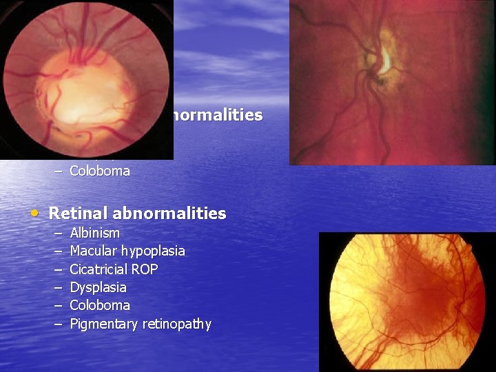
The Eye • Optic nerve abnormalities – Hypoplasia – Atrophy – Coloboma • Retinal abnormalities – – – Albinism Macular hypoplasia Cicatricial ROP Dysplasia Coloboma Pigmentary retinopathy
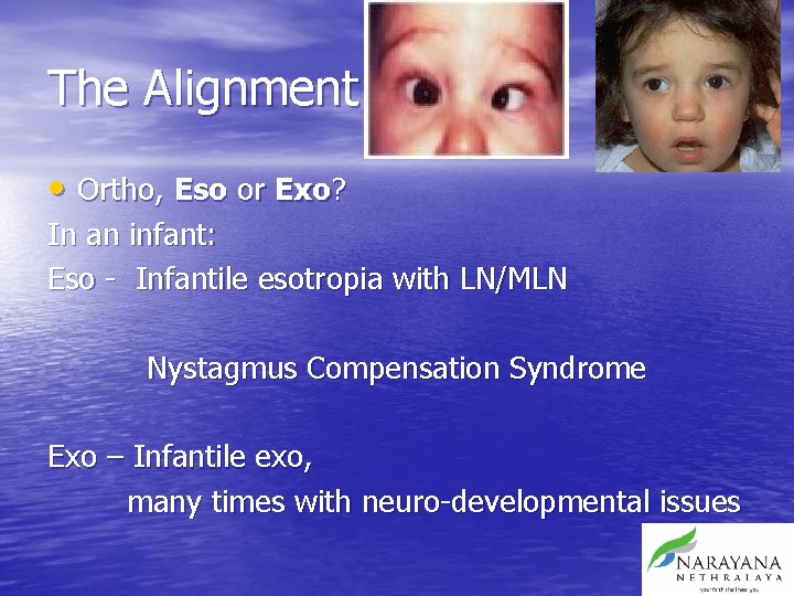
The Alignment • Ortho, Eso or Exo? In an infant: Eso - Infantile esotropia with LN/MLN Nystagmus Compensation Syndrome Exo – Infantile exo, many times with neuro-developmental issues
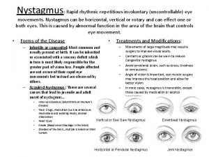
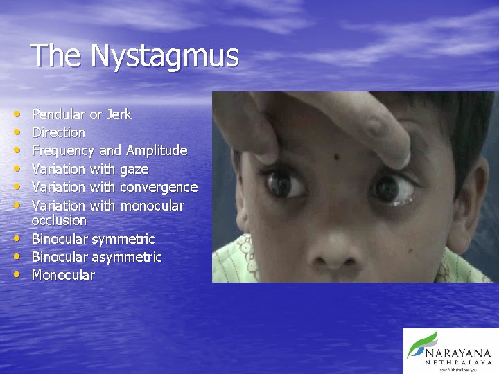
The Nystagmus • • • Pendular or Jerk Direction Frequency and Amplitude Variation with gaze Variation with convergence Variation with monocular occlusion Binocular symmetric Binocular asymmetric Monocular
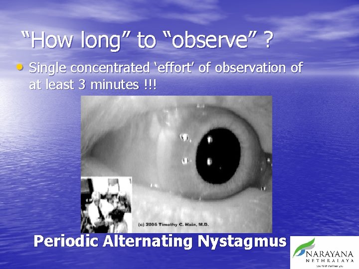
“How long” to “observe” ? • Single concentrated ‘effort’ of observation of at least 3 minutes !!! Periodic Alternating Nystagmus
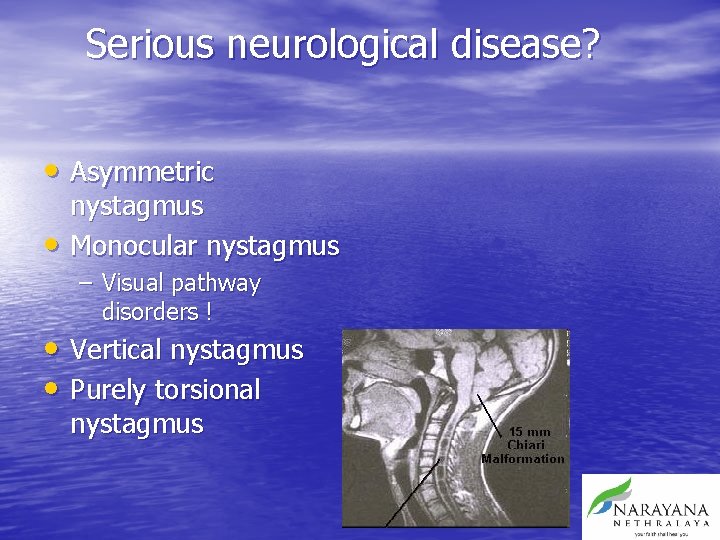
Serious neurological disease? • Asymmetric • nystagmus Monocular nystagmus – Visual pathway disorders ! • Vertical nystagmus • Purely torsional nystagmus
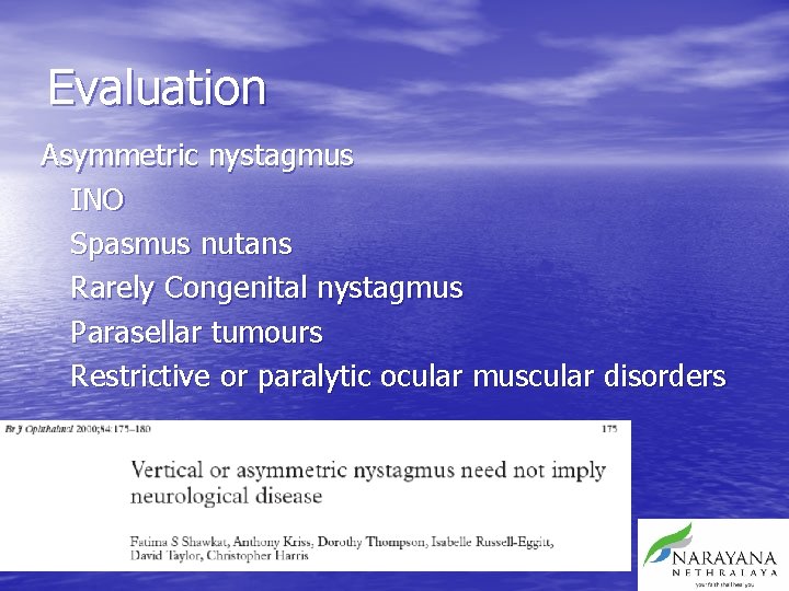
Evaluation Asymmetric nystagmus INO Spasmus nutans Rarely Congenital nystagmus Parasellar tumours Restrictive or paralytic ocular muscular disorders
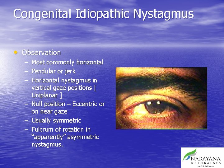
Congenital Idiopathic Nystagmus • Observation – – – Most commonly horizontal Pendular or jerk Horizontal nystagmus in vertical gaze positions [ Uniplanar ] – Null position – Eccentric or on near gaze – Usually symmetric – Fulcrum of rotation in “apparently” asymmetric nystagmus.
Congenital Idiopathic Nystagmus • Typically 3 phases of development [ Dr. Robert Reinecke] – – – Phase 1 - Broad triangular wave form [ 3 -6 mths] Phase 2 - low amp pendular waveform [6 -24 months] Phase 3 -Typical jerk nystagmus [24 -36 months] • Historically: – No oscillopsia – Invariably improves with age
Spasmus nutans • Head nodding • Anomalous head position • Monocular/asymmetric nystagmus – “ Shimmering” • RULE OUT CNS TUMOUR [ glioma ]
Latent nystagmus/ Manifest Latent Nystagmus Probably the only cause of Infantile nystagmus which does not need Electrophysiologic study or Neuro imaging
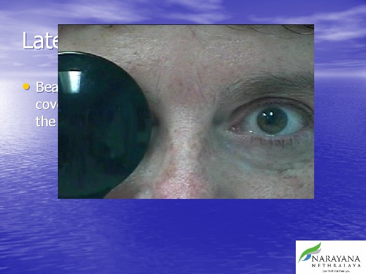
Latent nystagmus • Beats away from the covered eye [ towards the fixing eye ]
Anomalous Head Position • Null point – Beware PAN – Wandering Null point • Usually in an eccentric gaze position • Head is positioned AWAY from the null point – i. e. Null point to left, face turn to right • Mostly lateral turn, occasionally vertical and cyclovertical head turns
Electrophysiology • ERG, EOG and VER • Would probably be indicated in most situations as an initial ‘workup’ • May allow to avoid neuroimaging
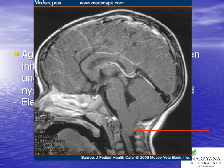
Neuro imaging • Again, would probably be required as an initial workup, unless there is unequivocally ophthalmic cause of nystagmus evident on examination and Electrophysiology
TREATMENT • Drug treatment • Optical treatment • Chemodenervation • Surgical treatment
Drug Therapy - Specific • Pendular Nystagmus – Gabapentin and Memantine • PAN – Baclofen • Superior Oblique Myokymia – Carbemazipine, Gabapentin
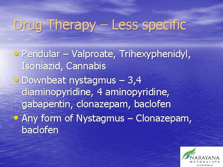
Drug Therapy – Less specific • Pendular – Valproate, Trihexyphenidyl, Isoniazid, Cannabis • Downbeat nystagmus – 3, 4 diaminopyridine, 4 aminopyridine, gabapentin, clonazepam, baclofen • Any form of Nystagmus – Clonazepam, baclofen
Optical treatment CORRECT REFRACTIVE ERROR
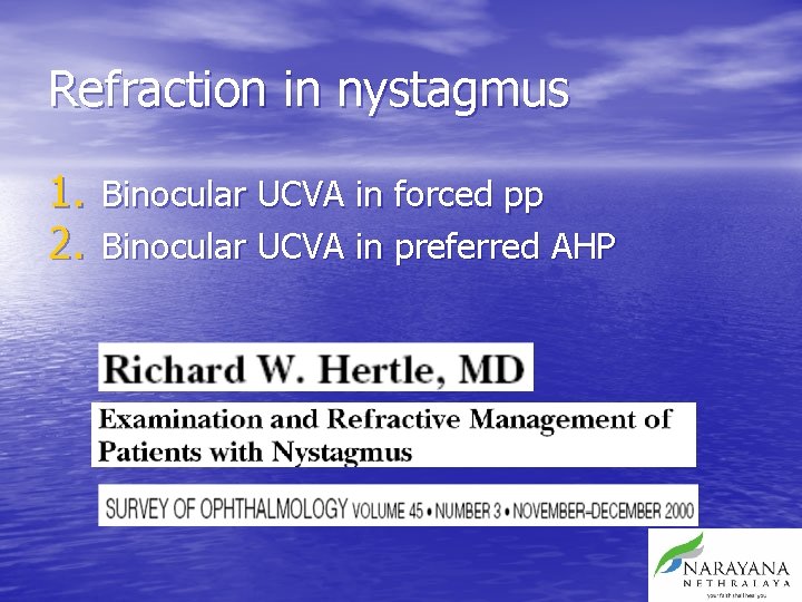
Refraction in nystagmus 1. Binocular UCVA in forced pp 2. Binocular UCVA in preferred AHP
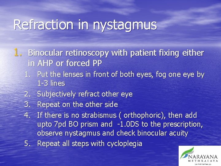
Refraction in nystagmus 1. Binocular retinoscopy with patient fixing either in AHP or forced PP 1. Put the lenses in front of both eyes, fog one eye by 1 -3 lines 2. Subjectively refract other eye 3. Repeat on the other side 4. If there is no strabismus ( orthophoric), then add upto 7 pd BO prism and -1. 0 DS to the prescription, observe nystagmus and check binocular acuity 5. Repeat all steps with cycloplegia
Factors which can be improved • Visual acuity – VA, contrast sensitivity, colour, motion sensitivity, gaze angle • Anomalous Head Position – Congenital nystagmus, acquired nystagmus, convergence damping, adduction null in LN/MLN • Oscillopsia – Acquired nystagmus, decompensated congenital nystagmus • Hypo accommodation • Photophobia
Refractive Correction • In children upto 10 years, full cycloplegic refraction • In adults, subjective, try to push over time if there is a difference in sub and obj refraction
Amblyopia therapy • May significantly decrease or eliminate MLN …… LN • Periods of occlusion have to be very prolonged in patients with LN • Alternatively fogging or penalisation may have to be used
Optical treatment • To direct the null point centrally – Prisms placed with apex directed towards the null point. – Large power prisms may have to be used. – Fresnels – May degrade vision
Optical treatment • To stabilize visual image on the retina – High plus spectacle with high minus contact lens[ -58 & +32 ] – Entire 30 deg field focussed to centre of eye, and CL refocuses to the retina. – Image remains stable irrespective of eye movement !!
Optical treatment • To induce convergence – Base out prisms bilaterally – Induce a convergence – Useful only if there is a convergence null – May have to compensate with a -1. 0 sph for induced accommodation
Chemodenervation • Botox – 2. 5 – 5 units into all horizontal recti – Retrobulbar injection of 25 – 30 units
Chemodenervation • Useful to reduce amplitude of nystagmus • Has been shown to improve foveation time and improve visual acuity slightly. • More useful in neurological acquired nystagmus, particularly in oculopalatal myoclonus • RB injection effect lasts for several weeks
Chemodenervation • Complications include – Ptosis – Diplopia – Filamentary keratitis
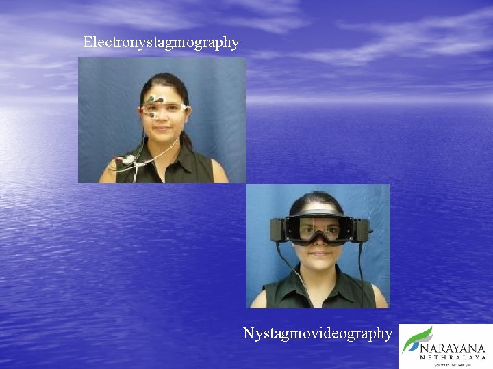
Electronystagmography Nystagmovideography
Surgical principles • Decrease the amplitude of nystagmus – Maximal recession of horizontal muscles – Tenotomy • Increase foveation time – Tenotomy • Broaden the null zone
• Rotate the null zone – – – Anderson Goto Kestenbaum Parks’ modification of Kestenbaum Augmented Kestenbaum • 40% • 60% • Induce an attempt to converge – Artificial divergence surgery
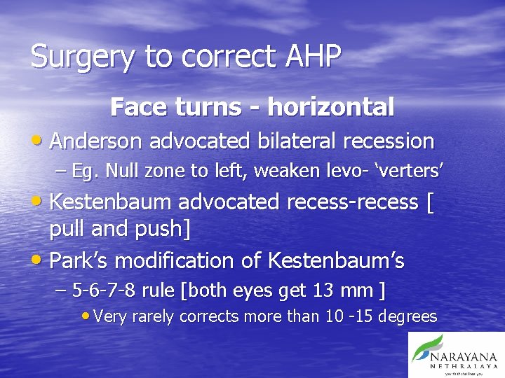
Surgery to correct AHP Face turns - horizontal • Anderson advocated bilateral recession – Eg. Null zone to left, weaken levo- ‘verters’ • Kestenbaum advocated recess-recess [ pull and push] • Park’s modification of Kestenbaum’s – 5 -6 -7 -8 rule [both eyes get 13 mm ] • Very rarely corrects more than 10 -15 degrees
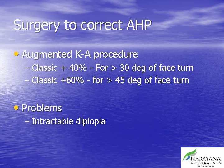
Surgery to correct AHP • Augmented K-A procedure – Classic + 40% - For > 30 deg of face turn – Classic +60% - for > 45 deg of face turn • Problems – Intractable diplopia
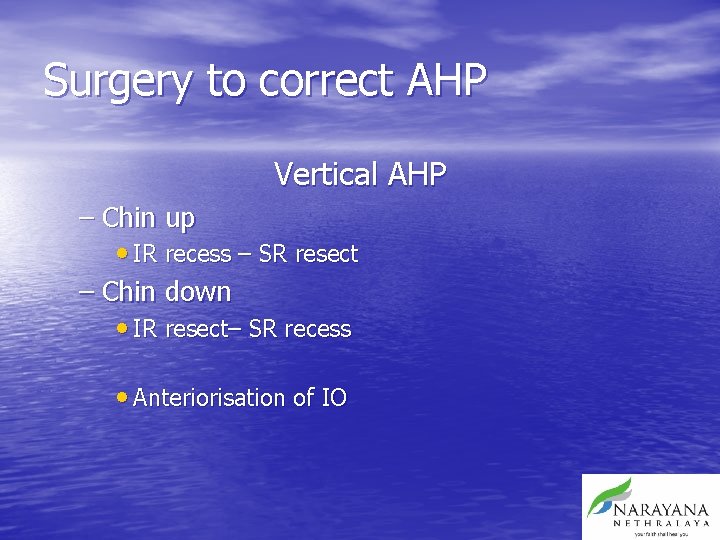
Surgery to correct AHP Vertical AHP – Chin up • IR recess – SR resect – Chin down • IR resect– SR recess • Anteriorisation of IO
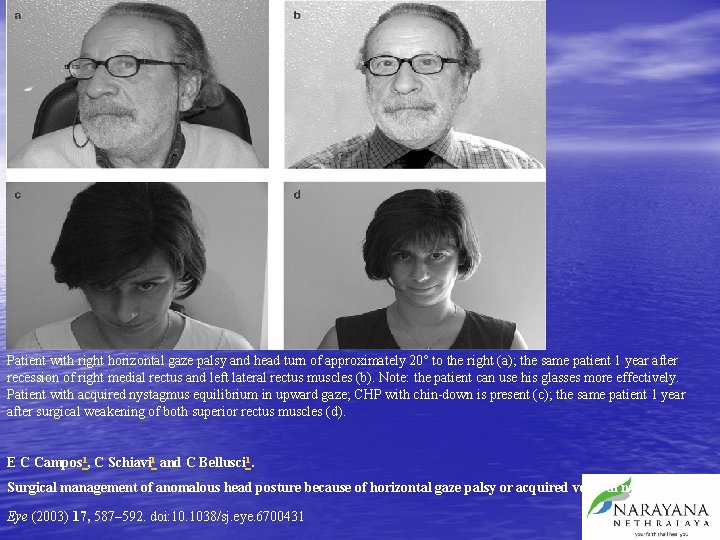
Patient with right horizontal gaze palsy and head turn of approximately 20° to the right (a); the same patient 1 year after recession of right medial rectus and left lateral rectus muscles (b). Note: the patient can use his glasses more effectively. Patient with acquired nystagmus equilibrium in upward gaze; CHP with chin-down is present (c); the same patient 1 year after surgical weakening of both superior rectus muscles (d). E C Campos 1, C Schiavi 1 and C Bellusci 1. Surgical management of anomalous head posture because of horizontal gaze palsy or acquired vertical nystagmus Eye (2003) 17, 587– 592. doi: 10. 1038/sj. eye. 6700431
Surgery to correct AHP Cyclovertical AHP • As an adaptation to torsional nystagmus • Surgery to recreate the torsional direction ‘created’ by the patient’s head tilt • Several methods – Strengthen or weaken obliques – Slanting recti insertions – Vertical recti slanting
Surgery • Other problems – Management of co existent strabismus with nystagmus – Acquiring of a new head position - PAN – Creating a new strabismus
Surgery primarily designed to improve vision • Artificial divergence – Bimedial recession – Unilateral recess-resect to XT • 4 – muscle retro equatorial recession – 10 mm MR and 12 mm LR – Ideal for PAN – May induce an exotropia
Dell’Osso & Hertle • Based on the principle of enthesial proprioceptive input to nystagmus at the insertion of the horizontal recti • • • Dell'Osso LF. Extraocular muscle tenotomy, dissection, and suture: A hypothetical therapy for congenital nystagmus. J Pediatr Ophthalmol Strab 1998; 35: 232 -3. Hertle RW, Dell'Osso LF, Fitz. Gibbon EJ, Thompson D, Yang D, Mellow SD. Horizontal rectus tenotomy in patients with congenital nystagmus. Results in 10 adults. Ophthalmology 2003; 110: 2097 -105. Hertle RW, Dell'Osso LF, Fitz. Gibbon EJ, Thompson D, Yang D, Mellow SD. Horizontal rectus muscle tenotomy in patients with infantile nystagmus syndrome: a pilot study. JAAPOS 2004; 8: 539 -48.
Summary • Evaluation of nystagmus is multidisciplinary • However, it is possible to improve the quality of life with drugs/optical devices/surgical procedures • No single procedure has shown to be consistently predictive of success • This does not mean we cannot try.
Thank you