
Keloid NEUROSENSORY SYSTEM ANATOMY PHYSIOLOGY OF EYES EXTERNAL



- Slides: 81
Keloid
NEUROSENSORY SYSTEM
ANATOMY & PHYSIOLOGY OF EYES EXTERNAL STRUCTURES EYELIDS CONJUNCTIVA PALBEBRAL BULBAR LACRIMAL APPARATUS LACRIMAL GLAND, DUCTS & PASSAGES 6 EXTRAOCULAR MUSCLES Levator palpebrae muscle
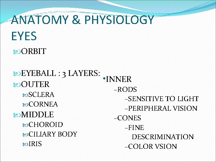
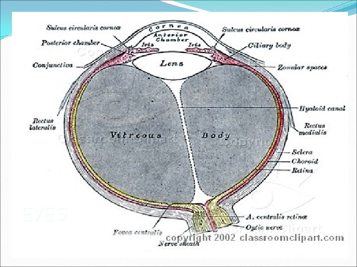
ANATOMY & PHYSIOLOGY EYES ORBIT EYEBALL : 3 LAYERS: • INNER OUTER SCLERA CORNEA MIDDLE CHOROID CILIARY BODY IRIS –RODS –SENSITIVE TO LIGHT –PERIPHERAL VISION –CONES –FINE DESCRIMINATION –COLOR VSION
EYES
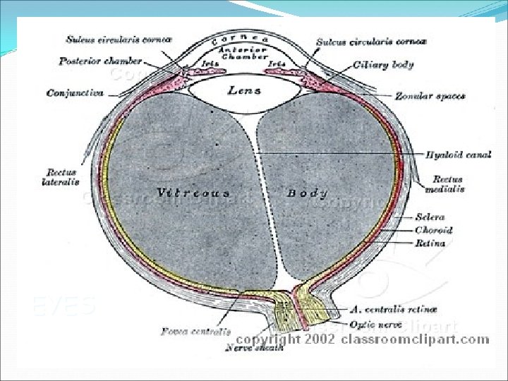
ANATOMY & PHYSIOLOGY EYES LENS – FOCUS IMAGE FLUIDS OF THE EYE: AQUEOUS HUMOR ANTERIOR & POSTERIOR CHAMBERS ANTERIOR EYE CAVITY NUTRIENTS TO LENS & CORNEA INTRAOCULAR PRESSURE MAINTENANCE 20 -25 mm. Hg VITREOUS HUMOR POSTERIOR EYE CAVITY TRANSPARENCY & FORM OF THE EYE
EYES
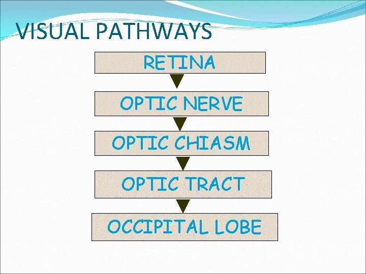
VISUAL PATHWAYS RETINA OPTIC NERVE OPTIC CHIASM OPTIC TRACT OCCIPITAL LOBE
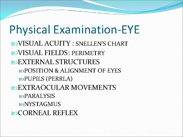
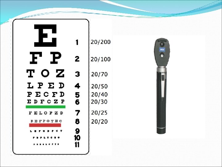
Physical Examination-EYE VISUAL ACUITY : SNELLEN’S CHART VISUAL FIELDS: PERIMETRY EXTERNAL STRUCTURES POSITION & ALIGNMENT OF EYES PUPILS (PERRLA) EXTRAOCULAR MOVEMENTS PARALYSIS NYSTAGMUS CORNEAL REFLEX
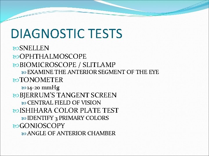
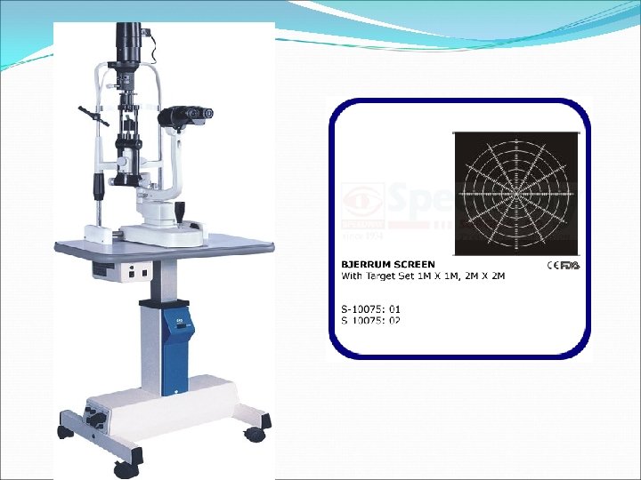
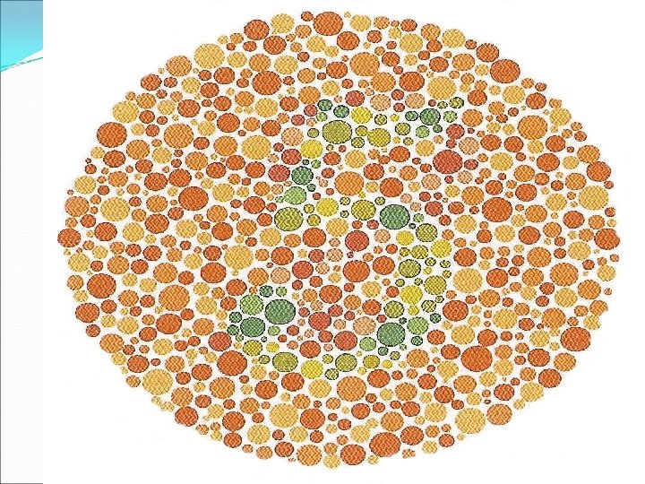
DIAGNOSTIC TESTS SNELLEN OPHTHALMOSCOPE BIOMICROSCOPE / SLITLAMP EXAMINE THE ANTERIOR SEGMENT OF THE EYE TONOMETER 14 -20 mm. Hg BJERRUM’S TANGENT SCREEN CENTRAL FIELD OF VISION ISHIHARA COLOR PLATE TEST IDENTIFY 3 PRIMARY COLORS GONIOSCOPY ANGLE OF ANTERIOR CHAMBER
PLANNING FOR HEALTH PROMOTION CARE OF THE EYES • PERSISTENT REDNESS EYEDROPS, DISCOURAGED • CONTINUED DISCOMFORT & PAIN ESP PRINTED MATTER: 14 INCHES AWAY FOLLOWING INJURY • CHILDREN: CROSSING OF EYES TV: 10 -12 FT AWAY • BLURRED SPOTS BEFORE THE EYES READ WITHVISION/ ILLUMINATION: 100 -150 WATTS • GROWTH ON THE EYE/ OPACITIES LIGHT FROM BEHIND • CONTINUAL DISCHARGE, CRUSTING OR TEACH ABOUT DANGER SIGNALS OF VISUAL TEARING DISORDER • PUPIL IRREGULARITIES
DISORDERS - EYE INJURIES & TRAUMA GLAUCOMA INFECTIONS DETACHMENT OF THE RETINA CATARACT REFRACTIVE ERRORS
INJURIES & TRAUMA EMERGENCY: TREAT THE PATIENT, LEAVE THE EYE ALONE, EXCEPT IN CHEMICAL INJURY - FLUSH EYES STAT FOREIGN BODIES: FLUSH WITH WATER FOR 15 MIN WHILE GOING TO THE DOCTOR; DON’T TOUCH CORNEA
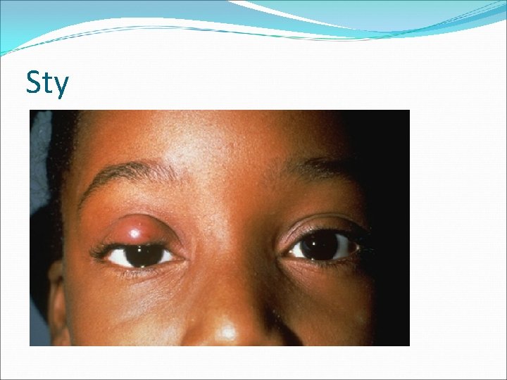
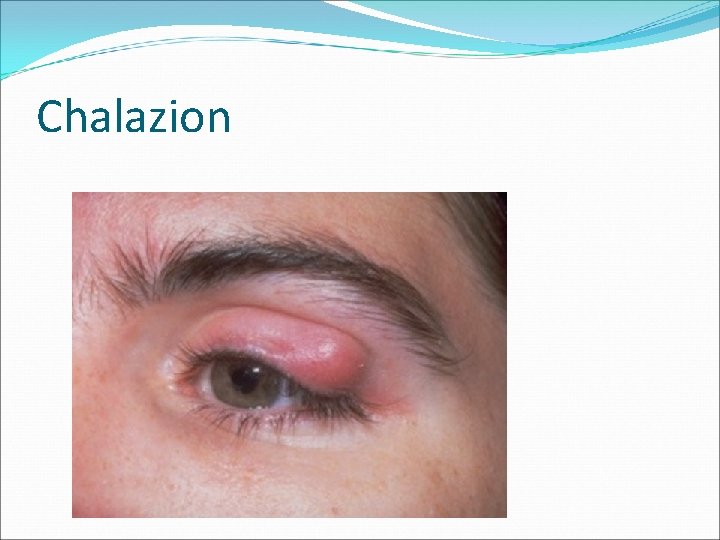
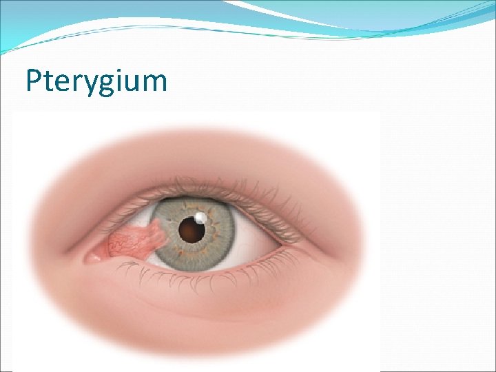
INFECTIONS HORDEOLUM/ STY -Zeis gland in the follicle CHALAZION –meibomian glands CONJUNCTIVITIS – pink eye bacterial infection, allergy, trauma UVEITIS - iris KERATITIS - cornea PTERYGIUM – triangular fold From white of the eye to the cornea
Conjunctivitis
Sty
Chalazion
Pterygium
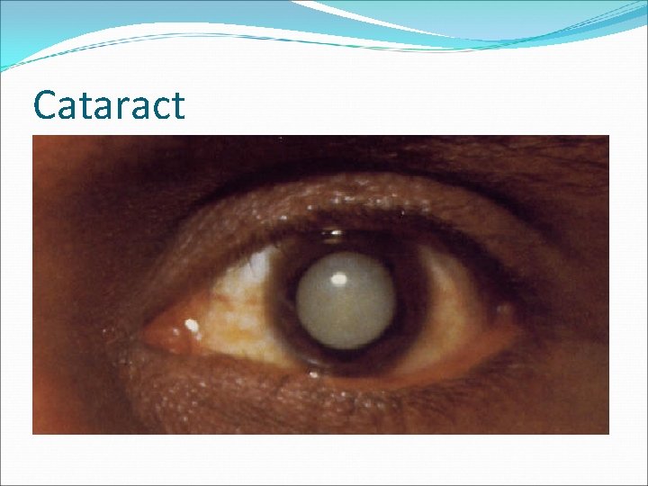
CATARACT Opacity of the lens & its capsule which interferes with transparency S/SX: Dimness in visual acuity Rapid & marked refraction error CLASSIFICATION: Primary/ senile Secondary/ traumatic Congenital
Cataract
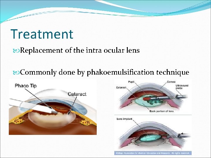
Treatment Replacement of the intra ocular lens Commonly done by phakoemulsification technique
EYE SURGERY NURSING CARE PRE-OP Orient to new environment Teach deep breathing & how to close eyes without squeezing Eye antibiotics preop Mydiatrics if ordered
EYE SURGERY NURSING CARE POST-OP Reorient patient to his surroundings Prevent increase in IOP & stress on the suture line
Contd…. ACTIVITIES THAT INCREASE IOP: • Coughing • Vomiting • Bending • Stooping • Promote comfort of the patient: mild analgesic to control pain
EYE SURGERY NURSING CARE POST-OP COMPLICATIONS: Observe & treat complications • NAUSEA & VOMITING • Antiemetics • Cold the compress • Promote rehab of the patient • HEMORRHAGE • Encourage the patient to become • Sudden pain of thewith eye him when he first independentwalk • PROLAPSE OF THE IRIS become ambulatory • Most common postop complication • Health teachings • Can precipitate glaucoma
EYE SURGERY HEALTH TEACHINGS: 1 -4 wks : dark glasses; temporary corrective lenses 6 -8 wks: permanent lenses It will take time to learn distances & climb stairs Color slightly changed Use one eye at a time unless with contact lens Decreased peripheral vision
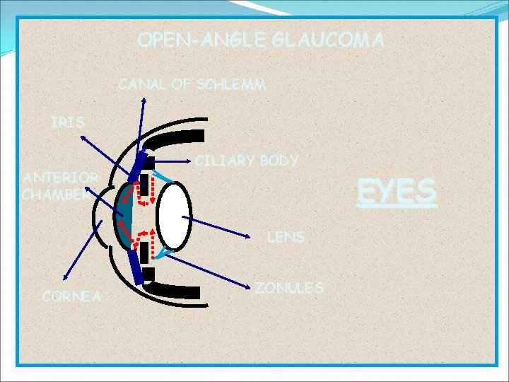
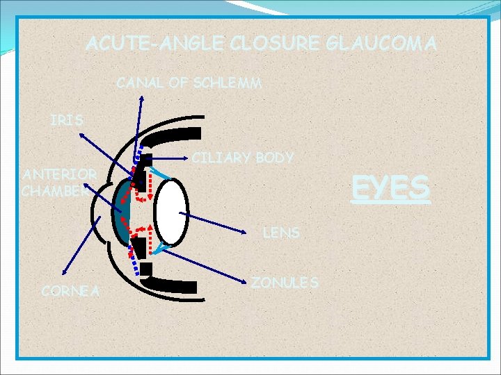
GLAUCOMA INCREASED IOP PROGRESSIVE LOSS OF PERIPHERAL VISION CAUSE: OBSTRUCTION TO CIRCULATION OF AQUEOUS HUMOR TYPES: 1. CHRONIC/ SIMPLE/ OPEN-ANGLE 2. ACUTE ANGLE CLOSURE 3. Congenital 4. Secondary – trauma, uveitis, postop 5. Absolute – uncontrolled- enucleation
OPEN-ANGLE GLAUCOMA CANAL OF SCHLEMM IRIS ANTERIOR CHAMBER CILIARY BODY LENS CORNEA ZONULES EYES
ACUTE-ANGLE CLOSURE GLAUCOMA CANAL OF SCHLEMM IRIS ANTERIOR CHAMBER CILIARY BODY LENS CORNEA ZONULES EYES
OPEN ANGLE GLAUCOMA S/SX: Loss of peripheral vision (tunnel) Difficulty in adjusting to darkness Failure to detect changes in color Headache, pain behind the eyeball Halos Nausea & vomiting
OPEN ANGLE GLAUCOMA MANAGEMENT: Conservative : Miotics : pupillary constriction draw iris smooth muscle away from the canal Acetazolamide : decrease aqueous production Fluid restriction
Definitive management Principle: improve drainage of aqueous • Iridocleisis-anterior chamber & subconjunctival space • Corneoscleral trephening – junction of cornea & sclera • Trabeculotomy • Laser therapy to meshwork
Acute Angle Glaucoma CAUSE: Pupillary dilation by mydiatrics Abnormal anterior displacement of iris S/SX: Severe eye pain Nausea & vomiting Blurred vision Colored halos around lights Dilated pupils Increased IOP
MANAGEMENT: • Miotics • Azetazolamide • Osmotic agents – glycerol • Surgery - iridectomy
GLAUCOMA NURSING CARE – SURGERY PRE-OP Explain that vision lost cannot be restored, but further loss can be prevented POST-OP Flat 24 H- prevent iris prolapse Narotics or sedatives Liquid diet until 1 st dressing Turn to unoperative site
LONG TERM CARE: • No restriction on the use of the eyes • No fluid restriction; exercise permitted • Medical follow up needed for life
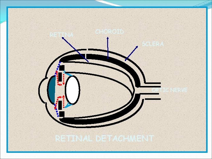
RETINA CHOROID SCLERA OPTIC NERVE RETINAL DETACHMENT
RETINAL DETACHMENT Fluid accumulation Tumor CAUSE: Myopic degeneration Trauma Aphakia S/SX: Floating spots or opacities before the eye Casts shadows on the retina Bright. Flashes of light Progressive constriction of vision in 1 eye
Management Conservative : • Quiet in bed with eyes covered • Head: positioned so that retinal holes lower • Photocoagulation – small burn to retina • Cryotherapy – cold probe to freeze retina Surgical: • Scleral buckling- sealing break & reattaching
RETINAL DETACHMENT POST-OP NURSING CARE: Cover eyes Area of detachment, dependent Mydiatrics Discharge instructions: No strenuous exercises & acivity x 6 mos Contact sports restricted No sudden jarring head motion No restriction with use of eyes
REFRACTIVE ERRORS REFRACTION – bending of light rays ACCOMMODATION – ability to adjust from near to far vision ADAPTATION – ability to see light from darkness COMMON ERRORS: Myopia Hyperopia Presbyopia • Astigmatism • Blindness
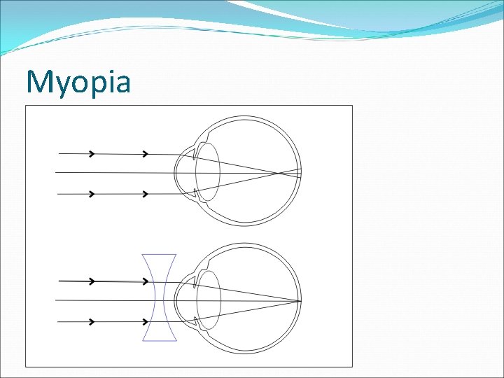
myopia NEAR-SIGHTED Long A-P dimension of the eyeball Light rays focus infront of the retina Good vision for near distances Concave lenses
Myopia
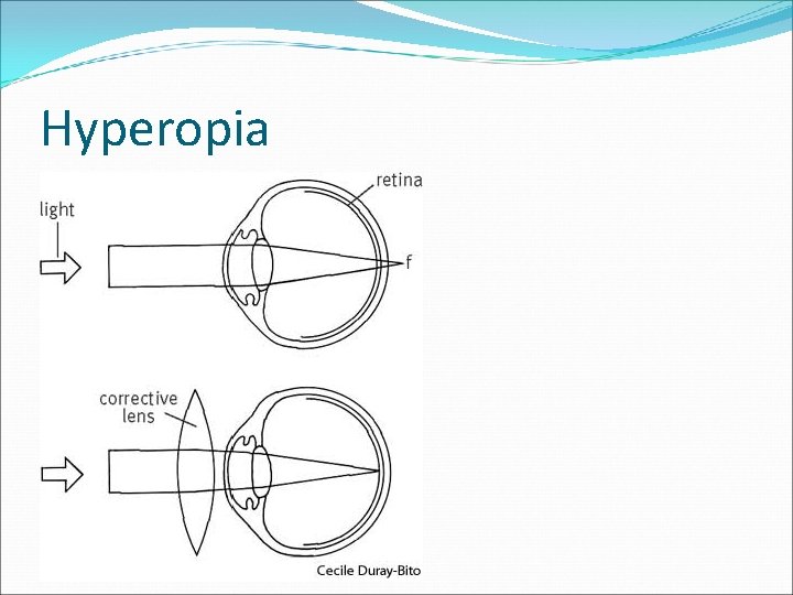
hyperopia FAR-SIGHTED Eyeball A-P dimension too short Light rays focus behind the retina Good vision for far distances Convex lenses
Hyperopia
presbyopia FARSIGHTEDNESS OF OLD AGE Gradual loss of accommodation Loss of lens elasticity Inability to read without holding the material more than 13 ft from the eye Bifocal lenses
ASTIGMATISM Asymmetry or irregular curvature of the cornea Cylindrical lenses BLINDNESS Vision: 20/200
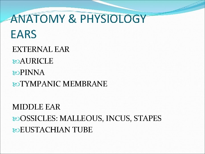
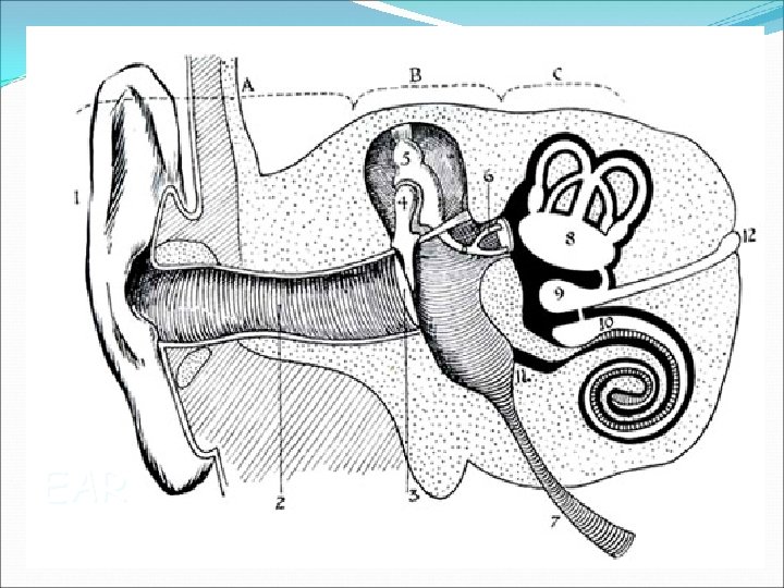
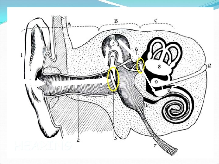
ANATOMY & PHYSIOLOGY EARS EXTERNAL EAR AURICLE PINNA TYMPANIC MEMBRANE MIDDLE EAR OSSICLES: MALLEOUS, INCUS, STAPES EUSTACHIAN TUBE
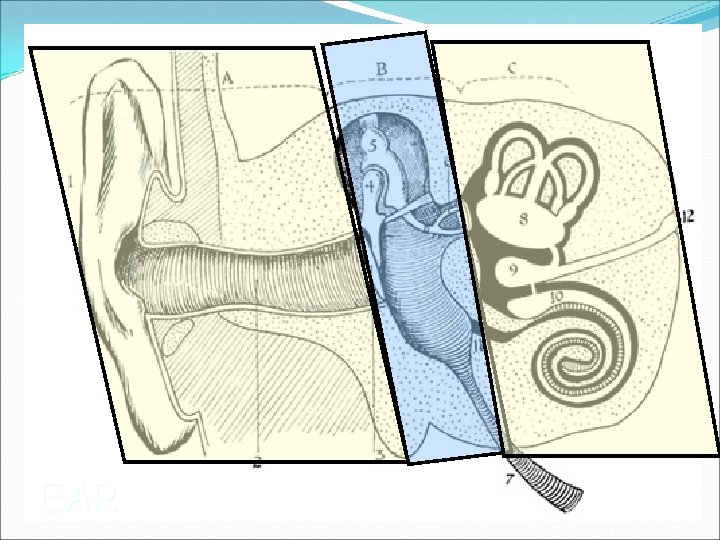
EAR
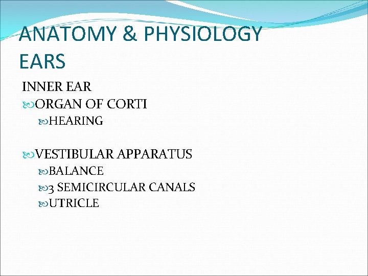
ANATOMY & PHYSIOLOGY EARS INNER EAR ORGAN OF CORTI HEARING VESTIBULAR APPARATUS BALANCE 3 SEMICIRCULAR CANALS UTRICLE
EAR
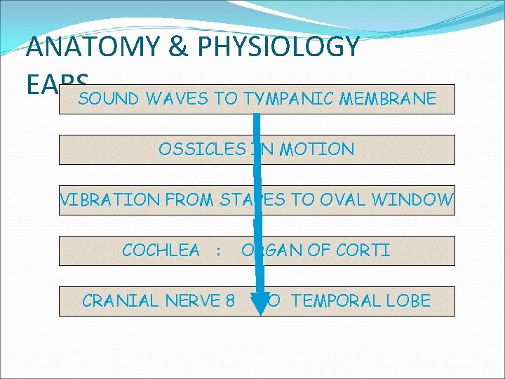
ANATOMY & PHYSIOLOGY EARSSOUND WAVES TO TYMPANIC MEMBRANE OSSICLES IN MOTION VIBRATION FROM STAPES TO OVAL WINDOW COCHLEA : CRANIAL NERVE 8 ORGAN OF CORTI TO TEMPORAL LOBE
HEARING
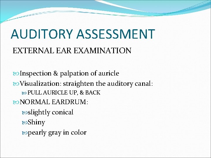
AUDITORY ASSESSMENT EXTERNAL EAR EXAMINATION Inspection & palpation of auricle Visualization: straighten the auditory canal: PULL AURICLE UP, & BACK NORMAL EARDRUM: slightly conical Shiny pearly gray in color
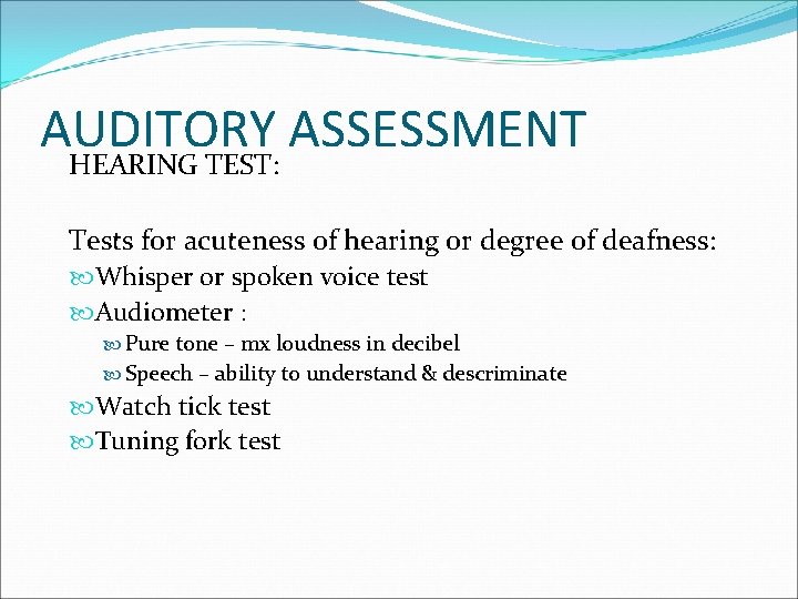
AUDITORY ASSESSMENT HEARING TEST: Tests for acuteness of hearing or degree of deafness: Whisper or spoken voice test Audiometer : Pure tone – mx loudness in decibel Speech – ability to understand & descriminate Watch tick test Tuning fork test
AUDITORY ASSESSMENT HEARING TEST: Test to localize cause of deafness: Rinne’s Weber’s
Auditory assessment WEBER’S • • Tuning fork top midline of the head Sound heard: normal ear vs affected ear Better in affected ear: conductive Better in normal ear : sensorineural
AUDITORY ASSESSMENT TEST FOR VESTIBULAR FUNCTON CALORIC TEST Check direction of nystagmus COWS ( cold-opposite; warm-same side of stimulated ear) ROTATION (BARANY) TEST Rotating chair Nystagmus is opposite to the direction of rotation
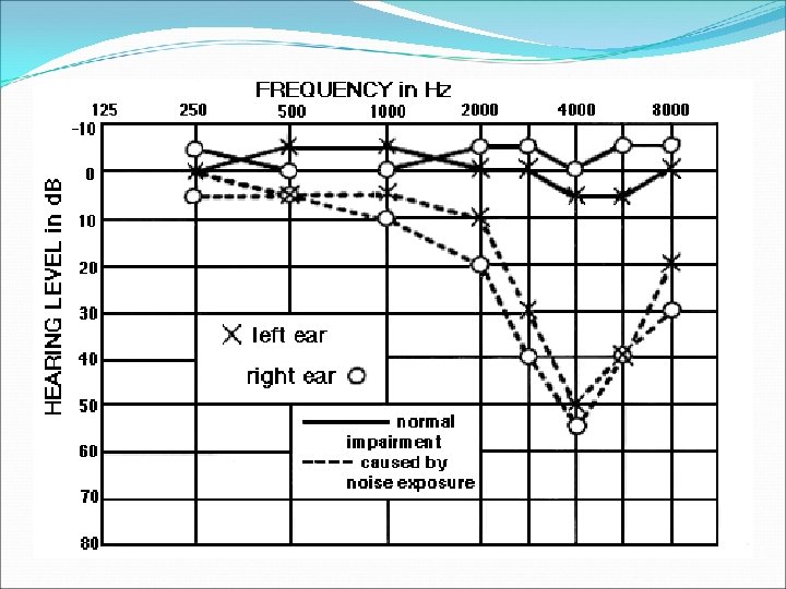
HEALTH PROMOTION EAR PROTECTION Noise over 70 decibels is potentially damaging to hearing Most common & impt type of occupational hearing is caused by LOUD NOISE
GENERAL EAR CARE Ear is self-cleaning Cerumen-lubricant; traps dirt Cleanse the external ear reached by vision
NURSING INTERVENTIONS EAR DROPS Warm After adm’n, head should remain tilted SOFTENING & REMOVING IMPACTED CERUMEN Few drops of hydrogen peroxide/ warm glycerine Irrigate the ear
NURSING INTERVENTIONS EAR IRRIGATION To clean the external canal Remove impated cerumen Caloric test Apply antiseptic solutions Remove foreign bodies
COMMON EAR PROBLEMS 1. OTOSCLEROSIS 2. MENIERE’S DISEASE 3. HEARING IMPAIRMENT
OTOSCLEROSIS Normal bone is replaced by spongy bone Ankylosis of the footplate of the stapes Impaired vibration system
OTOSCLEROSIS ASSESSMENT Gradual hearing loss Difficulty hearing a whisper Own voice is loud Paracusis : hear better in loud environment Rinne’s test: bone conduction better
OTOSCLEROSIS PLANNING & IMPLEMENTATION Hearing aid Surgery – primary form of tx Stapedectomy Stapes mobilization operation Fenestration operation : new window is created
EAR SURGERY PRE-OP CARE; Hair shampoo Inform client: Head still during surgery Post op: get out of bed with assistance avoid nose blowing until 1 week
EAR SURGERY POST OP Promote comfort & safety Promote psychological well-being Prevent complications
Complications • Facial nerve involvement • Facial paralysis, facial weakness • Inability to show teeth, wrinkle forehead, raise eyebrows or close eyes • Meningitis – bacterial • Report signs & symptoms • Bleeding
MENIERE’S DSE Chronic Increase in endolymphatic pressure ASSESSMENT: Tinnitus Unilateral hearing loss Vertigo
MENIERE’S DSE PLANNING & IMPLEMENTATION CONSERVATIVE: palliative Bed rest Meds Sedative : Phenobarbital Antihistamine Antiemetics Low salt diet
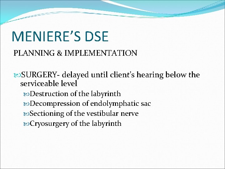
MENIERE’S DSE PLANNING & IMPLEMENTATION SURGERY- delayed until client’s hearing below the serviceable level Destruction of the labyrinth Decompression of endolymphatic sac Sectioning of the vestibular nerve Cryosurgery of the labyrinth
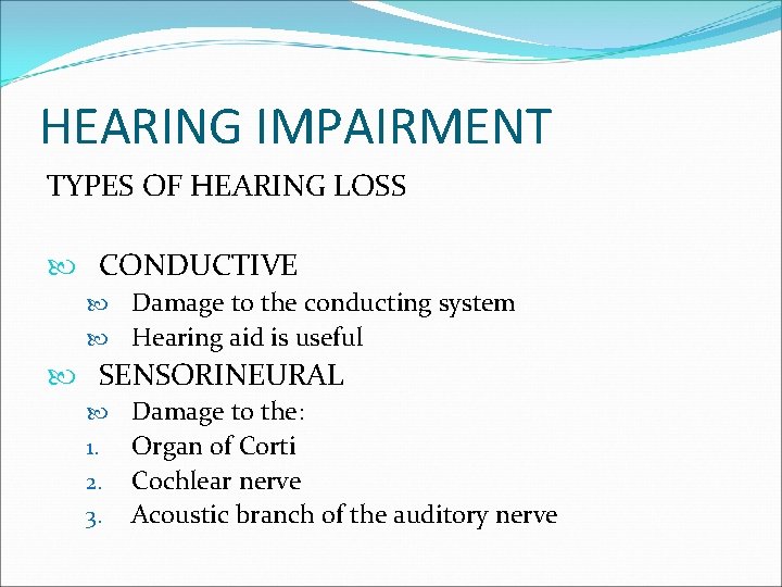
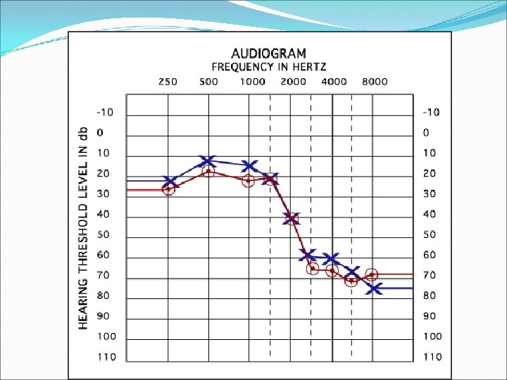
HEARING IMPAIRMENT TYPES OF HEARING LOSS CONDUCTIVE Damage to the conducting system Hearing aid is useful SENSORINEURAL 1. 2. 3. Damage to the: Organ of Corti Cochlear nerve Acoustic branch of the auditory nerve
COMMUNICATING WITH HEARINGIMPAIRED CLIENTS Avoid use of gestures without speech Do not shout Speak distinctly & as close to the client Use short phrases Do not communicate with someone else in front of a hearing-impaired client Hearing impairment goes with visual problems in elderly
SOUND AMPLIFICATION TYPES OF HEARING AIDS; Post-auricular Body-type In-the ear model Select hearing aid that has cotrollable volume & is properly fitted
Thank You