2 PART 1 Cells The Living Units Power


- Slides: 57

2 PART 1 Cells: The Living Units Power. Point® Lecture Presentations prepared by Leslie Hendon University of Alabama, Birmingham © 2014 Pearson Education, Inc.

Overview of Cells • Cells are the smallest living units in the body • Obtain nutrients • Make molecules needed to survive • Dispose of wastes • Maintain shape of cell • Replicate © 2014 Pearson Education, Inc.

Overview of Cells • Organelles • Subunits of cells with specific functions • Most cells contain the same basic organelles • Not all cells have all organelles in the same abundance © 2014 Pearson Education, Inc.

Overview of Cells • Cells have three main components • Plasma membrane—the outer boundary • Cytoplasm—contains most organelles • Nucleus—controls cellular activities © 2014 Pearson Education, Inc.

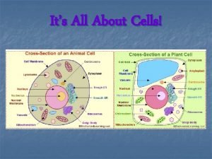
Figure 2. 1 Structure of a generalized cell. Chromatin Nucleolus Nuclear envelope Nucleus Plasma membrane Smooth endoplasmic reticulum Cytosol Mitochondrion Lysosome Rough endoplasmic reticulum Centrioles Centrosome matrix Ribosomes Golgi apparatus Cytoskeletal elements Microtubule Intermediate filaments © 2014 Pearson Education, Inc. Secretion being released from cell by exocytosis Peroxisome

The Plasma Membrane • Plasma membrane defines the extent of the cell • Separates the intracellular fluid within cell from extracellular fluid outside and between cells • Structure of membrane • Fluid mosaic model (lipid bilayer) • Types of membrane proteins • Integral proteins—firmly imbedded in, or attached to lipid bilayer • Short chains of carbohydrates attach to integral proteins • Peripheral proteins—attach to membrane surface • Support plasma membrane from the cytoplasmic side © 2014 Pearson Education, Inc.

Figure 2. 2 The plasma membrane according to the fluid mosaic model. Polar head of phospholipid molecule Nonpolar tail of phospholipid molecule Cholesterol Extracellular fluid (watery environment) Glycolipid Glycoprotein Carbohydrate of glycocalyx Bimolecular lipid layer containing proteins Outward-facing layer of phospholipids Inward-facing layer of phospholipids Cytoplasm (watery environment) Integral Filament of Peripheral proteins cytoskeleton proteins © 2014 Pearson Education, Inc.

The Plasma Membrane • Functions—relate to location at the interface of cell’s exterior and interior • Provides barrier against substances outside cell • Some plasma membranes act as receptors • Determines which substances enter or leave the cell • Membrane is selectively permeable © 2014 Pearson Education, Inc. Membrane Structure

Membrane Transport Extracellular fluid • Simple diffusion— tendency of molecules to move down their concentration gradient Lipidsoluble solutes Water molecules Lipid bilayer Cytoplasm • Osmosis— diffusion of water molecules across a membrane © 2014 Pearson Education, Inc. Simple diffusion of fat-soluble molecules directly through the phospholipid bilayer down their concentration gradient Osmosis, diffusion of water through the lipid bilayer

Membrane Transport Mechanisms • Facilitated diffusion— movement of molecules down their concentration gradient through an Lipid bilayer integral protein • Active transport— integral proteins move molecules across the plasma membrane against their concentration gradient © 2014 Pearson Education, Inc. Water soluble solutes Extracellular fluid Solute ATP Cytoplasm Facilitated diffusion An integral protein that spans the plasma membrane enables the passage of a particular solute across the membrane. Active transport Some transport proteins use ATP as an energy source to actively pump substances across the plasma membrane against their concentration gradient.

Endocytosis • Mechanism by which particles enter cells • Phagocytosis—“cell eating” • Pinocytosis—“cell drinking” • Receptor-mediated endocytosis • Plasma proteins bind to certain molecules • Invaginates and forms a coated pit • Pinches off to become a coated vesicle • This is the method by which insulin, other hormones, enzymes, and low-density lipoproteins enter cells © 2014 Pearson Education, Inc.

Figure 2. 4 The three types of endocytosis. Phagocytosis The cell engulfs a large particle by forming projecting pseudopods (“false feet”) around it and enclosing it within a membrane sac called a phagosome. The phagosome then combines with a lysosome, and its contents are digested. Vesicle may or may not be protein-coated but has receptors capable of binding to microorganisms or solid particles. Receptors Phagosome Pinocytosis Infolding of the plasma membrane carries a drop of extracellular fluid containing solutes into the cell in a tiny membrane-bound vesicle. No receptors are used, so the process is nonspecific. Vesicle Receptor recycled to plasma membrane © 2014 Pearson Education, Inc. Receptor-mediated endocytosis Extracellular substances bind to specific receptor proteins in regions of protein-coated pits, enabling the cell to ingest and concentrate specific substances in protein-coated vesicles. The ingested substance may simply be released inside the cell or combined with a lysosome to digest contents. Receptors are recycled to the plasma membrane in vesicles.

Exocytosis • Exocytosis—a mechanism that moves substances out of the cell • Substance is enclosed in a vesicle • The vesicle migrates to the plasma membrane Photomicrograph of a secretory vesicle releasing its contents by exocytosis (110, 000 x) © 2014 Pearson Education, Inc. Extracellular fluid Secretory vesicle Plasma membrane SNARE (t-SNARE) Vesicle SNARE (v-SNARE) Molecule to be secreted Cytoplasm Fused v- and t-SNAREs The process of exocytosis Figur 1 The molecule to be secreted migrates to the plasma membrane in a membrane-bound vesicle. 2 At the plasma membrane, proteins at the vesicle surface (v-SNARES, v for “vesicle”) bind with t-SNAREs (plasma membrane proteins, t for “target”). Fusion pore formed 3 The vesicle and plasma membrane fuse and a pore opens up. 4 Vesicle contents are released to the cell exterior.

The Cytoplasm • Cytoplasm • Lies internal to plasma membrane • Consists of cytosol, organelles, and inclusions • Cytosol • Jelly-like fluid in which other cellular elements are suspended • Consists of water, ions, and enzymes © 2014 Pearson Education, Inc.

Cytoplasmic Organelles • Ribosomes—constructed of proteins and ribosomal RNA; not surrounded by a membrane • Site of protein synthesis • Assembly of proteins is called translation • Are the “assembly line” of the manufacturing plant • Free ribosomes function within the cytosol • Ribosomes attached to ER • Make proteins to renew plasma membrane • Make proteins that are exported from the cell © 2014 Pearson Education, Inc.

Cytoplasmic Organelles • Endoplasmic reticulum—system of membrane-walled envelopes and tubes throughout cytoplasm • Rough ER—ribosomes stud the external surfaces • Smooth ER—consists of tubules in a branching network • No ribosomes are attached; therefore no protein synthesis © 2014 Pearson Education, Inc.

Figure 2. 6 The endoplasmic reticulum (ER) and ribosomes. Nucleus Smooth ER Nuclear envelope Rough ER Ribosomes Cisterns Diagrammatic view of smooth and rough ER © 2014 Pearson Education, Inc. Electron micrograph of smooth and rough ER (18, 000 x)

Cytoplasmic Organelles • Golgi apparatus—a stack of 3 to 10 discshaped envelopes • Sorts products of rough ER and sends them to proper destination • Products of rough ER move through the Golgi from the convex (cis) to the concave (trans) side • Is the “packaging and shipping” division of the manufacturing plant © 2014 Pearson Education, Inc.

Figure 2. 7 Golgi apparatus. Transport vesicle from rough ER Cis face— “receiving” side of Golgi apparatus New vesicles forming Cisterns New vesicles forming Transport vesicle from trans face Secretory vesicle Trans face—“shipping” side of Golgi apparatus Many vesicles in the process of pinching off from the Golgi apparatus © 2014 Pearson Education, Inc. Transport vesicle at the trans face Electron micrograph of the Golgi apparatus (25, 000 x)

Figure 2. 8 The sequence of events from protein synthesis on the rough ER to the final distribution of these proteins. 1 Proteincontaining vesicles pinch off rough ER and migrate to fuse with membranes of Golgi apparatus. Rough ER ER membrane Phagosome Plasma membrane Proteins in cisterns Pathway C: Lysosome containing acid hydrolase enzymes 2 Proteins are modified within the Golgi compartments. Vesicle becomes lysosome 3 Proteins are then packaged within different vesicle types, depending on their ultimate destination. Golgi apparatus Pathway A: Vesicle contents destined for exocytosis © 2014 Pearson Education, Inc. Secretory vesicle Secretion by exocytosis Pathway B: Vesicle membrane to be incorporated into plasma membrane Extracellular fluid

Cytoplasmic Organelles • Lysosomes—membrane-walled sacs containing digestive enzymes • Digest unwanted substances • Are the cell’s “demolition crew” Lysosomes Figure 2. 9 Electron micrograph of lysosomes (27, 000 ), artificially colored. © 2014 Pearson Education, Inc. Light green areas are regions where materials are being digested.

Cytoplasmic Organelles • Mitochondria—surrounded by double-walled membrane • Generate most of the cell’s energy • “Power plant” of the cell • Release energy stored in chemical bonds and transfer energy to produce ATP © 2014 Pearson Education, Inc.

Cytoplasmic Organelles • Mitochondria—continued • Cells with high energy requirements have more mitochondria—e. g. , muscle cells • Most complex organelle • Contain some maternally inherited DNA • Believed to have arisen from bacteria © 2014 Pearson Education, Inc.

Figure 2. 10 Mitochondria. Outer mitochondrial membrane Ribosome Mitochondrial DNA Inner mitochondrial membrane Cristae Matrix Enzymes © 2014 Pearson Education, Inc.

Peroxisomes • Peroxisomes—membrane-walled sacs of oxidase enzymes • Enzymes neutralize free radicals and break down poisons • Break down long chains of fatty acids • Are numerous in the liver and kidneys • Are the “toxic waste removal system” © 2014 Pearson Education, Inc.

Cytoplasmic Organelles • Cytoskeleton—“cell skeleton”—an elaborate network of rods • Contains three types of rods: • Microtubules—cylindrical structures made of proteins • Microfilaments—filaments of contractile protein actin • Intermediate filaments—protein fibers © 2014 Pearson Education, Inc.

Figure 2. 11 a Cytoskeletal elements. Microfilaments Strands made of spherical protein subunits called actins Actin subunit 7 nm Microfilaments form the blue network surrounding the pink nucleus in this photo. © 2014 Pearson Education, Inc.

Figure 2. 11 a Cytoskeletal elements. Intermediate filaments Tough, insoluble protein fibers constructed like woven ropes Fibrous subunits 10 nm Intermediate filaments form the purple batlike network in this photo. © 2014 Pearson Education, Inc.

Figure 2. 11 c Cytoskeletal elements. Microtubules Hollow tubes of spherical protein subunits called tubulins Tubulin subunits 25 nm Microtubules appear as gold networks surrounding the cells’ pink nuclei in this photo. © 2014 Pearson Education, Inc.

Cytoplasmic Organelles • Centrosomes and centrioles • Centrosome—a spherical structure in the cytoplasm • Composed of centrosome matrix and centrioles • Centrioles—paired cylindrical bodies • Act in forming cilia • Necessary for karyokinesis (nuclear division) © 2014 Pearson Education, Inc.

Cytoplasmic Inclusions • Temporary structures • Not present in all cell types • May consist of pigments, crystals of protein, and food stores • Lipid droplets—found in liver cell and fat cells • Glycosomes—store sugar in the form of glycogen © 2014 Pearson Education, Inc.

The Nucleus • The nucleus— • Control center of the cell • DNA directs the cell’s activities • Provides instructions for protein synthesis • Nucleus is approximate 5 µm in diameter © 2014 Pearson Education, Inc.

Figure 2. 13 The nucleus. Surface of nuclear envelope Nucleus Nuclear pores Nuclear envelope Fracture line of outer membrane Chromatin (condensed) Nucleolus Nuclear pore complexes. Each pore is ringed by protein particles. Cisterns of rough ER © 2014 Pearson Education, Inc. Nuclear lamina. The netlike lamina composed of intermediate filaments formed by lamins lines the inner surface of the nuclear envelope.

The Nucleus • Nuclear envelope—two parallel membranes separated by fluid-filled space • Nuclear pores penetrate the nuclear envelope • Pores allow large molecules to pass in and out of the nucleus • Nucleolus—“little nucleus”—in the center of the nucleus • Contains parts of several chromosomes • Site of ribosome subunit assembly © 2014 Pearson Education, Inc.

Chromatin and Chromosomes • DNA double helix is composed of four subunits: • Thymine (T), adenine (A), cytosine (C), and guanine (G) • DNA is packed with protein molecules • DNA plus the proteins form chromatin • Each cluster of DNA and histone proteins is a nucleosome © 2014 Pearson Education, Inc.

Figure 2. 14 Molecular structure of DNA. Hydrogen bond Nucleotides Sugar-phosphate backbone Deoxyribose sugar Phosphate Adenine (A) Thymine (T) Cytosine (C) Guanine (G) © 2014 Pearson Education, Inc.

Chromatin and Chromosomes • Extended chromatin • Is the active region of DNA where DNA’s genetic code is copied onto m. RNA in transcription • Condensed chromatin • Tightly coiled nucleosomes • Inactive form of chromatin • Chromosomes—highest level of organization of chromatin • Contains a long molecule of DNA • A typical human cell contains 46 chromosomes © 2014 Pearson Education, Inc.

Figure 2. 15 Chromatin and chromosome structure. 1 DNA double helix (2 -nm diameter) Histones 2 Extended chromatin structure with nucleosomes Linker DNA Nucleosome (10 -nm diameter; eight histone proteins wrappted by two winds of the DNA double helix) 3 Condensed chromatin; a tight helical fiber (30 -nm diameter) 4 Looped domain structure (300 -nm diameter) 5 Chromatid (700 -nm diameter) 6 Chromosome in metaphase (at midpoint of cell division) consists of two sister chromatids © 2014 Pearson Education, Inc.

The Cell Life Cycle • The cell life cycle is the series of changes a cell undergoes • Interphase • G 1 phase—growth 1, or Gap 1, phase • The first part of interphase • Cell metabolically active • Makes proteins and grows rapidly • Variable in length from hours to YEARS (egg cell) • Centrioles begin to replicate near the end of G 1 © 2014 Pearson Education, Inc.

The Cell Life Cycle • S (synthetic) phase—DNA replicates itself • Ensures that daughter cells receive identical copies of the genetic material (chromatin extended) • G 2 phase—growth 2, or Gap 2 • Centrioles finish copying themselves • Enzymes needed for cell division are synthesized in G 2 • During S (synthetic) and G 2 phases, cell carries on normal activities © 2014 Pearson Education, Inc.

Figure 2. 16 The cell cycle. G 1 checkpoint (restriction point) S Growth and DNA synthesis G 1 Growth M G 2 Growth and final preparations for division G 2 checkpoint © 2014 Pearson Education, Inc.

The Cell Life Cycle • Cell division • M (mitotic) phase—cells divide during this stage • Follows interphase (G 1, S, and G 2) Cell division involves: • Mitosis—division of the nucleus during cell division • Chromosomes are distributed to the two daughter nuclei • Cytokinesis—division of the cytoplasm • Occurs after the nucleus divides © 2014 Pearson Education, Inc.

The Stages of Mitosis • Prophase—the first and longest stage of mitosis • Early prophase—chromatin threads condense into chromosomes • Chromosomes are made up of two threads called chromatids (sister chromatids) • Chromatids are held together by the centromere • Centriole pairs separate from one another • The mitotic spindle forms © 2014 Pearson Education, Inc.

The Stages of Mitosis • Prophase—continued • Late prophase—centrioles continue moving away from each other • Nuclear membrane fragments © 2014 Pearson Education, Inc.

Figure 2. 17 Mitosis is the process of nuclear division in which the chromosomes are distributed to two daughter nuclei. Together with cytokinesis, it produces two identical daughter cells. (1 of 2) Interphase Centrosomes (each has two centrioles) Early Prophase Plasma membrane Late Prophase Early mitotic spindle Aster Nucleolus Nuclear envelope Chromatin © 2014 Pearson Education, Inc. Chromosome consisting of two sister chromatids Polar microtubule Fragments of nuclear Spindle envelope pole Centromere Kinetochore microtubule

The Stages of Mitosis • Metaphase—the second stage of mitosis • Chromosomes cluster at the middle of the cell • Centromeres are aligned along the equator • Anaphase—the third and shortest stage of mitosis • Centromeres of chromosomes split © 2014 Pearson Education, Inc.

Figure 2. 17 Mitosis is the process of nuclear division in which the chromosomes are distributed to two daughter nuclei. Together with cytokinesis, it produces two identical daughter cells. (2 of 2) Metaphase Anaphase Nucleolus forming Nuclear Contractile ring envelope at cleavage forming furrow Spindle Metaphase plate © 2014 Pearson Education, Inc. Telophase Cytokinesis Daughter chromosomes

The Stages of Mitosis • Telophase begins as chromosomal movement stops • Chromosomes at opposite poles of the cell uncoil • Resume threadlike extended-chromatin form • A new nuclear membrane forms • Cytokinesis completes the division of the cell into two daughter cells Mitosis © 2014 Pearson Education, Inc.

Developmental Aspects of Cells • Specialized functions of cells relate to: • Shape of cell • Arrangement of organelles © 2014 Pearson Education, Inc.

Cellular Diversity • Cells that connect body parts or cover organs • Fibroblast—makes and secretes protein component of fibers • Erythrocyte—concave shape provides surface area for uptake of the respiratory gases • Epithelial cell—hexagonal shape allows maximum number of epithelial cells to pack together © 2014 Pearson Education, Inc.

Figure 2. 18 a Cellular diversity. Erythrocytes Fibroblasts Epithelial cells Cells that connect body parts, form linings, or transport gases © 2014 Pearson Education, Inc.

Cellular Diversity • Cells that move organs and body parts • Skeletal and smooth muscle cells • Elongated and filled with actin and myosin • Contract forcefully Skeletal muscle cell Smooth muscle cells Cells that move organs and body parts © 2014 Pearson Education, Inc.

Cellular Diversity • Cells that store nutrients • Fat cell—shape is produced by large fat droplet in its cytoplasm • Cells that fight disease • Macrophage—moves through tissue to reach infection sites Macrophage Fat cell Cell that stores nutrients © 2014 Pearson Education, Inc. Cell that fights disease

Cellular Diversity • Cells that gather information • Neuron—has long processes for receiving and transmitting messages Nerve cell Cell that gathers information and controls body functions © 2014 Pearson Education, Inc.

Cellular Diversity • Cells of reproduction • Sperm (male)—possesses long tail for swimming to the egg for fertilization Sperm Cell of reproduction © 2014 Pearson Education, Inc.

Aging • Aging—a complex process caused by a variety of factors • Free radical theory • Damage from by-products of cellular metabolism • Radicals build up and damage essential molecules of cells • Mitochondrial theory • A decrease in production of energy by mitochondria weakens and ages our cells © 2014 Pearson Education, Inc.

Aging • Aging—continued • Genetic theory proposes that aging is programmed by genes • Telomeres—“end caps” on chromosomes • Telomerase—prevents telomeres from degrading © 2014 Pearson Education, Inc.
 Rough er
 Chapter 3 cells the living units
 Chapter 3 cells the living units
 Power traiangle
 Waters view
 Tubular reabsorption
 Thyroid gland
 How are mitosis and meiosis similar
 Why dna is more stable than rna
 Red blood cells and white blood cells difference
 Prokaryotic v. eukaryotic cells
 Similarities between plant and animal cells venn diagram
 Prokaryotic cell
 The organelle trail
 Masses of cells form and steal nutrients from healthy cells
 Younger cells cuboidal older cells flattened
 Prokaryotic cells vs eukaryotic cells
 Is a red blood cell prokaryotic or eukaryotic
 Chapter 8 cellular reproduction cells from cells
 Cells and life lesson 1 answer key
 Glabella botox
 When units manufactured exceed units sold:
 Difference between enzyme and protein
 Cell to tissue to organ to organ system to organism
 Are eukaryotic cells living or nonliving
 Venn diagram of living things and nonliving things
 Is tomato living or nonliving
 Living non living dead
 The smallest living unit of all living things is
 Lesson 5: introduction to generators
 Define work done
 Power for living
 Hình ảnh bộ gõ cơ thể búng tay
 Slidetodoc
 Bổ thể
 Tỉ lệ cơ thể trẻ em
 Chó sói
 Chụp tư thế worms-breton
 Bài hát chúa yêu trần thế alleluia
 Các môn thể thao bắt đầu bằng tiếng bóng
 Thế nào là hệ số cao nhất
 Các châu lục và đại dương trên thế giới
 Công thức tính thế năng
 Trời xanh đây là của chúng ta thể thơ
 Mật thư anh em như thể tay chân
 Làm thế nào để 102-1=99
 độ dài liên kết
 Các châu lục và đại dương trên thế giới
 Thể thơ truyền thống
 Quá trình desamine hóa có thể tạo ra
 Một số thể thơ truyền thống
 Cái miệng bé xinh thế chỉ nói điều hay thôi
 Vẽ hình chiếu vuông góc của vật thể sau
 Thế nào là sự mỏi cơ
 đặc điểm cơ thể của người tối cổ
 Ví dụ về giọng cùng tên
 Vẽ hình chiếu đứng bằng cạnh của vật thể
 Phối cảnh
