
Chapter 3 Part A Cells The Living Units

- Slides: 36
Chapter 3 : Part A Cells: The Living Units
Cell Theory The cell is the smallest structural and functional living unit Organismal functions depend on individual and collective cell functions Biochemical activities of cells are dictated by their specific sub cellular structures called organelles
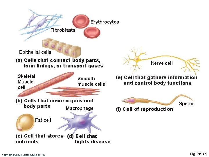
Cell Diversity Over 200 different types of human cells Types differ in size, shape, subcellular components, and functions
Erythrocytes Fibroblasts Epithelial cells (a) Cells that connect body parts, form linings, or transport gases Skeletal Muscle cell Smooth muscle cells (b) Cells that move organs and body parts Macrophage Nerve cell (e) Cell that gathers information and control body functions (f) Cell of reproduction Sperm Fat cell (c) Cell that stores (d) Cell that nutrients fights disease Copyright © 2010 Pearson Education, Inc. Figure 3. 1
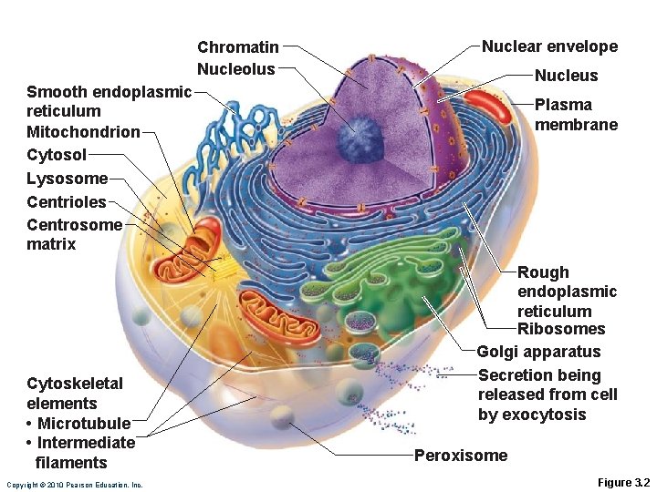
Generalized Cell All cells have some common structures and functions Human cells have four basic parts: ◦ ◦ Plasma membrane - flexible outer boundary Cytoplasm - intracellular fluid Organelles - multiple functions Nucleus - control center
Chromatin Nucleolus Nuclear envelope Nucleus Smooth endoplasmic reticulum Mitochondrion Cytosol Lysosome Centrioles Centrosome matrix Cytoskeletal elements • Microtubule • Intermediate filaments Copyright © 2010 Pearson Education, Inc. Plasma membrane Rough endoplasmic reticulum Ribosomes Golgi apparatus Secretion being released from cell by exocytosis Peroxisome Figure 3. 2
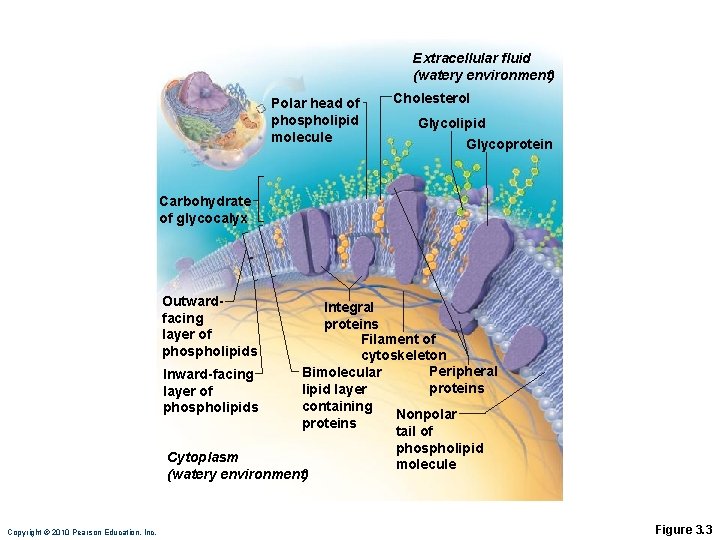
Plasma Membrane Bimolecular layer of lipids and proteins in a constantly changing fluid mosaic Plays a dynamic role in cellular activity Separates intracellular fluid (ICF) from extracellular fluid (ECF) ◦ Interstitial fluid (IF) = ECF that surrounds cells
Extracellular fluid (watery environment) Polar head of phospholipid molecule Cholesterol Glycolipid Glycoprotein Carbohydrate of glycocalyx Outwardfacing layer of phospholipids Integral proteins Filament of cytoskeleton Peripheral Bimolecular Inward-facing proteins lipid layer of containing phospholipids Nonpolar proteins tail of phospholipid Cytoplasm molecule (watery environment) Copyright © 2010 Pearson Education, Inc. Figure 3. 3
Membrane Lipids Phospholipids (lipid bilayer) ◦ Phosphate heads: polar and hydrophilic ◦ Fatty acid tails: nonpolar and hydrophobic (Review Fig. 2. 16 b)
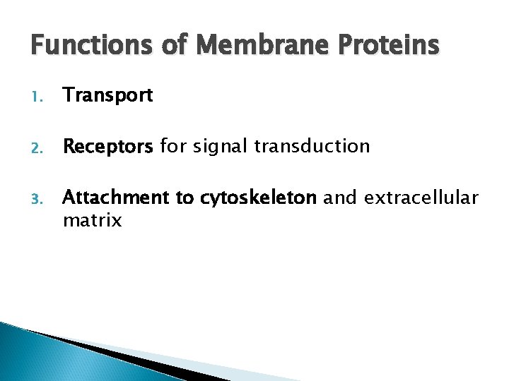
Functions of Membrane Proteins 1. Transport 2. Receptors for signal transduction 3. Attachment to cytoskeleton and extracellular matrix
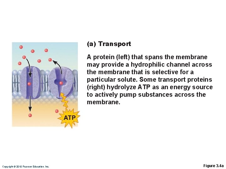
(a) Transport A protein (left) that spans the membrane may provide a hydrophilic channel across the membrane that is selective for a particular solute. Some transport proteins (right) hydrolyze ATP as an energy source to actively pump substances across the membrane. Copyright © 2010 Pearson Education, Inc. Figure 3. 4 a
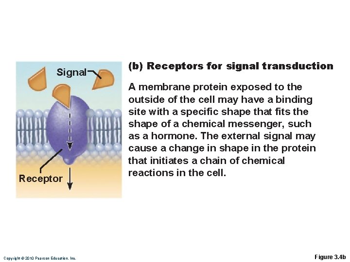
Signal Receptor Copyright © 2010 Pearson Education, Inc. (b) Receptors for signal transduction A membrane protein exposed to the outside of the cell may have a binding site with a specific shape that fits the shape of a chemical messenger, such as a hormone. The external signal may cause a change in shape in the protein that initiates a chain of chemical reactions in the cell. Figure 3. 4 b
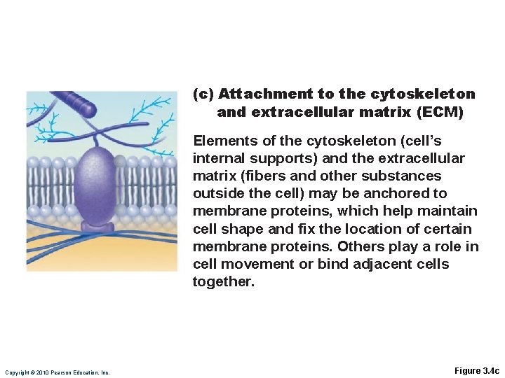
(c) Attachment to the cytoskeleton and extracellular matrix (ECM) Elements of the cytoskeleton (cell’s internal supports) and the extracellular matrix (fibers and other substances outside the cell) may be anchored to membrane proteins, which help maintain cell shape and fix the location of certain membrane proteins. Others play a role in cell movement or bind adjacent cells together. Copyright © 2010 Pearson Education, Inc. Figure 3. 4 c
Functions of Membrane Proteins 4. Enzymatic activity 5. Intercellular joining 6. Cell-cell recognition
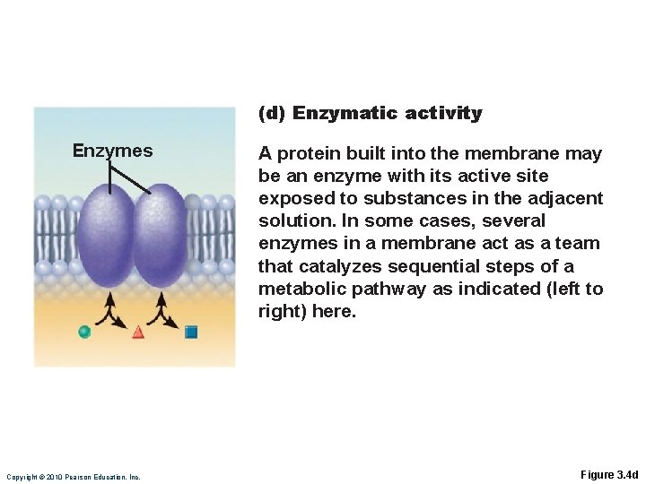
(d) Enzymatic activity Enzymes Copyright © 2010 Pearson Education, Inc. A protein built into the membrane may be an enzyme with its active site exposed to substances in the adjacent solution. In some cases, several enzymes in a membrane act as a team that catalyzes sequential steps of a metabolic pathway as indicated (left to right) here. Figure 3. 4 d
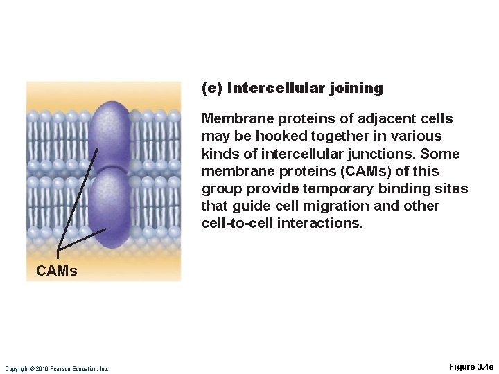
(e) Intercellular joining Membrane proteins of adjacent cells may be hooked together in various kinds of intercellular junctions. Some membrane proteins (CAMs) of this group provide temporary binding sites that guide cell migration and other cell-to-cell interactions. CAMs Copyright © 2010 Pearson Education, Inc. Figure 3. 4 e
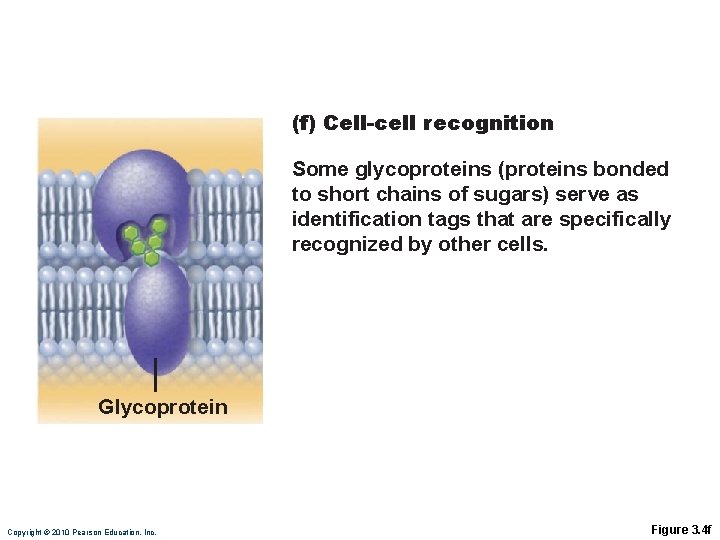
(f) Cell-cell recognition Some glycoproteins (proteins bonded to short chains of sugars) serve as identification tags that are specifically recognized by other cells. Glycoprotein Copyright © 2010 Pearson Education, Inc. Figure 3. 4 f
Membrane Transport Plasma membranes are selectively permeable Some molecules easily pass through the membrane; others do not
Types of Membrane Transport Passive processes ◦ No cellular energy (ATP) required ◦ Substance moves down its concentration gradient Active processes ◦ Energy (ATP) required ◦ Occurs only in living cell membranes
Passive Processes What determines whether or not a substance can passively permeate a membrane? 1. Lipid solubility of substance 2. Channels of appropriate size 3. Carrier proteins PLAY Animation: Membrane Permeability
Passive Processes Simple diffusion Carrier-mediated facilitated diffusion Channel-mediated facilitated diffusion Osmosis
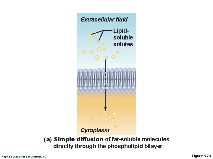
Passive Processes: Simple Diffusion Nonpolar lipid-soluble (hydrophobic) substances diffuse directly through the phospholipid bilayer Diffusion is the movement of solutes from a solution of higher concentration to that of a lower concentration PLAY Animation: Diffusion
Extracellular fluid Lipidsoluble solutes Cytoplasm (a) Simple diffusion of fat-soluble molecules directly through the phospholipid bilayer Copyright © 2010 Pearson Education, Inc. Figure 3. 7 a
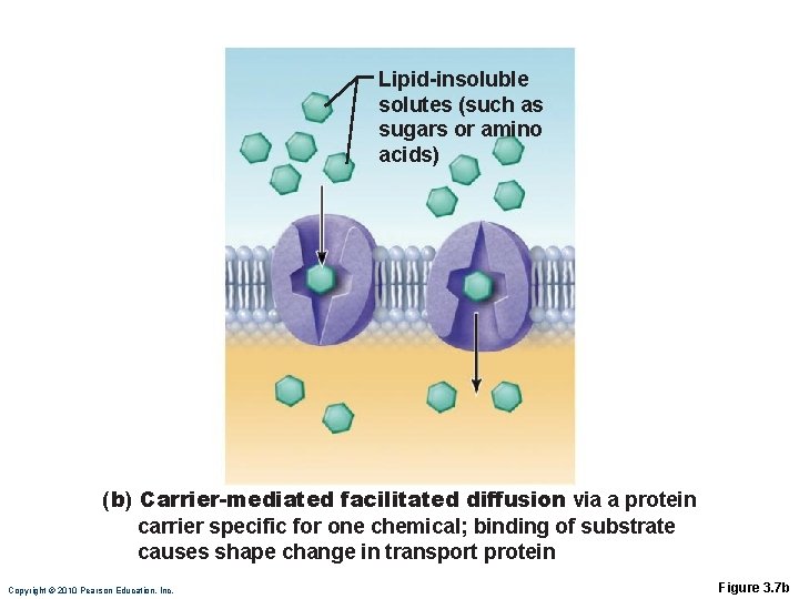
Passive Processes: Facilitated Diffusion Certain lipophobic molecules (e. g. , glucose, amino acids, and ions) use carrier proteins or channel proteins, both of which: ◦ Exhibit specificity (selectivity) ◦ Are saturable; rate is determined by number of carriers or channels ◦ Can be regulated in terms of activity and quantity
Facilitated Diffusion Using Carrier Proteins Transmembrane integral proteins transport specific polar molecules (e. g. , sugars and amino acids) Binding of substrate causes shape change in carrier
Lipid-insoluble solutes (such as sugars or amino acids) (b) Carrier-mediated facilitated diffusion via a protein carrier specific for one chemical; binding of substrate causes shape change in transport protein Copyright © 2010 Pearson Education, Inc. Figure 3. 7 b
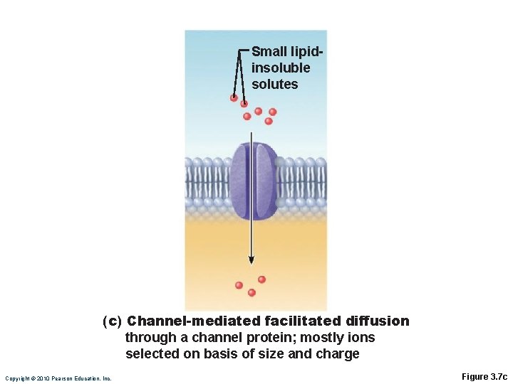
Facilitated Diffusion Using Channel Proteins Aqueous channels formed by transmembrane proteins selectively transport ions or water Two types: ◦ Leakage channels Always open ◦ Gated channels Controlled by chemical or electrical signals
Small lipidinsoluble solutes (c) Channel-mediated facilitated diffusion through a channel protein; mostly ions selected on basis of size and charge Copyright © 2010 Pearson Education, Inc. Figure 3. 7 c
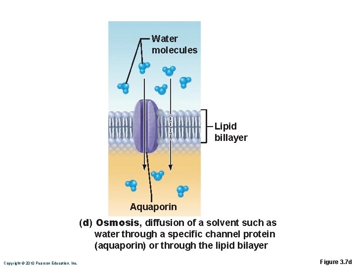
Passive Processes: Osmosis Movement of solvent (water) from a solution of low concentration to that of a higher concentration, across a selectively permeable membrane Water diffuses through plasma membranes: ◦ Through the lipid bilayer ◦ Through water channels called aquaporins (AQPs)
Water molecules Lipid billayer Aquaporin (d) Osmosis, diffusion of a solvent such as water through a specific channel protein (aquaporin) or through the lipid bilayer Copyright © 2010 Pearson Education, Inc. Figure 3. 7 d
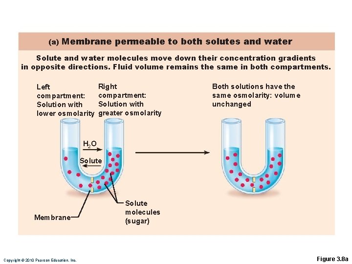
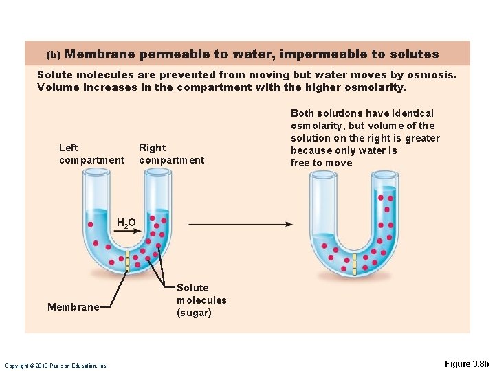
Passive Processes: Osmosis Water concentration is determined by solute concentration because solute particles displace water molecules Osmolarity: The measure of total concentration of solute particles When solutions of different osmolarity are separated by a membrane, osmosis occurs until equilibrium is reached
(a) Membrane permeable to both solutes and water Solute and water molecules move down their concentration gradients in opposite directions. Fluid volume remains the same in both compartments. Left compartment: Solution with lower osmolarity Right compartment: Solution with greater osmolarity Both solutions have the same osmolarity: volume unchanged H 2 O Solute Membrane Copyright © 2010 Pearson Education, Inc. Solute molecules (sugar) Figure 3. 8 a
(b) Membrane permeable to water, impermeable to solutes Solute molecules are prevented from moving but water moves by osmosis. Volume increases in the compartment with the higher osmolarity. Left compartment Right compartment Both solutions have identical osmolarity, but volume of the solution on the right is greater because only water is free to move H 2 O Membrane Copyright © 2010 Pearson Education, Inc. Solute molecules (sugar) Figure 3. 8 b
Importance of Osmosis When osmosis occurs, water enters or leaves a cell Change in cell volume disrupts cell function PLAY Animation: Osmosis
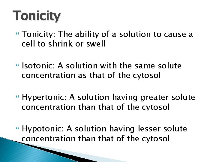
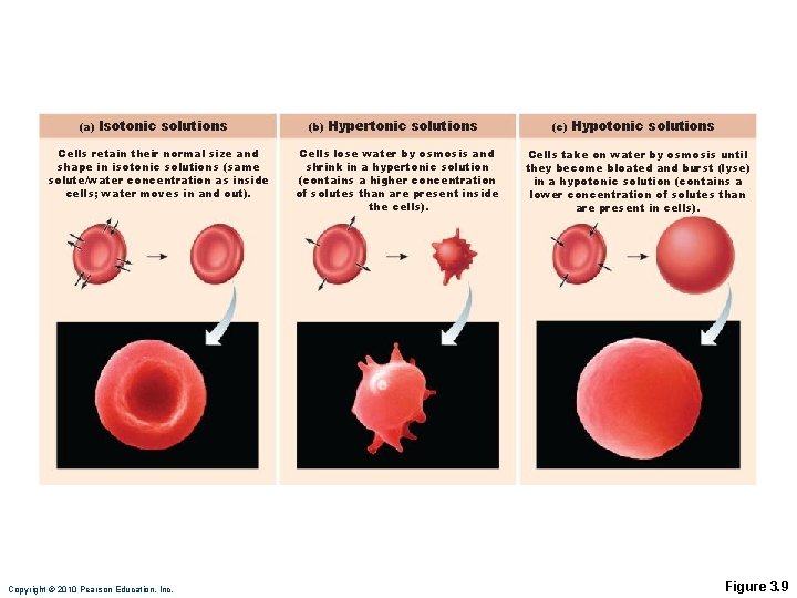
Tonicity Tonicity: The ability of a solution to cause a cell to shrink or swell Isotonic: A solution with the same solute concentration as that of the cytosol Hypertonic: A solution having greater solute concentration that of the cytosol Hypotonic: A solution having lesser solute concentration that of the cytosol
(a) Isotonic solutions Cells retain their normal size and shape in isotonic solutions (same solute/water concentration as inside cells; water moves in and out). Copyright © 2010 Pearson Education, Inc. (b) Hypertonic solutions Cells lose water by osmosis and shrink in a hypertonic solution (contains a higher concentration of solutes than are present inside the cells). (c) Hypotonic solutions Cells take on water by osmosis until they become bloated and burst (lyse) in a hypotonic solution (contains a lower concentration of solutes than are present in cells). Figure 3. 9