TOPIC PRESENTATION AORTIC ANEURYSMS Anatomy and Physiology of


 is a relatively new method of treating aortic")


 or")


 Aneu. Rx® device - Modular bifurcated stent graft composed of a nitinol exoskeleton")


 �AD �Mutations in TGFBR 1 and TGFBR 2 � 4")
 �AD �COL 3 A 1 mutation �Abnormal type")
 �")


 2 x")


")

- Slides: 90
TOPIC PRESENTATION AORTIC ANEURYSMS
Anatomy and Physiology of Aorta � Aortic Aneurysms – Thoracic and Abdominal Ø Etiology & Pathogenesis Ø Clinical Manifestations Ø Diagnosis Ø Natural History Ø Management § Surgical repair § Endovascular repair § Medical management �
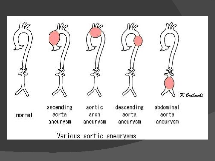
Anatomy & Physiology • Largest artery of body • Thoracic and Abdominal part • Aortic root • Ascending aorta • Aortic arch • Descending aorta • Supra-renal part • Infra-renal part
� � � � Elastic conduit. Biochemical properties due to elastin and collagen in media and adventitia. Nonlinear pressure-diameter relationship. In healthy adults diameter not exceed 40 mm and taper gradually distally. Rate of aortic expansion 0. 9 mm in men and 0. 7 mm in women per decade. Factors affecting aortic diameter-age, sex, body size and BP. Ageing decreases the curve steep § Increase in collagen to elastin ratio § Distortion of medial elastic fibres § Deposition of collagen and calcification § Atherosclerotic changes
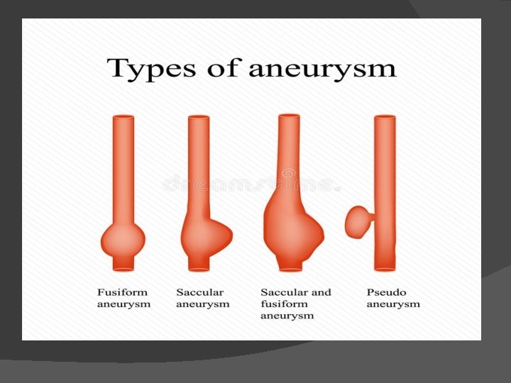
Aortic Aneurysm � Second most frequent disease of aorta after atherosclerosis � Pathological segment of aortic dilatation that expands and can eventually rupture. � Greater than 50% of expected diameter. � Classification Morphology. – Size, Shape, Location
Crawford classification scheme based on the anatomic extent of the aneurysm
Abdominal aortic aneurysms Defined by abdominal aorta greater than 3 cm in diameter � Most common form of aortic aneurysms. � Infra-renal 80% � 10% para-renal or visceral. � Male 5 x females. �
Abdominal aortic aneurysms � Risk factors�Age �Smoking �Emphysema �Hypertension �Hyperlipidemia �Family history �Genetics
Pathogenesis � � � Chronic aortic wall inflammation. Inflm causes infiltration of media with leukocytes, microphages, mast cells, lymphocytes etc. Increased local expression of proteases. Imbalance between degenerative and reparative processes and more degradation of structural connective tissue occurs. Mechanical failure of medial elastin and adventitial collagen. Aneurysmal dilatation and rupture.
Pathogenesis
Clinical Features � Develops insidiously over many years. � Usually asymptomatic � Most are incidental finding on imaging studies. � Physical examination – pulsatile epigastric or periumbilical mass. Insensitive-only 30 -40% � AAA associated with other aneurysms � 85% of femoral artery aneurysm � 60% of popliteal artery aneurysm � 25% of thoracic aorta aneurysm
� Risk of distal thromboembolism, rapid expansion or rupture. � Acute hemorrhage, severe abdominal pain, hypotension and sudden death � Periaortic hematoma with severe backache radiating to flank and groin. � Acute limb ischemia and claudication � Rupture has very high mortality � 30 -50% die before hospitalization � 30 -40% after hospitalization but before surgery �Post operative mortality is 40 -50% �Mortality is less in EVAR as compared to OSR. (Eur J Vasc surg, 2016)
v The annual risk of rupture based upon aneurysm size : (The Society for Vascular Surgery practice guidelines - Journal of Vascular Surgery, 2018) � Less than 4. 0 cm in diameter = less than 1 in 200 � 4. 0 to 4. 9 cm in diameter = between 1 in 200 and 1 in 20 � 5. 0 to 5. 9 cm in diameter = between 1 in 20 and 1 in 10 � 6. 0 to 6. 9 cm in diameter = between 1 in 10 and 2 in 10 � 7. 0 to 7. 9 cm in diameter = between 2 in 10 and 4 in 10 � 8. 0 cm or more in diameter = between 3 in 10 and 5 in 10
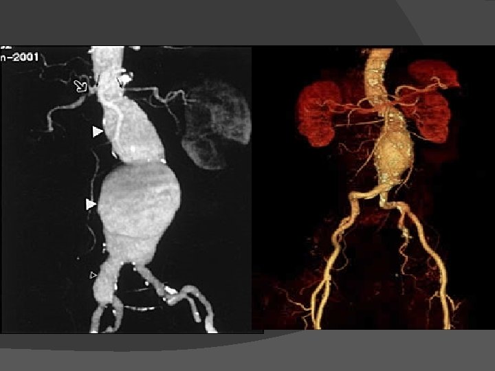
Diagnostic imaging � Abdonimal USG �Non-invasive, inexpensive, free of radiation and contrast free � CT angiogram �More accurate than USG �Relationship of AAA with other vessels �Coexisting mural thrombosis, calcification, occlusive atherosclerosis �Enhance visualization of AAA before EVAR � MRA- Magnetic resonance angiography
Diagnostic imaging
Screening Done using USG � Incidence � 1 per 1000 in males over 60 years of age � 7 per 1000 in males over 70 years of age � 10% in males over 60 years of age with risk factors like smoking, F/H, HTN, atherosclerosis � Impact of screening-early diagnosis � 50% reduction in rupture cases � 50% reduction in overall aneurysm related mortality. � In females controversial with no clear survival benefit (MASS � study)
The Society of Vascular Surgery recommendations for one time screening of AAA • All males age >65 years • All males age >55 years with F/H of AAA • All females of age >65 years with F/H of AAA or H/o tobacco use
Surveillance � SVS surveillance imaging strategy � 2. 5 -2. 9 cm every 5 years � 3. 0 -3. 4 cm every 3 years � 3. 5 -4. 5 cm every 12 months � 4. 5 -5. 4 cm every 6 months USG is used for small size aneurysms while for large size aneurysm >4. 5 cm CT is preferred.
Medical management � Most medical therapy based for Risk reduction �smoking cessation �statin therapy �physical exercise �ACE inhibitors or Losartan (Hackam et al) �C. pneumoniae infection associated with an increased rate of AAA expansion and roxithromycin decreases the rate of aneurysm expansion. (Lindholt et al) �Doxycycline-MMP 9 inhibitor �Experimental therapy-monoclonal antibodies against the proinflammatory protienases, elastase etc
Medical management � Many AAA associated with other Co-morbid diseases � CAD leading cause of early and late mortality after AAA repair. � CKD, COPD, Stroke and DM may influence morbidity and mortality. � A significantly lower incidence of myocardial infarction among patients undergoing EVAR as compared with open repair � EVAR should be considered inpatients with intermediate to high cardiac risk.
Preoperative cardiac evaluation for patients undergoing aneurysm repair (ACC/AHA 2017 Guidelines on perioperative cardiovascular evaluation and care for noncardiac surgery: executive summary. Circulation 2017; 116: 1971 -96. )
Preoperative cardiac evaluation for patients undergoing aneurysm repair (ACC/AHA 2017 Guidelines on perioperative cardiovascular evaluation and care for noncardiac surgery: executive summary. Circulation 2017; 116: 1971 -96. )
Preoperative cardiac evaluation for patients undergoing aneurysm repair (ACC/AHA 2017 Guidelines on perioperative cardiovascular evaluation and care for noncardiac surgery: executive summary. Circulation 2017; 116: 1971 -96. )
Surgery � Indications �Symptomatic AAA (tenderness or abdominal or back pain, evidence for embolization, rupture) �Asymptomatic aneurysm size ≥ 5. 5 cm �AAA that has expanded by more than 0. 5 cm within a six-month interval
Surgery � � � OSR via transperitoneal or left retroperitoneal approach Occlusion by tube or bifurcated graft Excluded aneurysm is not resected to prevent aorto-enteric fistula OSR mortality 4 to 8% OSR post op complications 10 to 30% (cardiac, pulmonary, renal, colonic ischemia) OSR late complications 15 to 30% (incision related, perianstomosic aneurysm, graft infection, graft-enteric fistula and graft limd occlusions with LL ischemia
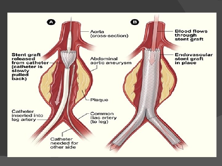
EVAR � Endovascular aneurysm repair (EVAR) is a relatively new method of treating aortic aneurysms � Performed by inserting graft components folded and compressed within a delivery sheath through the lumen of an access vessel, usually the common femoral artery � Upon deployment, the endograft expands, contacting the vessel wall proximally and distally to exclude the aortic aneurysm sac from aortic blood flow and pressure
EVAR-Preop planning and imaging � To define the anatomy � To determine the feasibility of endovascular repair � To choose the size and configuration of endograft components � CT angiography with 3 -D reconstruction preferred because in 2 D CT- aortic diameter measurements sometimes overestimated if the aorta is angulated and the longitudinal axis is not perpendicular to the imaging plane � MR angiography in setting of renal dysfunction. But MRA fails to depict vessel wall calcification, which has implications for vascular access
EVAR-Preop planning an imaging � Aortic neck diameter �Aortic diameter at the lowest renal artery �Endograft diameter required = 15 to 20% more of measured aortic neck diameter �Over-sizing - kinking of the device, incomplete expansion of graft, inadequate seal, a nidus for thrombus formation or endoleak �Under sizing- inadequate seal
EVAR-Preop planning an imaging � Aortic neck angulation �The angle formed between points connecting the lowest renal artery, the origin of the aneurysm, and the aortic bifurcation �Greater angle-difficulties in implantation, kinking, endoleak, and the potential for distal device migration �Ideally, the aortic neck angle should be < 600
EVAR-Preop planning an imaging � Aortic neck length �The distance from the lowest renal artery to the origin of the aneurysm �Should be at least 10 to 15 mm to provide an adequate proximal landing zone for endograft fixation
EVAR-Preop planning an imaging � Iliac artery morphology �No significant stenosis , mural thrombus, calcification and tortuosity �CIA is the preferred distal attachment site �When EIA is used for distal fixation the hypogastric artery needed to be embolized to prevent back bleeding into the aneurysm sac �EIA diameter of 7 mm is needed to allow safe passage of the endograft delivery sheath �Diffuse narrowing or significant calcification - an iliac conduit can be created
v Ideal case for EVAR o Proximal neck length >10 mm o Aortic neck angulation ≤ 60 o Iliac artery diameter >7 mm and < 15 mm with Minimal to moderate tortuosity, No mural thrombus at attachment sites, Minimal calcification o No associated occlusive disease mesenteric
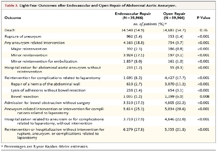
v The main advantages over open repair � � � � Avoidance of long incisions in the thorax or abdomen No cross-clamping of the aorta Less blood loss Lower incidence of visceral, renal, and spinal cord ischemia Less peri-operative mortality Earlier return to normal activity Recent trails have shown mortality benefit of EVAR over OSR
EVAR 1 Trial design: Patients with an AAA randomized to endovascular repair (n = 626) vs open repair (n = 626). Median follow-up was 6 years. Results (p = 0. 72) (p = 0. 73) 9 per 100 person-years 7. 5 • • 7. 7 • 6 30 -day operative mortality: 1. 8% with endovascular repair vs. 4. 3% with open repair All-cause death rate: 7. 5/100 person-yrs vs. 7. 7/100 person-yrs, respectively Aneurysm-related death rate: 1. 0/100 personyrs vs. 1. 2/100 person-yrs, respectively Conclusions 3 1. 0 0 All-cause death all mortality Endovascular repair 1. 2 Aneurysm-related death aneurysm mortality Open repair • Among patients with abdominal aortic aneurysm, endovascular repair associated with lower operative mortality with similar long-term allcause mortality • Similar overall frequency of complications after endovascular repair
EVAR 2 Trial design: Patients with an AAA ineligible for surgery were randomized to endovascular repair (n = 197) vs. medical management (n = 207). Median follow -up was 3. 1 years. (p = 0. 97) 24 (p = 0. 02) 21. 0 22. 1 Results • • per 100 person-years • 12 7. 3 3. 6 0 All-cause death all mortality rate Endovascular repair Aneurysm-related aneurysm mortality death rate No repair 30 -day operative mortality: 7. 3% with endovascular repair Rupture rate in the no repair group: 12. 4/100 person-yrs All-cause death rate: 21. 0/100 person-yrs with endovascular repair vs. 22. 1/100 person -yrs with no repair Conclusions • In patients with abdominal aortic aneurysm, ineligible for open surgery repair, endovascular repair is associated with relatively similar overall mortality • Aneurysm-related death significantly reduced from endovascular repair
DREAM Trial design: Patients with an AAA were randomized to endovascular repair (n = 173) vs. open repair (n = 178). Median follow-up was 6. 4 years. Results 90 (p = 0. 97) (p = 0. 03) • % 82 80 70 60 • 69 70 Survival survival Endovascular repair 70 Freedom from reintervention Open repair • • Survival: 69% with endovascular repair vs. 70% open repair (p = 0. 97) Freedom from reintervention: 70% vs. 82% (p = 0. 03), respectively In the endovascular repair group, common causes for reintervention were thromboocclusive disease, endoleak, and graft migration In the open repair group, a common cause for reintervention was treatment of incisional hernia Conclusions • Among patients with abdominal aortic aneurysm, endovascular repair results in similar long-term survival as open repair; however, this approach results in the need for significantly more reinterventions
OVER Trial design: Patients with unruptured AAA were randomized to endovascular repair (EVAR) or open surgical repair. Patient follow-up was a mean of 1. 8 years. (p = 0. 13) 20 20 % 10 (p = 0. 14) 9. 8 % 10 7. 0 2. 7 0 Mortality at 2 years Open repair (n = 437) 0 1. 4 MI at 1 year EVAR (n = 444) Results • 30 -day mortality ↑ with open repair (2. 3% vs. 0. 2%, p = 0. 006); no difference at 2 years (9. 8% vs. 7. 0%, p = 0. 13) • MI: 2. 7% vs. 1. 4%, p = 0. 14; stroke: 0. 9% vs. 1. 6%, p = 0. 38 • Procedural time, duration of hospital stay ↓ in EVAR arm (p < 0. 001) Conclusions • long-term outcomes similar with EVAR and open repair for unruptured AAA; length of stay and procedural time was shorter for EVAR
Review of Evidence EVAR vs OSR
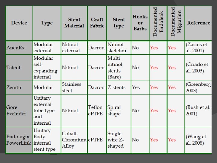
� Endografts �Fabric-covered stent �Significant variations in endovascular graft design �Three types of components are common to all: Ø A delivery system Ø Main body device Ø Extensions (limb)
(A) Aneu. Rx® device - Modular bifurcated stent graft composed of a nitinol exoskeleton and polyester lining. Distal and proximal extension cuffs are available. (B) The Gore Excluder -Modular bifurcated device, with a nitinol exoskeleton and a polytetrafluoroethylene graft. It has proximal barbs to anchor into the proximal infrarenal aorta. (C) The Powerlink®system- A unibody device made of polytetrafluoroethylene and a cobalt chromium alloy endoskeleton. It has a long main body and sits on the anatomic bifurcation (D) The Zenith® device-The bare proximal stents allow for suprarenal fixation. The device also has barbs to allow for more secure attachment into the suprarenal aorta. It is modular bifurcated with a stainless steel exoskeleton.
Stepwise procedure v v v v Gaining vascular access Placement of arterial guidewires and sheaths Imaging to confirm aortoiliac anatomy Main body deployment Gate cannulation (bifurcated graft) Iliac limb deployment Graft ballooning Completion imaging
EVAR complications Deployment related Device related Failed deployment Structural failure Bleeding Endoleaks Hematoma Limb occlusion/stent-graft kink Lymphocoel Sac enlargement/proximal neck dilatation Infection Embolization Stent migration AAA rupture Perforation Infection Arterial rupture Buttock/leg claudication Dissection
EVAR complications v Systemic Ø Cardiac Ø Pulmonary Ø Renal insufficiency, contrast-induced neuropathy Ø Deep vein trombosis Ø Pulmonary embolism Ø Coagulopathy Ø Bowel ischemia Ø Spinal cord ischemia
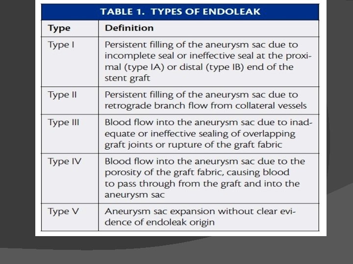
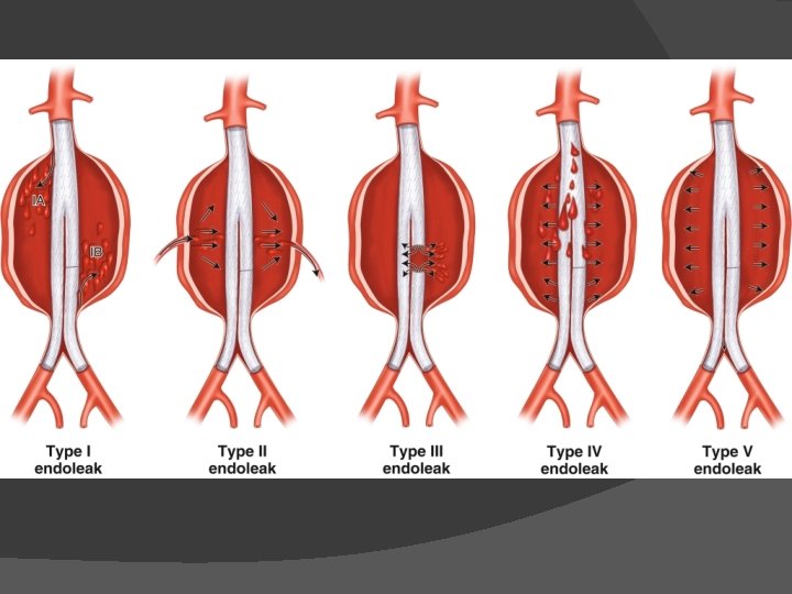
endoleak � Endoleak is a term that describes the presence of persistent flow of blood into the aneurysm sac after device placement � Five types of endoleaks are described
Type Endovascular Open surgery Type I Moulding balloon angioplasty Giant Palmaz stent Stent-graft cuff/ extension External banding of aneurysm neck Surgical conversion Type II Trans-arterial embolisation Translumbar injection 30% decrease Type III Angioplasty/ stenting of junctions Secondary graft placement Open conversion Type V Co-axial graft (‘re-lining’) Evacuation of hygroma Open conversion
Thoracic aortic aneurysm Estimated incidence 5 to 10 per 100, 000 � Aortic root and ascending aorta-60% � Descending aorta-35% � Thoracoabdominal aortic aneurysm-10% � Aortic arch <10% �
� Risk factors�Age �Smoking �Emphysema �Hypertension �Hyperlipidemia �Family history �Genetics � Genetic and heritable disorders are important cause of TAA especially of root and ascending aorta
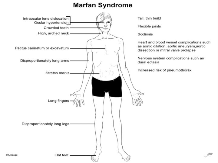
� Marfan syndrom �AD �FBN 1 gene mutation �Abnormal fibrillin-1 �Excess free TGF-β �Interaction with AT receptor and upregulation �Current reccomendation-maximal dose of beta- blocker or ARB treatment to reduce AA growth rate �Dutch Marfan Trial - losartan+beta blocker vs beta- blocker alone
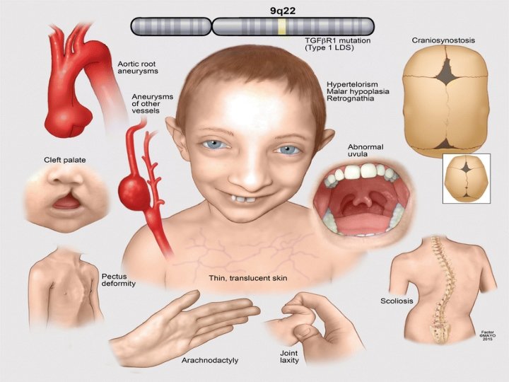
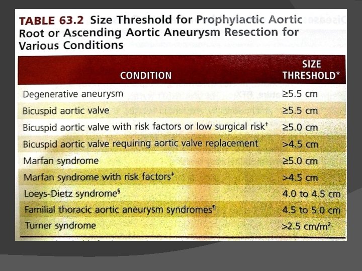
� Loeys-Dietz syndrom (LDS) �AD �Mutations in TGFBR 1 and TGFBR 2 � 4 types of LDS �More aggressive vascular phenotype than MFS �Aortic aneurysms and dissection more common than MFS and relatively younger age �Current recommendation-surgery if aortic root >4 -4. 5 cm
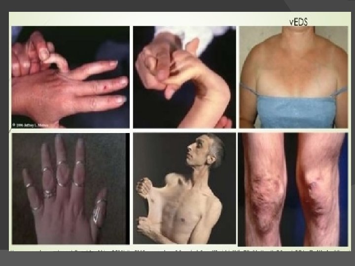
� Vascular Ehlers-Danlos syndrom (v. EDS) �AD �COL 3 A 1 mutation �Abnormal type 3 procollagen synthesis �Medium sized vessels �Aortic root involvement less common. Mostly descending and abdominal aorta and its branches �Vessels are very fragile so high risk of sudden rupture and difficult surgical repair
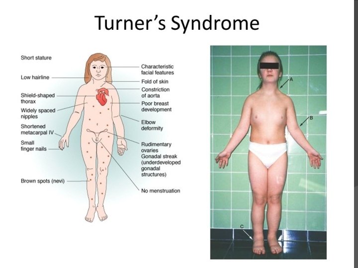
Turner syndrome � Complete or partial loss of second sex chromosome (XO, Xp) � 50 to 75% CV defects � BAV 30% � Coarctation 12% � Ascending aorta dilatation 33% � Patient with TS+CV defect-100 x increased risk of dissection � TS without CV defect every 5 -10 years � TS with CV defect every 1 -2 years
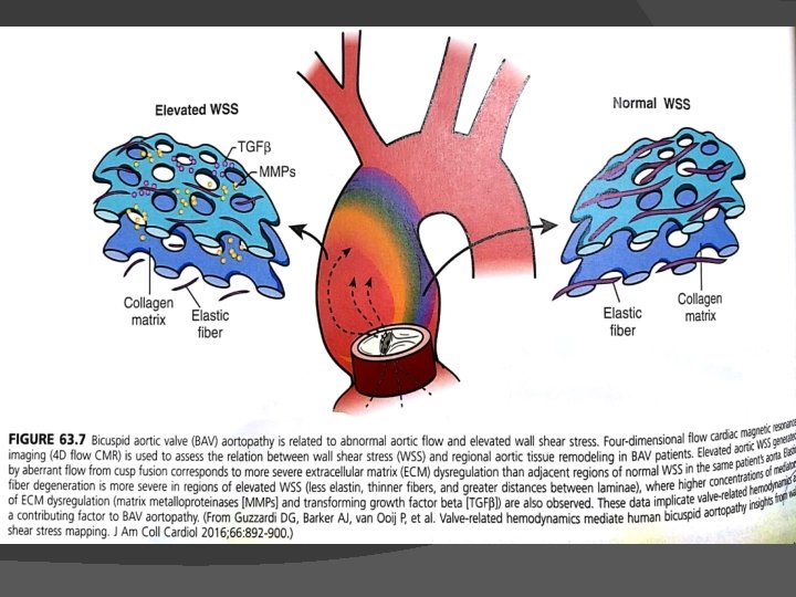
Bicuspid aortic valve disease � 1% of general population � Ascending aortic aneurysm, coarctation of aorta, aortic disssection � BAV exhibits abnormal leaflet folding, wrinkling and doming causing turbulance even in absence of stenotic or regurgitant lesion � BAV compared to TAV aneurysm exhibit increased apoptosis, MMP-2 activity, TGF-β abnormalities, protien kinase-C pathway � Ascending TAA function � May develop later after AVR may develop independent of valve
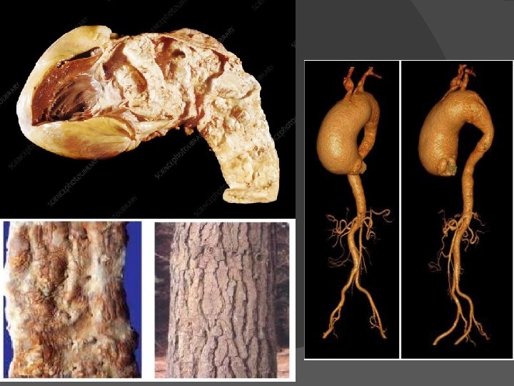
� Syphilis and aortitis �Tertiary stage of syphilis �Latent period of 10 to 25 years �Rare due to antibiotic treatment �Mostly ascending aorta and arch �Tree bark or wrinkled appearance
Other infectious aortitis �Bacterial and fungal � Non infectious aortitis �Gaint cell arteritis, idiopathic aortitis, Ig. G 4 disease, vasculitides � Aortic dissection �Mostly descending aorta, pseudo aneurysm � Degenerative aneurysm �atherosclerotic �
Clinical manifestation Most are asymptomatic and discovered incidentally � Local mass effect, progressive AR, systemic emboliozation � Persistent chest or back pain � Compression of SVC, trachea or bronchus, esophagus � Rupture � Infection �
Diagnosis � Chest X-ray �Insensitive � TTE and TEE � CT angiogram � MRA
Natural history Factors affecting aneurysmal growth �Etiology-MFS, BAV �Location-descending aorta (2 mm/yr) 2 x of root and ascending aorta (1 mm/year) �Size-larger the aneurysm size faster the growth � Ascending aorta size >6 cm risk of aortic events-15. 6% � Descending aorta aneurysm risk of aortic events � 5. 0 cm-5. 5% � 5. 5 cm-7. 2% � 6. 0 cm-9. 3% � 7. 0 cm-15. 4% �
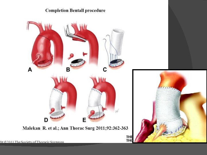
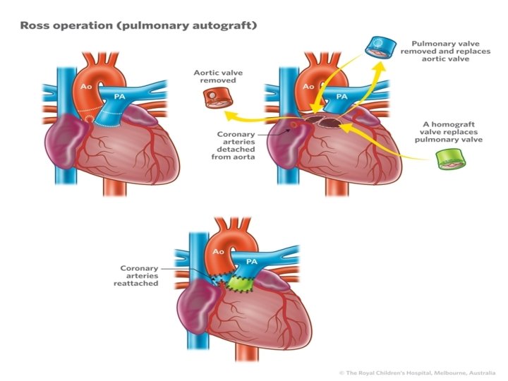
Surgery � Ascending aorta �Opening the ascending aorta and placement of a prosthetic graft with or without concomitant AVR �Modified Bentall procedure �David procedure �Yacoub procedure �Ross procedure
David procedure
Yacoub procedure
Surgery � Arch of aorta �Difficult surgically because of its branches. �Sx recommended when diameter >55 mm �Hemiarch resection �Extended arch resection �Elephant trunk procedure
Hemi and Extended Arch Resection
Elephant trunk procedure
Descending thoracic aneurysm-TEVAR � TEVAR refers to the percutaneous placement of a stent graft in the descending thoracic or thoracoabdominal aorta in patients with aortic aneurysms � The thoracic aorta is of larger caliber than that of the infrarenal aorta so needs larger diameter stent grafts � There is high force of blood flow in the thoracic aorta so requires a longer seal zone (20 mm both proximal and distal) � Hybrid approach- for proximal aneurysm
CHALLENGES OF ENDOVASCULAR REPAIR OF THE AORTIC ARCH � Seal Zone �Seal zone-a length of > 25 mm that is free of excess calcification and thrombus is required. Difficult to get due to curve part and various branching �Increase chances for type I endoleak and graft migration �Hybrid arch repair �
� Device Durability �High pulsatoion of the aortic arch, subjecting the stents to more significant fatigue loading conditions � 2 to 3 times higher than seen in the descending thoracic and abdominal aorta �Branch vessel motion relative to the aorta and respiration-graft wear, stent fracture, and stent kink �Research ongoing for new materials with long-term device durability
Aortic Valve �Current TEVAR systems large in diameter, and delivery system tips are relatively stiff �Increasing the risk of valve damage, �Endovascular devices and delivery systems that will limit valve interaction and be atraumatic to the valve when interactions do occur are required � Stroke �Stroke rates of > 6% �Emboli due to device, delivery system, and/or wire manipulation; air emboli released from the delivery system; and coverage of branched vessels. �Intentional coverage of the left subclavian artery with reanstomosis �
CONCLUSION • TEVAR is a reliable way of preventing aortic-related death in patients with thoracic aortic aneurysms and results in less major morbidity than OSR. • Midterm follow-up suggests that midterm survival after repair of the thoracic aorta is poor, especially after TEVAR, which is mainly due to non–aortic-related death. • Information from large prospectively collected registries lacking
• 42 nonrandomized studies - 5, 888 patients (38 comparative studies, 4 registries) • Conclusion: TEVAR may reduce early death, paraplegia, renal insufficiency, transfusions, reoperation for bleeding, cardiac complications, pneumonia, and length of stay compared with open surgery • Sustained benefits on survival have not been proven
THANK YOU.