Synaptic transmission in the Neuromuscular junction 1 Anatomy


 receptor. The nicotinic ACh receptor is a heteropentamer")


- Slides: 55
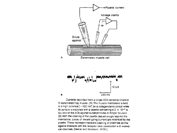
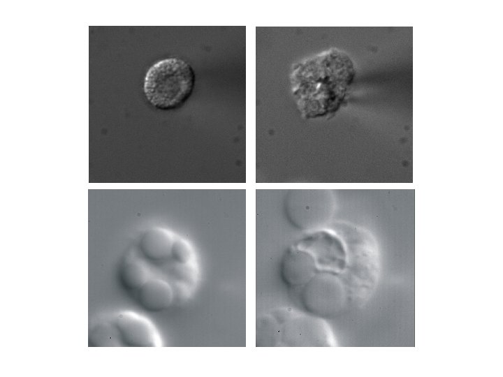
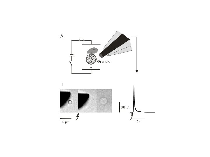
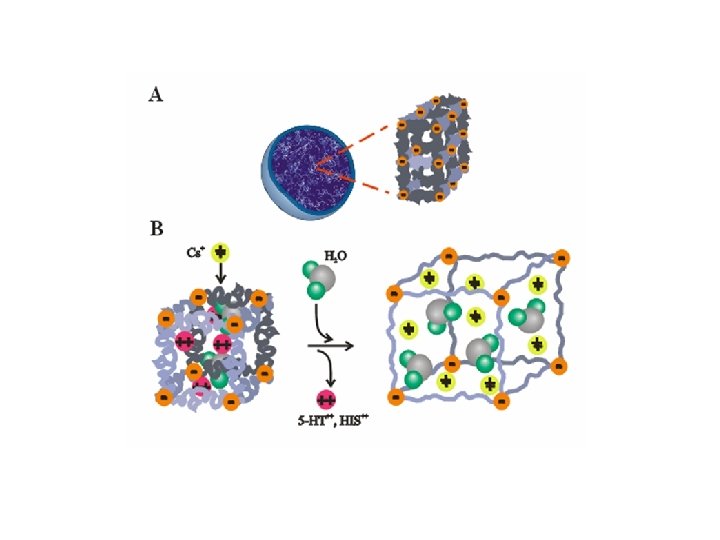
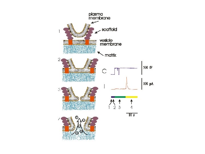
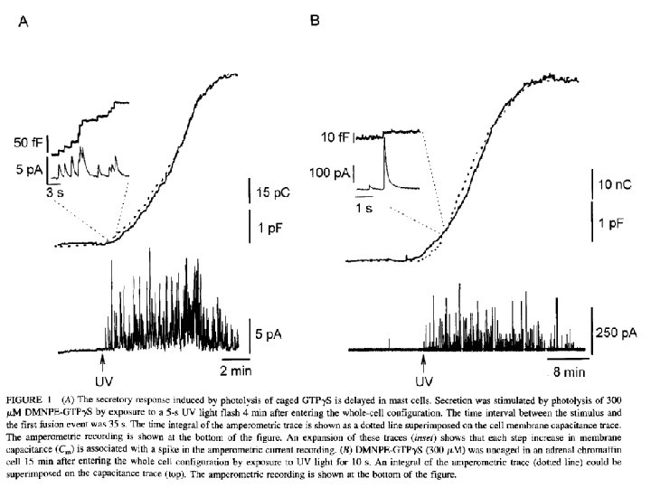
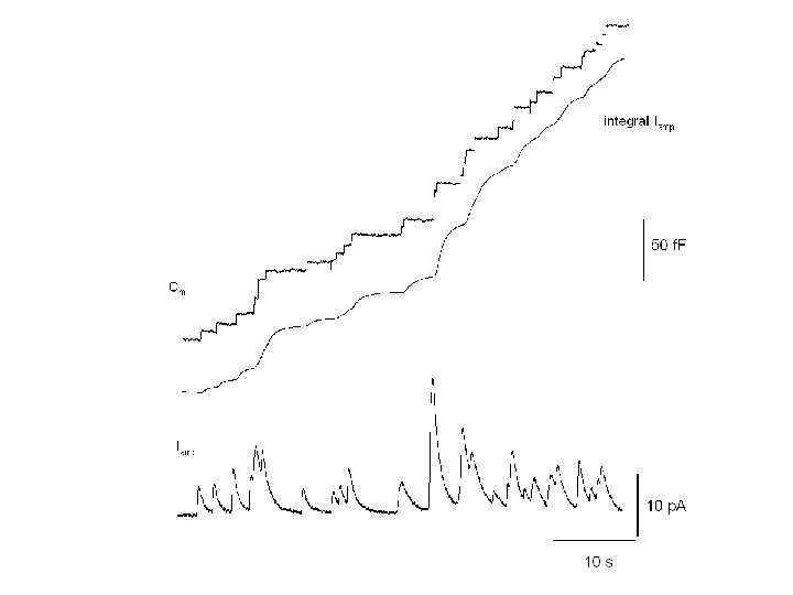
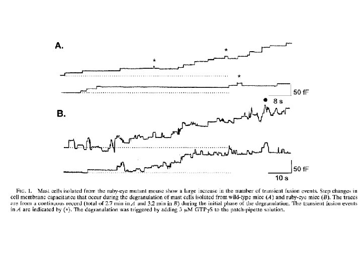
Synaptic transmission in the Neuromuscular junction 1. Anatomy and function of the neuromuscular junction 2. synaptic transmission 3. Quantal release and the Poisson distribution 4. Modern experiments Readings: 1. Katz B. Neural transmitter release: From quantal secretion to exocytosis and beyond-The Fenn Lecture. J Neurocytol. 2003 Jun; 32(5 -8): 437 -46. 2. Neher E, Sakmann B. Single-channel currents recorded from membrane of denervated frog muscle fibres. Nature. 1976 Apr 29; 260(5554): 799 -802. 3. Alvarez de Toledo G, Fernandez-Chacon R, Fernandez JM. Release of secretory products during transient vesicle fusion. Nature. 1993 Jun 10; 363(6429): 554 -8.
Keywords: Neurotransmitter release Nicotinic, Muscarinic Secretory vesicle, fusion pore Capacitance recordings SNARE proteins End plate potentials, MEPP’s Quantal release Poisson distribution Amperometry Ion exchange
Problems: 1. How many ACh molecules are stored into a synaptic vesicle that is only 50 nm in diameter and where ACh is stored at 150 m. M? Describe alternative mechanisms of storage. 2. Describe the sequence of events in neuromuscular transmission.
An electrical synapse consists of one or more gap junction channels permeable to small ions and molecules.
A chemical synapse. Synaptic transmission at a chemical synapse can be thought of as occurring in seven steps.
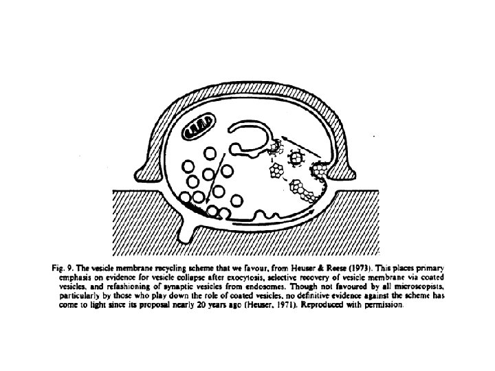
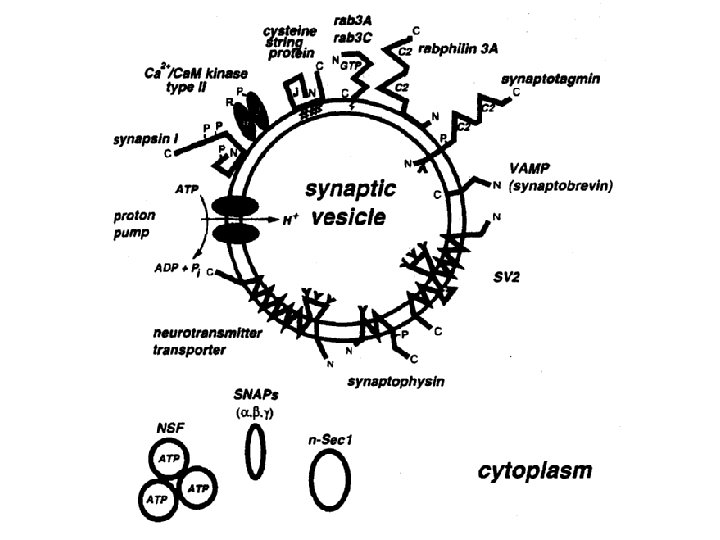
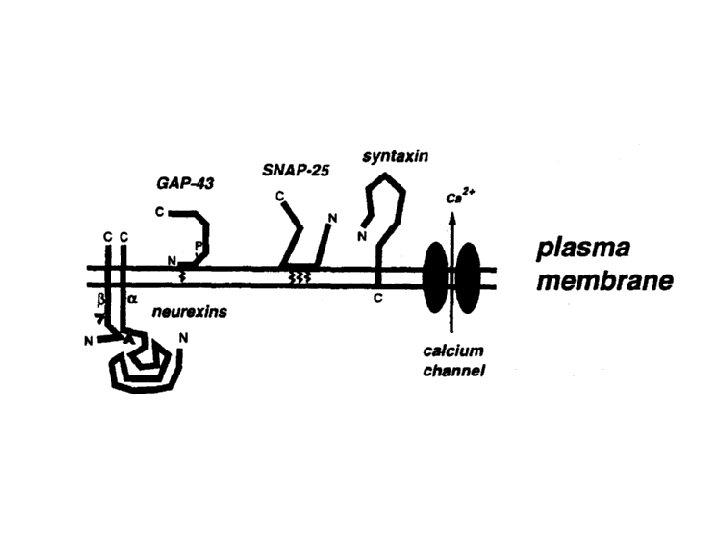
Synthesis and recycling of synaptic vesicles and their content.
Ionotropic and metabotropic acetylcholine receptors. A, This example illustrates a "nicotinic" acetylcholine receptor, which is a ligand gated channel on the postsynaptic membrane. In a skeletal muscle, the end result is muscle contraction. B, This example illustrates a "muscarinic" acetylcholine receptor, which is coupled to a heterotrimeric G protein. In a cardiac muscle, the end result is decreased heart rate. Note that the presynaptic release of ACh is very similar here and in A. ACh, acetylcholine; GTP, guanosine triphosphate.
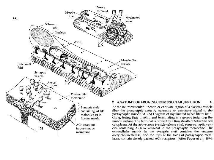
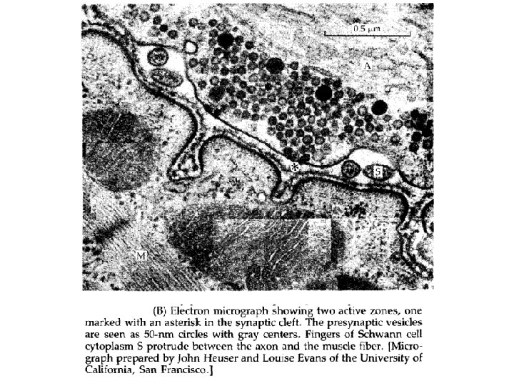
The vertebrate neuromuscular junction or motor end plate.
The effect of extracellular Ca 2+ and Mg 2+ on end plate potentials. The data, obtained by stimulating the motor neuron and monitoring the evoked subthreshold EPP, show that the EPP is stimulated by increasing levels of Ca 2+ but inhibited by increasing levels of Mg 2+. (Data from Dodge FA Jr, Rahaminoff R: Co operative action of calcium ions in transmitter release at the neuromuscular junction. J Physiol 193: 419 432, 1967. )
End plate currents obtained at different membrane potentials in a voltage clamp experiment. A, Two electrode voltage clamp is used to measure the end plate current in a frog muscle fiber. The tips of the two microelectrodes are in the muscle fiber. B, The six records represent end plate currents that were obtained while the motor nerve was stimulated and the postsynaptic membrane was clamped to Vm values of 120, 91, 68, 37, +24, +38 m. V. Notice that the peak current reverses from inward to outward as the holding potential shifts from 37 to +24 m. V. C, The reversal potential is near 0 m. V because the nicotinic ACh receptor has a poor selectivity for Na+ versus K+. (Data from Magleby KL, Stevens CF: The effect of voltage on the time course of end plate current. J Physiol 223: 151 171, 1972. )
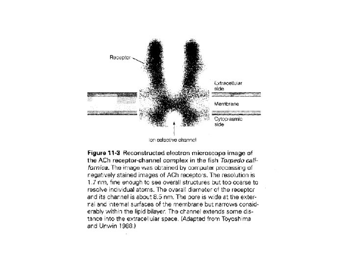
Structure of the nicotinic acetylcholine (ACh) receptor. The nicotinic ACh receptor is a heteropentamer with the subunit composition of a 2 bgd. These subunits are highly homologous to one another, and each has four membrane spanning segments (M 1 M 4).
The effect of extracellular Ca 2+ and Mg 2+ on end plate potentials. The data, obtained by stimulating the motor neuron and monitoring the evoked subthreshold EPP, show that the EPP is stimulated by increasing levels of Ca 2+ but inhibited by increasing levels of Mg 2+. (Data from Dodge FA Jr, Rahaminoff R: Co operative action of calcium ions in transmitter release at the neuromuscular junction. J Physiol 193: 419 432, 1967. )
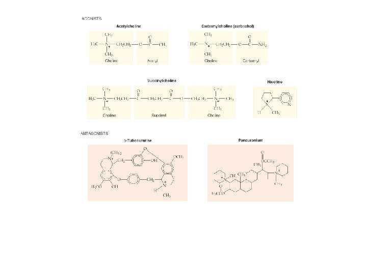
Pharmacology of the vertebrate neuromuscular junction. Many of the proteins that are involved in synaptic transmission at the mammalian neuromuscular junction are the targets of naturally occurring or synthetic drugs. The antagonists are shown as " " signs highlighted inred. The agonists are shown as "+" signs highlighted in green.
Model of synaptic vesicle fusion and exocytosis. ADP, adenosine diphosphate; ATP, adenosine triphosphate; NSF, N ethyl maleimide sensitive factor; SNAP 25, synaptosome associated protein 25 k. Da; a SNAP, soluble NSF attachment protein; SNARE, SNAP receptor.
A B
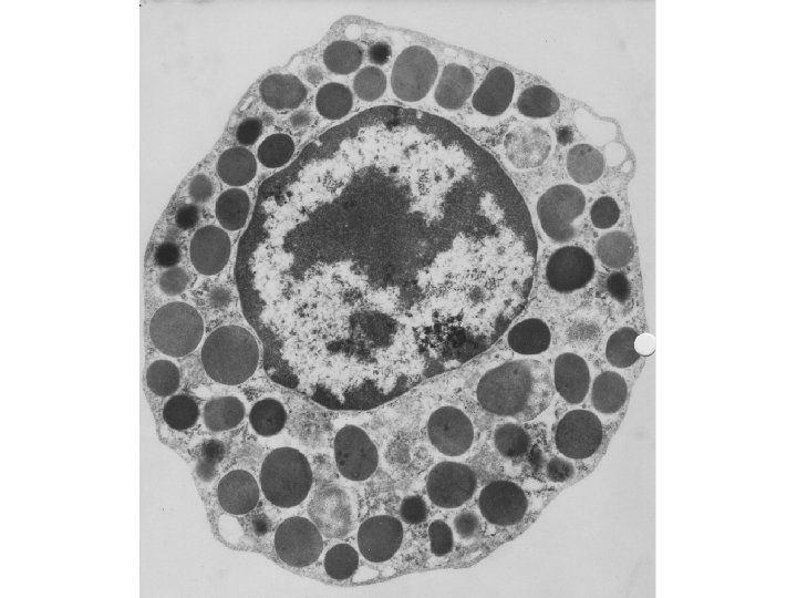
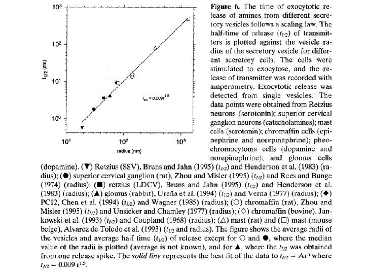
Detection of serotonin that is released from synaptic vesicles. A, The serotonin that is released from a synaptic terminal of a leech neuron can be detected electrochemically using a carbon fiber microelectrode. The current carried by the carbon fiber increases with the amount of serotonin that is released, reflecting the oxidation of serotonin molecules on the surface of the carbon fiber. B, The top panel shows the action potential recorded from the stimulated motor neuron. The middle panel shows the evoked serotonin release (measured as a current) at both a [Ca 2+]o of 5 m. M (high level of serotonin release) and a [Ca 2+]o, of 1 m. M (lower level or release). The bottom panel shows five consecutive trials at a [Ca 2+]o of 1 m. M and illustrates that the release of serotonin can occur in either small quanta or very large quanta. These two sizes of quanta correspond to small clear vesicles and large dense core vesicles, both of which can be observed by electron microscopy. (Data from Bruns D, Jahn R: Real time measurement of transmitter release from single synaptic vesicles. Nature 377: 62 65, 1995. )