Kharkov National Medical University Nervous Tissue Department of
- Slides: 52
Kharkov National Medical University Nervous Tissue Department of histology, cytology and embryology
Nervous Tissue consists of 2 types of cells • 1 - Neurons – main cells, specialized to • perception of sensory stimuli, • processing received information and • transmission it further to other neurons in form of nerve impulses • 2 - Neuroglia • they support, • nourish and • protect neurons
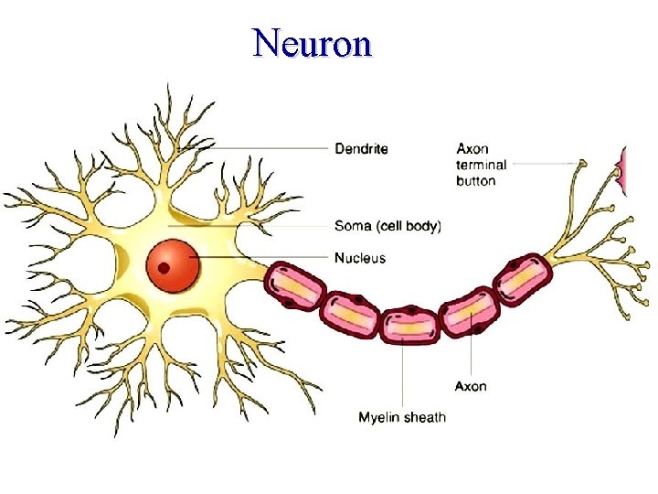
Neuron
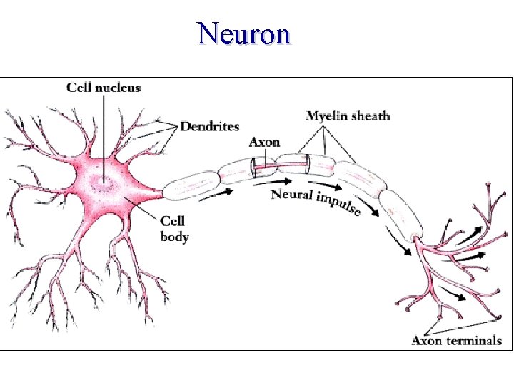
Neuron
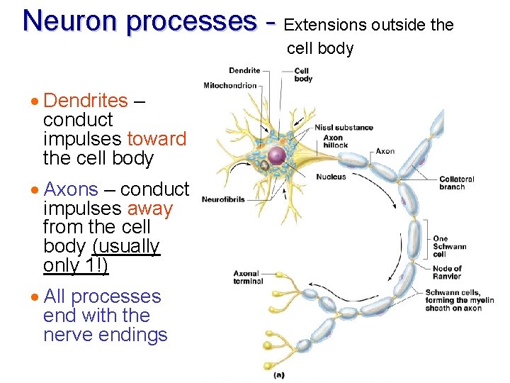
Neuron Structure · 1. Cell body = perikaryon = contains nucleus and is the metabolic center of the cell · 2. Processes – that extend from the cell body (dendrites and axon) · 3. Nerve endings (synapses, special receptors)
· Cell body has: · Nucleus with large nucleolus · Neurofibrils · “Nissl bodies” (chromophilic substance)
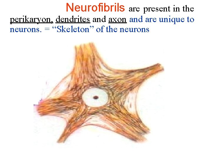
Neurofibrils are present in the perikaryon, dendrites and axon and are unique to neurons. = “Skeleton” of the neurons
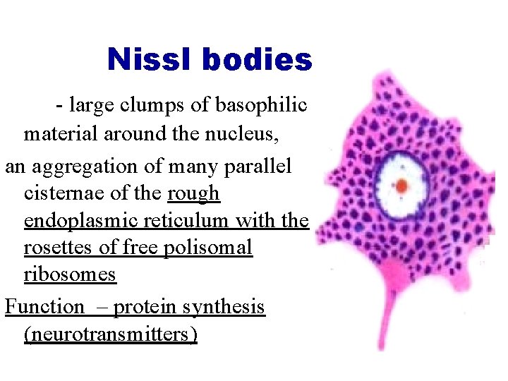
Nissl bodies - large clumps of basophilic material around the nucleus, an aggregation of many parallel cisternae of the rough endoplasmic reticulum with the rosettes of free polisomal ribosomes Function – protein synthesis (neurotransmitters)
Neuron processes - Extensions outside the cell body · Dendrites – conduct impulses toward the cell body · Axons – conduct impulses away from the cell body (usually only 1!) · All processes end with the nerve endings Slide 8
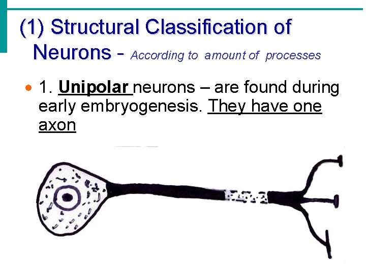
(1) Structural Classification of Neurons - According to amount of processes · 1. Unipolar neurons – are found during early embryogenesis. They have one axon
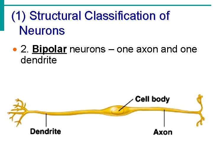
(1) Structural Classification of Neurons · 2. Bipolar neurons – one axon and one dendrite
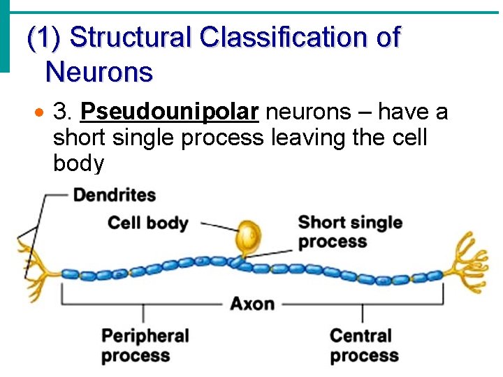
(1) Structural Classification of Neurons · 3. Pseudounipolar neurons – have a short single process leaving the cell body
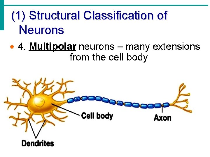
(1) Structural Classification of Neurons · 4. Multipolar neurons – many extensions from the cell body
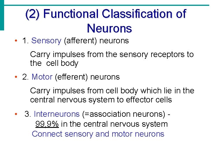
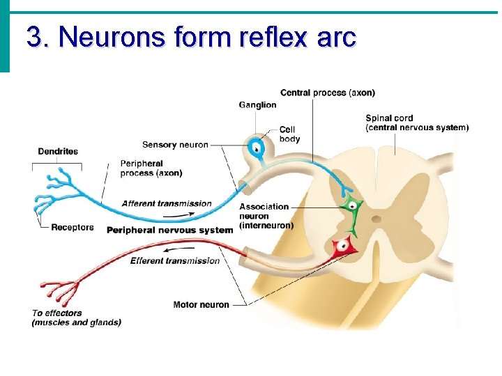
(2) Functional Classification of Neurons • 1. Sensory (afferent) neurons Carry impulses from the sensory receptors to the cell body • 2. Motor (efferent) neurons Carry impulses from cell body which lie in the central nervous system to effector cells • 3. Interneurons (=association neurons) 99, 9% in the central nervous system Connect sensory and motor neurons
3. Neurons form reflex arc
Supporting Cells (Neuroglia or Glia) = Macroglia + Microglia
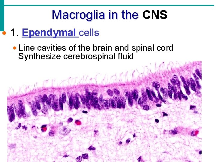
Macroglia in the CNS · 1. Ependymal cells · Line cavities of the brain and spinal cord Synthesize cerebrospinal fluid
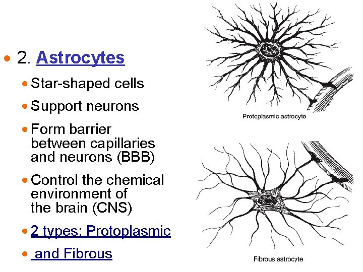
· 2. Astrocytes · Star-shaped cells · Support neurons · Form barrier between capillaries and neurons (BBB) · Control the chemical environment of the brain (CNS) · 2 types: Protoplasmic · and Fibrous
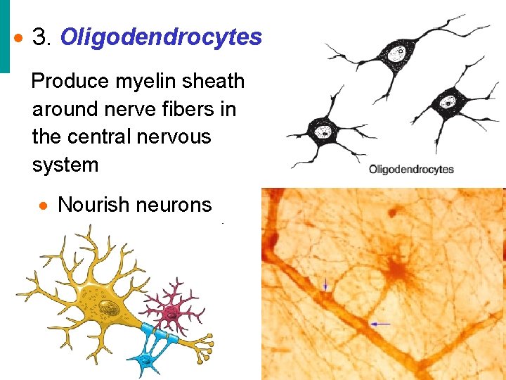
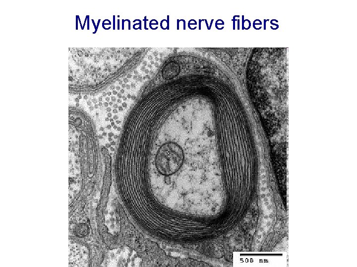
· 3. Oligodendrocytes Produce myelin sheath around nerve fibers in the central nervous system · Nourish neurons
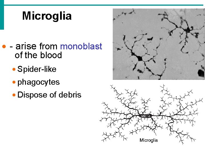
Microglia · - arise from monoblast of the blood · Spider-like · phagocytes · Dispose of debris
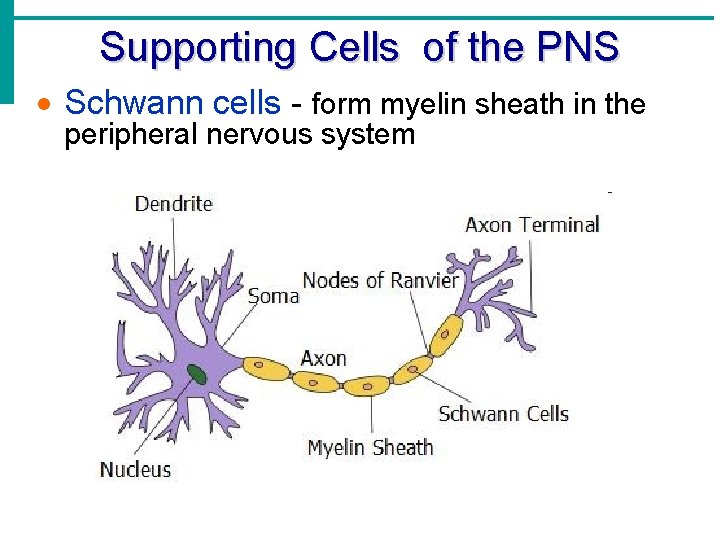
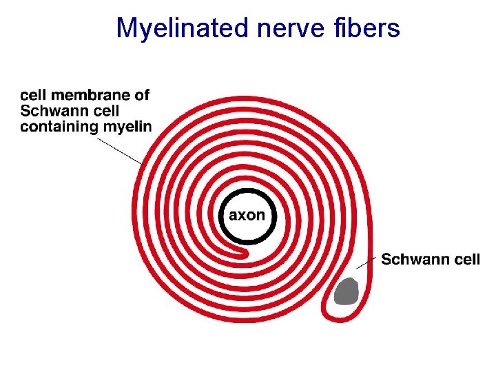
Supporting Cells of the PNS · Schwann cells - form myelin sheath in the peripheral nervous system
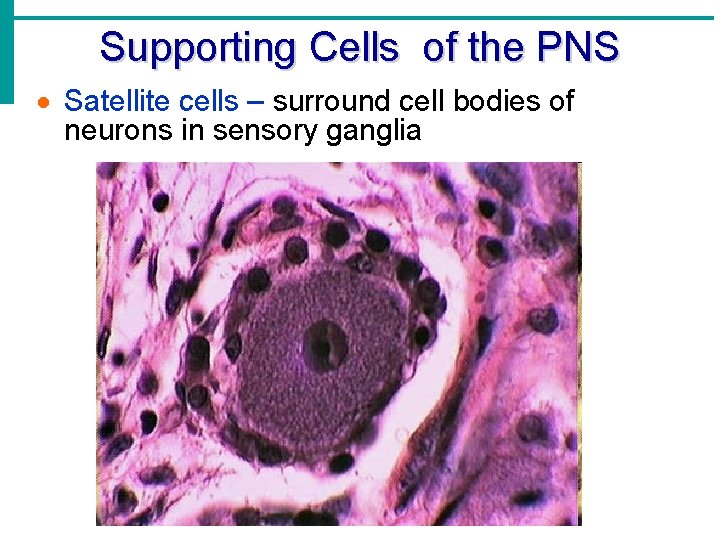
Supporting Cells of the PNS · Satellite cells – surround cell bodies of neurons in sensory ganglia
Nerve fibers 1. Unmyelinated 2. Myelinated
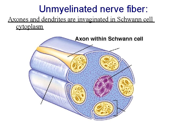
Unmyelinated nerve fiber: Axones and dendrites are invaginated in Schwann cell cytoplasm
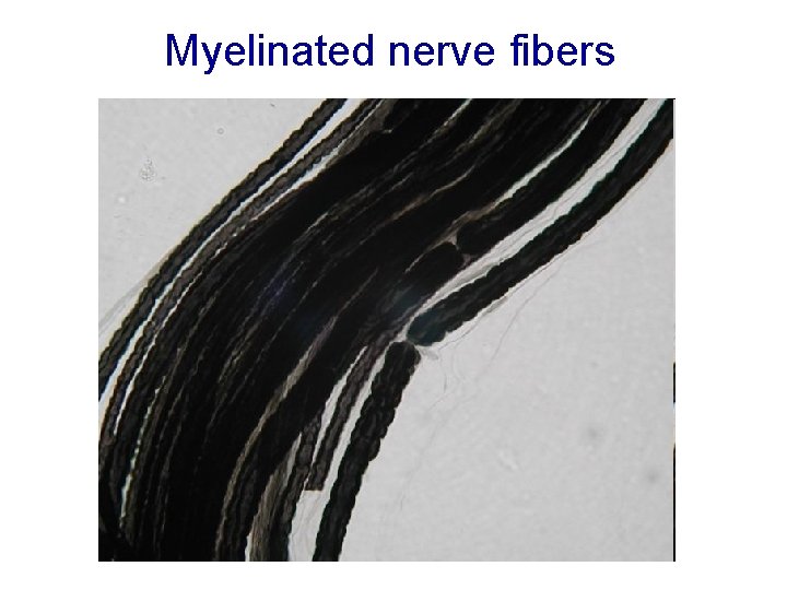
Myelinated nerve fibers
Myelinated nerve fibers
Myelinated nerve fibers
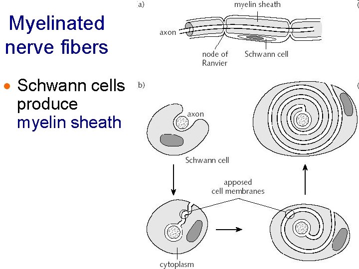
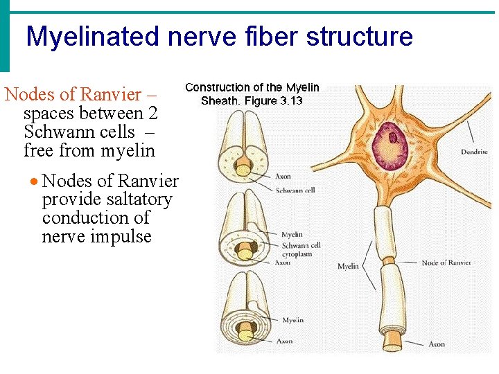
Myelinated nerve fibers · Schwann cells produce myelin sheath
Myelinated nerve fiber structure Nodes of Ranvier – spaces between 2 Schwann cells – free from myelin · Nodes of Ranvier provide saltatory conduction of nerve impulse
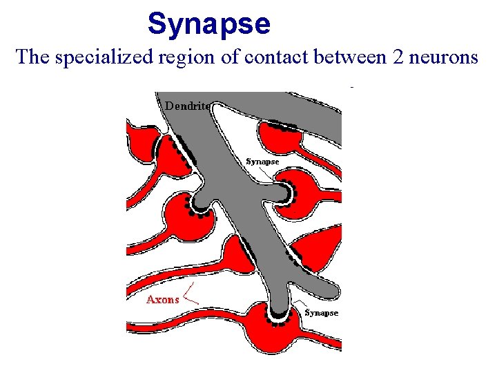
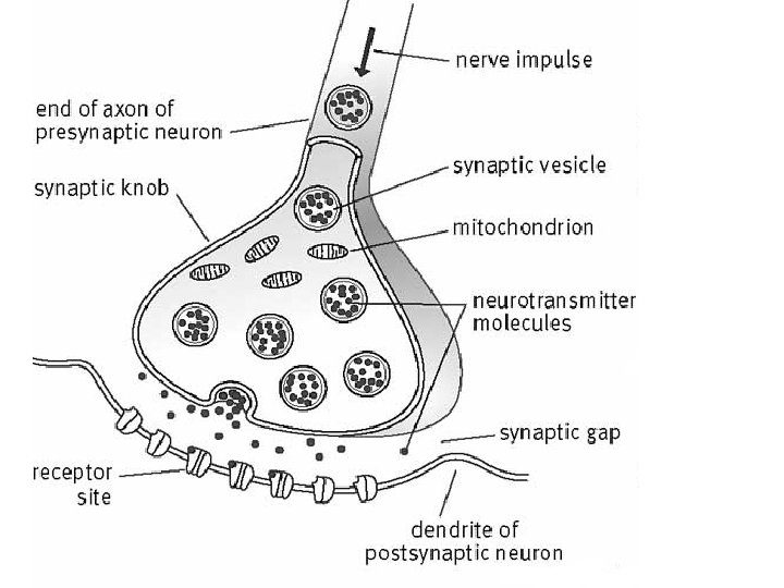
Synapse The specialized region of contact between 2 neurons
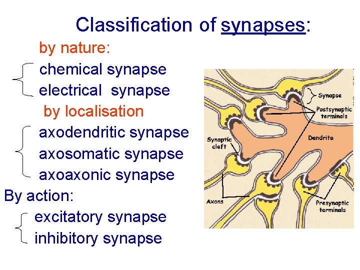
Classification of synapses: by nature: chemical synapse electrical synapse by localisation axodendritic synapse axosomatic synapse axoaxonic synapse By action: excitatory synapse inhibitory synapse
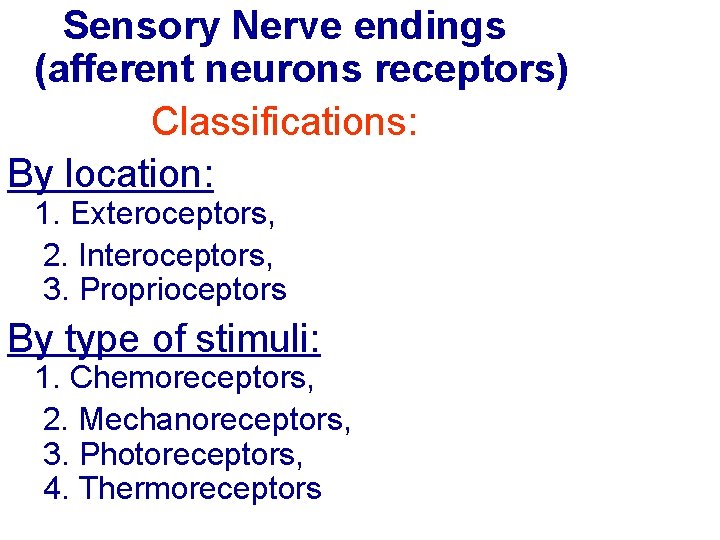
Sensory Nerve endings (afferent neurons receptors) Classifications: By location: 1. Exteroceptors, 2. Interoceptors, 3. Proprioceptors By type of stimuli: 1. Chemoreceptors, 2. Mechanoreceptors, 3. Photoreceptors, 4. Thermoreceptors
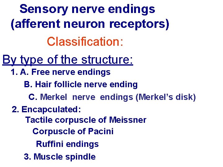
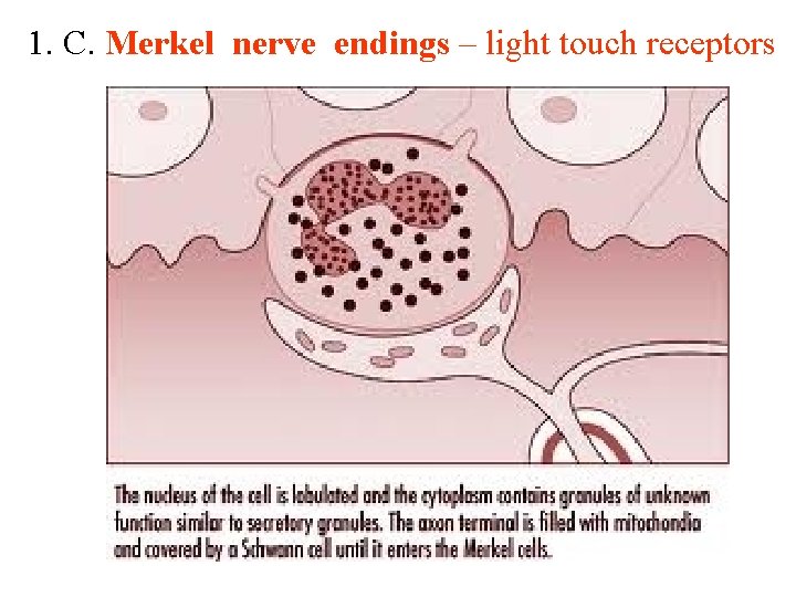
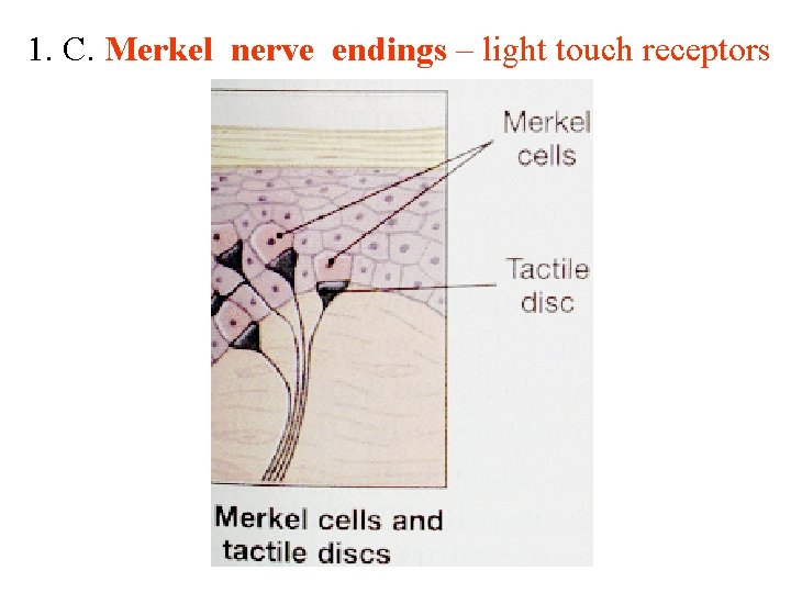
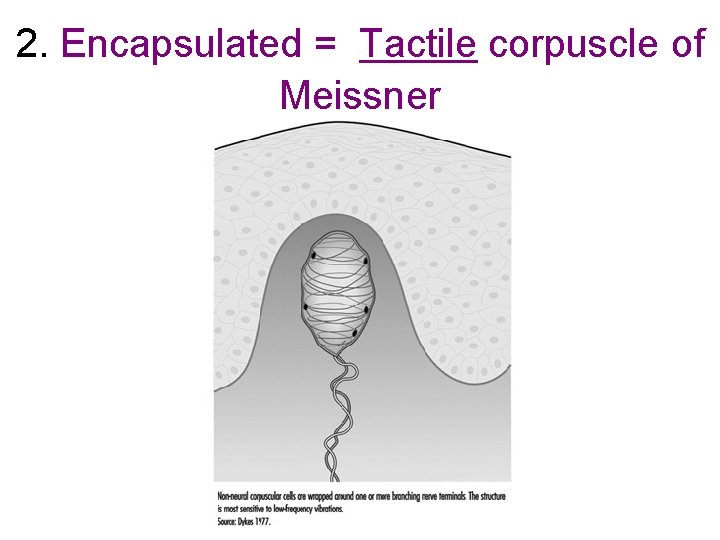
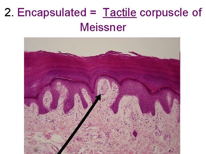
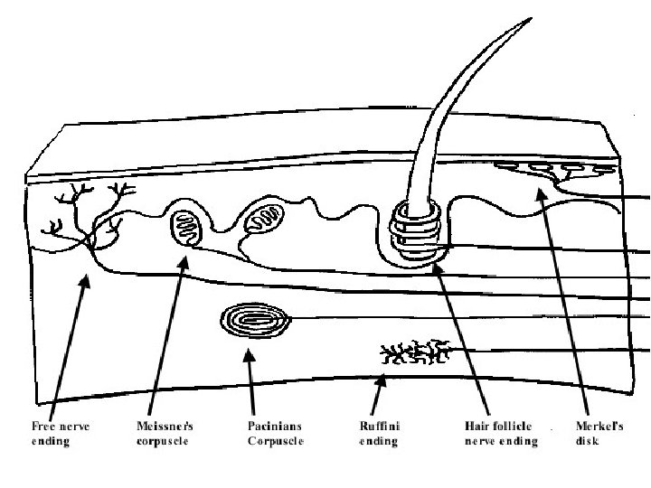
Sensory nerve endings (afferent neuron receptors) Classification: By type of the structure: 1. A. Free nerve endings B. Hair follicle nerve ending C. Merkel nerve endings (Merkel’s disk) 2. Encapculated: Tactile corpuscle of Meissner Corpuscle of Pacini Ruffini endings 3. Muscle spindle
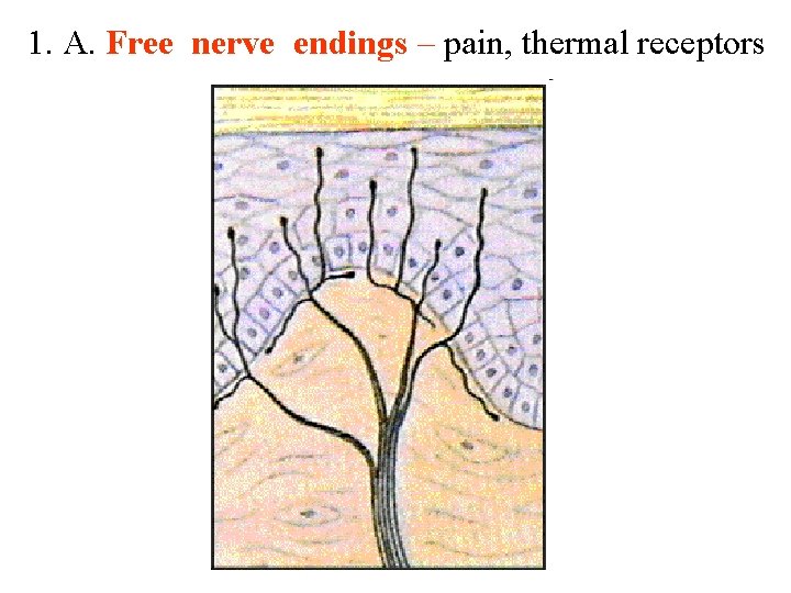
1. A. Free nerve endings – pain, thermal receptors
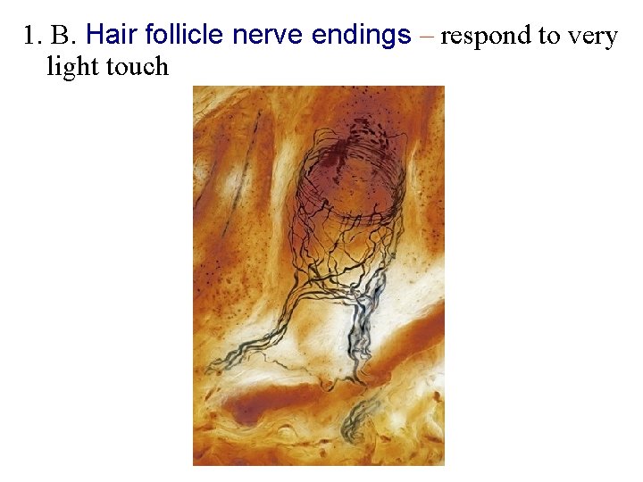
1. B. Hair follicle nerve endings – respond to very light touch
1. C. Merkel nerve endings – light touch receptors
1. C. Merkel nerve endings – light touch receptors
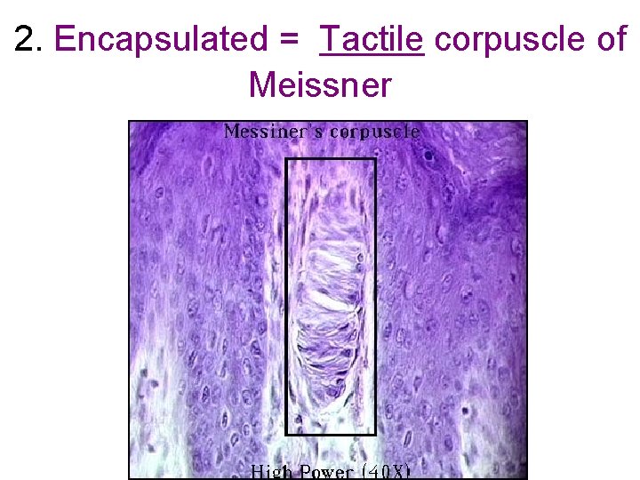
2. Encapsulated = Tactile corpuscle of Meissner
2. Encapsulated = Tactile corpuscle of Meissner
2. Encapsulated = Tactile corpuscle of Meissner
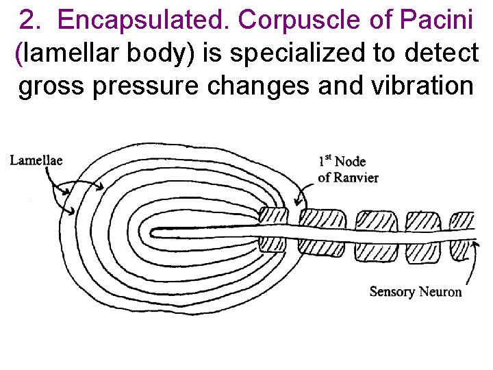
2. Encapsulated. Corpuscle of Pacini (lamellar body) is specialized to detect gross pressure changes and vibration
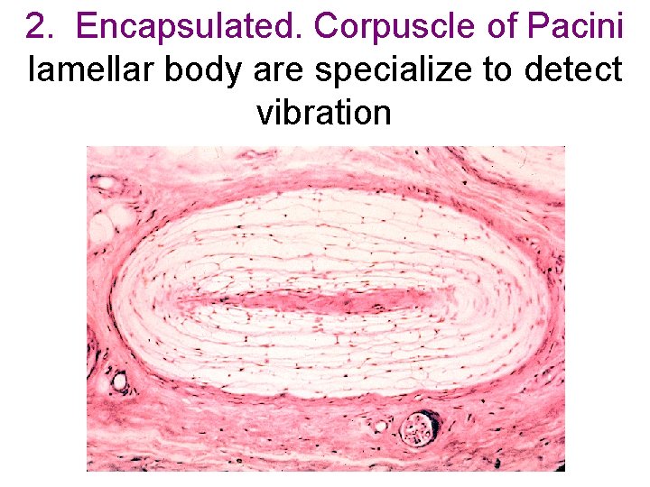
2. Encapsulated. Corpuscle of Pacini lamellar body are specialize to detect vibration
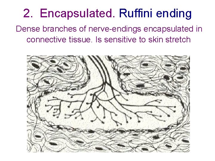
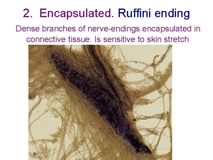
2. Encapsulated. Ruffini ending Dense branches of nerve-endings encapsulated in connective tissue. Is sensitive to skin stretch
2. Encapsulated. Ruffini ending Dense branches of nerve-endings encapsulated in connective tissue. Is sensitive to skin stretch
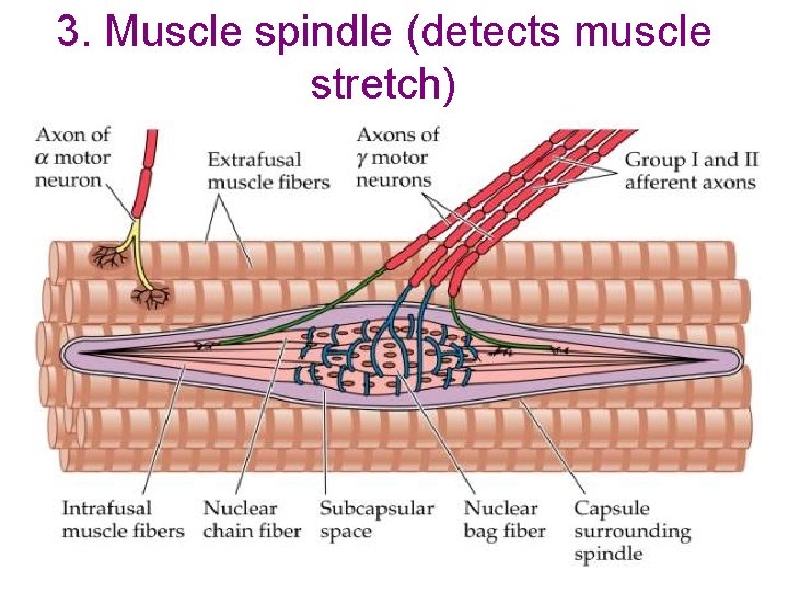
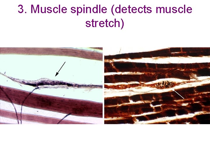
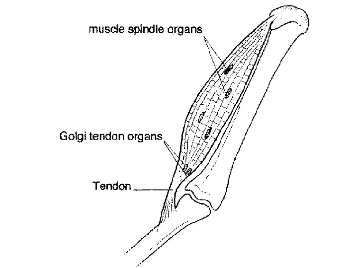
3. Muscle spindle (detects muscle stretch)
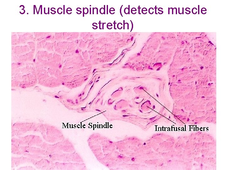
3. Muscle spindle (detects muscle stretch)
3. Muscle spindle (detects muscle stretch)
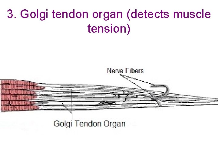
3. Golgi tendon organ (detects muscle tension)
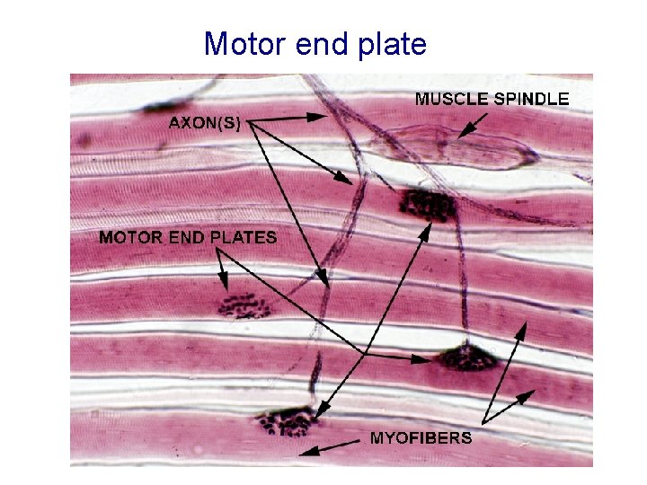
Motor end plate