The Neuromuscular Junction Neuromuscular Synapse Textbook of medical
 Textbook of medical physiology Guyton & Hall")
The Neuromuscular Junction ( Neuromuscular Synapse ) Textbook of medical physiology Guyton & Hall (13 th edition) UNIT II CHAPTER 6 & 7 Dr. Taha Sadig Ahmed

Objectives of the lecture At the end of the lecture the student should be able to: Know and describe the followings: The physiologic anatomy of the skeletal muscle and NM junction. The general mechanism of skeletal muscle contraction. Motor End Plate potential and how action potential and excitation- contraction coupling are generated in skeletal muscle. The molecular mechanism of skeletal muscle contraction & relaxation. Sliding filament mechanism. Drugs/ diseases affecting the neuromuscular transmission.

Chemical Signals One neuron will transmit info to another neuron or to a muscle or gland cell by releasing chemicals called neurotransmitters. l The site of this chemical interplay is known as the synapse. l An axon terminal (synaptic knob) will abut another cell, a neuron, muscle fiber, or gland cell. l This is the site of transduction – the conversion of an electrical signal into a chemical signal. l
")
Neuromuscular Junction (NMJ)

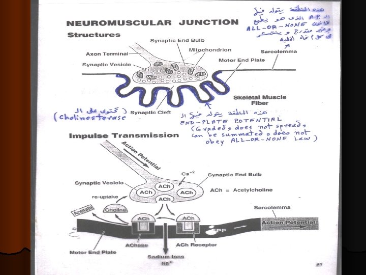
Physiologic Anatomy of the Neuromuscular Junction • Motor End Plate • Synaptic trough/ gutter • Presynaptic terminal • Postsynaptic terminal • Synaptic space/cleft • Subneural cleft • Acetylcholine (Ach) • Synaptic vesicles

The Neuromuscular junction consists of A/ Axon Terminal : contains around 300, 000 vesicles which contain the neurotransmitter acetylcholine (Ach). B/ Synaptic Cleft : 20 – 30 nm ( nanometer ) space between the axon terminal & the muscle cell membrane. It contains the enzyme cholinesterase which can destroy Ach. C/ Synaptic Gutter ( Synaptic Trough) It is the muscle cell membrane which is in contact with the nerve terminal. It has many folds called Subneural Clefts , which greatly increase the surface area , allowing for accomodation of large numbers of Ach receptors are located here.

The Neuromuscular junction consists of § Ø Ø The entire structure of axon terminal , synaptic cleft and synaptic gutter is called “ Motor End-Plate ”. Ach is synthesized locally in the cytoplasm of the nerve terminal , from active acetate (acetylcoenzyme A) and choline. Then it is rapidly absorbed into the synaptic vesicles and stored there. The synaptic vesicles themselves are made by the Golgi Apparatus in the nerve soma ( cell-body). Then they are carried by Axoplasmic Transport to the nerve terminal , which contains around 300, 000 vesicles.
 Ø Ø Ø Ach is synthesized locally in the cytoplasm of the")
Acetylcholine (1) Ø Ø Ø Ach is synthesized locally in the cytoplasm of the nerve terminal , from active acetate (acetylcoenzyme A) and choline. Then it is rapidly absorbed into the synaptic vesicles and stored there. The synaptic vesicles themselves are made by the Golgi Apparatus in the nerve soma ( cell-body). Then they are carried by Axoplasmic Transport to the nerve terminal , which contains around 300, 000 vesicles. Each vesicle is then filled with around 10, 000 Ach molecules.
 l l When a nerve impulse reaches the nerve terminal , it")
Acetylcholine (2) l l When a nerve impulse reaches the nerve terminal , it opens calcium channels calcium diffuses from the ECF into the axon terminal Ca++ releases Ach from vesicles by a process of EXOCYTOSIS One nerve impulse can release 125 Ach vesicles. The quantity of Ach released by one nerve impulse is more than enough to produce one End-Plate Potential.

Ach combines with its receptors in the subneural clefts. This opens sodium channels & sodium diffuses into the muscle causing a local, nonpropagated potential called the “ End-Plate Potential (EPP)”, . v This EPP triggers a muscle AP which spreads down inside the muscle to make it contract. v

l After ACh acts on the receptors , it is hydrolyzed by the enzyme Acetylcholinesterase (cholinesterase ) into Acetate & Choline. The Choline is actively reabsorbed into the nerve terminal to be used again to form ACh. This whole process of Ach release, action & destruction takes about 5 -10 ms.

Destruction of Ach 1425 – 2004

Drugs That Enhance or Block Transmission at the Neuromuscular Junction Drugs That Stimulate the Muscle Fiber by Ach-Like Action: l Methacholine, Carbachol, and Nicotine. They act for minutes or hours—are not destructed by cholinesterase. Drugs That Stimulate the NMJ by Inactivating Acetylcholinesterase: l Neostigmine, Physostigmine [inactivate acetylcholinesterase for l Diisopropyl fluorophosphate (nerve gas poison) [inactivates several hours] acetylcholinesterase for weeks -------can cause death because of respiratory muscle spasm]

Drugs That Enhance or Block Transmission at the Neuromuscular Junction Drugs That Block Transmission at the NMJ l Curare & Curariform like-drugs. Prevent passage of impulses from the nerve ending into the muscle by blocking the action of Ach on its receptors. act by competitive inhibition to Ach at its receptors & can not cause Depolarization. l Botulinum Toxin. Bacterial poison that decreases the quantity of Ach release by the nerve terminals.

Myasthenia Gravis l Auto-immune disease l Antibodies against Ach receptors destroy many of the receptors decreasing the EPP , or even preventing its formation weakness or paralysis of muscles ( depending on the severity of the disease ). l patient may die because of paralysis of respiratory muscles. l Treatment : Anti-cholinestersae drugs. These drugs inactivate the cholinesterase enzyme ( which destroys Ach) and thereby allow relatively large amounts of Ach to accumulate and act on the remaining healthy receptors good EPP is formed muscle contraction.

Myasthenia Gravis Ø Disease of adult females affects eyelid, extra ocular bulbar and proximal limb muscles. Ø Presents with ptosis, dysarthria, dysphagia, and proximal limb weakness in hands& feet.

-An autoimmune disorder - body form antibodies against Ach receptors. Patients have 20% of number of Ach receptors -the EPPs are too small to trigger action potentials & the muscles can not contract. .

Physiology of Skeletal Muscle Contraction
 Muscle RMP = -90 m. V (")
The Muscle Action Potential ( AP ) Muscle RMP = -90 m. V ( same as in nerves ). l Duration of AP = 1 -5 ms ( longer duration than nerve AP , which is usually about 1 ms ). l Conduction Velocity = 3 -5 m/s ( slower than big nerves ). l
 is covered by a cellmembrane called Sarcolemma. l Each")
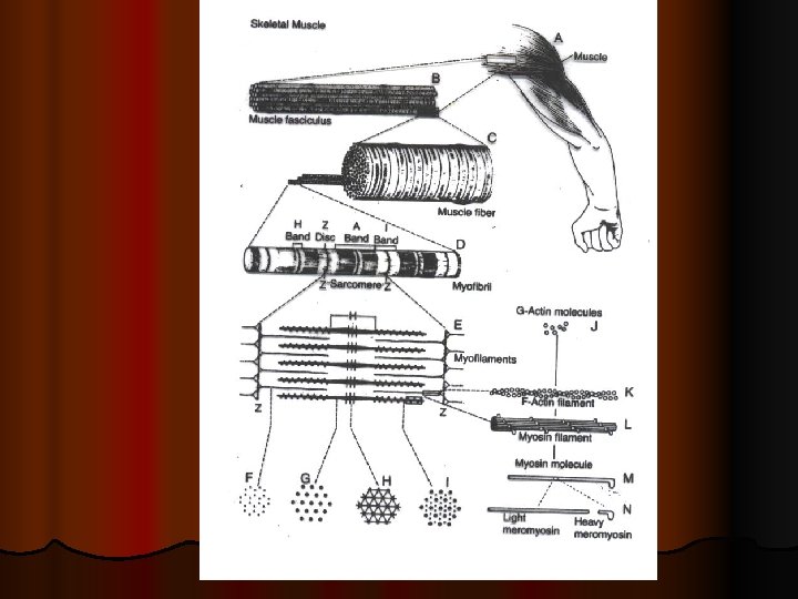
l Each muscle cell (fiber) is covered by a cellmembrane called Sarcolemma. l Each cell contains between a few hundreds to a few thousands Myofibrils.
 & Myosin (thick) filaments. l Each myofibril")
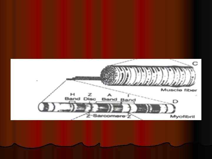
l Each Myofibril contains Actin filaments (thin) & Myosin (thick) filaments. l Each myofibril is striated: consisting of dark bands (called A-bands) and light (I-bands).

Sarcoplasm= matrix inside muscle fiber in which myofilaments susbended Sarcoplasmic reticulum= it is endoplasmic reticulum inside sarcoplasm full of Ca. T- tubules: extend from one side of muscle to other (function? ).

Sarcomere= contractile unit of muscle, it is the zone between two Z lines ( discs)=2 micrometer in length in resting state. Z discs (lines) = lines extend all way across myofibrils

The functional unit of a myofibril is the Sarcomere

Inside each sarcomere there are 3 bands: - I band = of actin only - H band= of myosin only - A band= formed of actin & myosin filaments
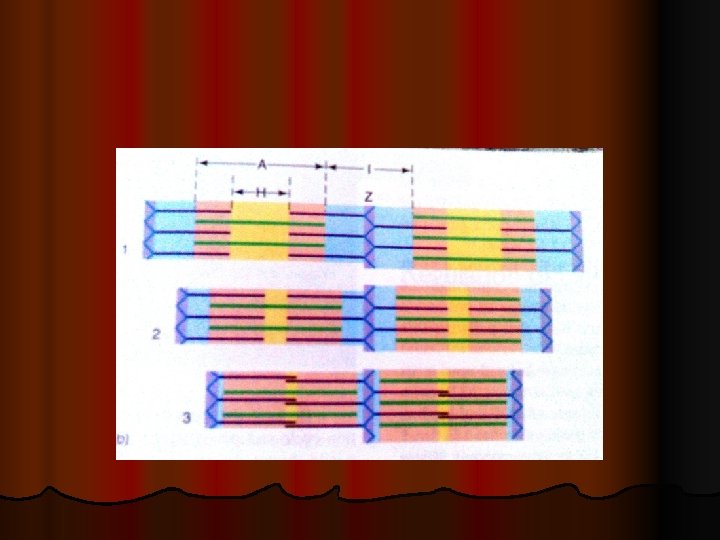
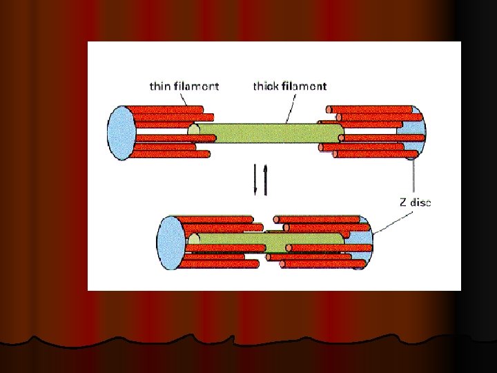

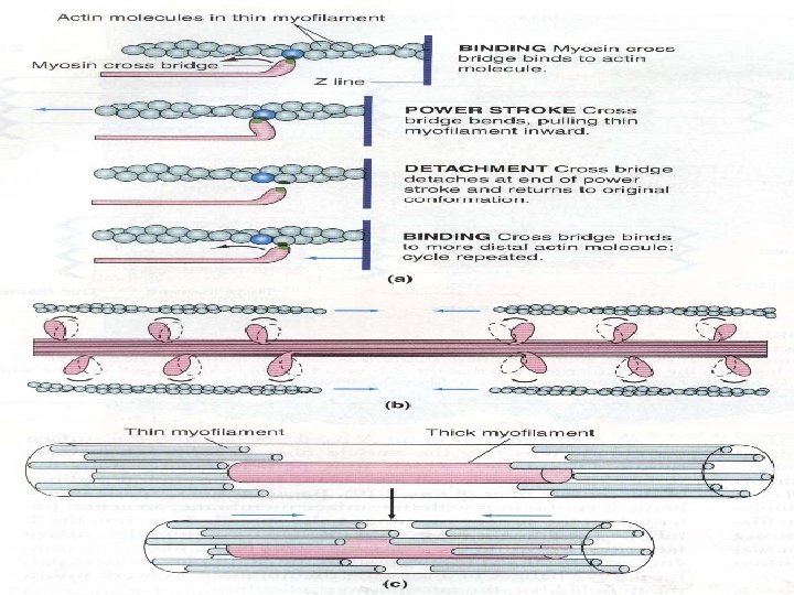
When contraction takes place Actin & Myosin slide upon each other , & the distance between two z-discs decreases : This is called Sliding Filament Mechanism Ø

From: http: //www. 3 dotstudio. com/zz. html

EM Evidence for Sliding Filaments

Muscle Contraction muscle proteins : a. Thick filament: Myosin b. Thin filament : 1. Actin 2. Troponin 3. Tropomyosin

Sarcomere filamentous proteins From: Alberts et al. . 1994 Molecular Biology of the Cell.
 -")
Thick filament: Myosin filament it has head + tail cross bridges (? ) - Head has ATP site -?

Thin filament Two F-Actin strands Groove between the 2 Factin strands

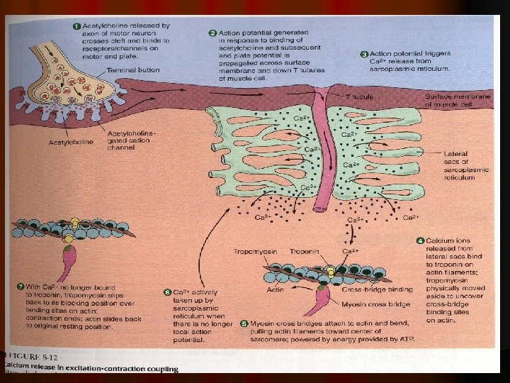
MOLECULAR MECHANISM OF MUSCLE CONTRACTION Excitation –contraction coupling

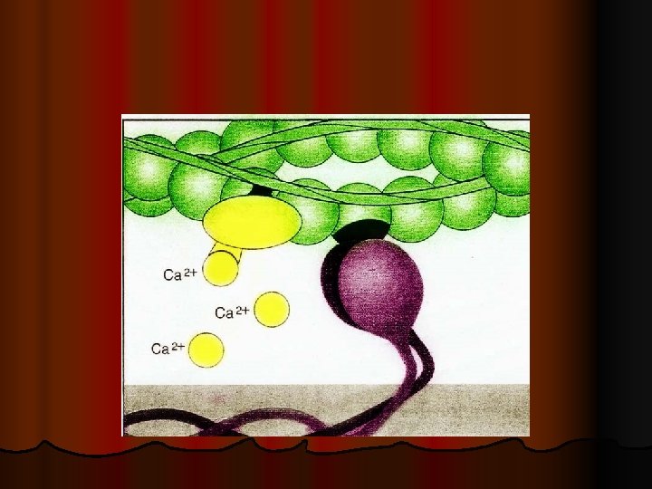
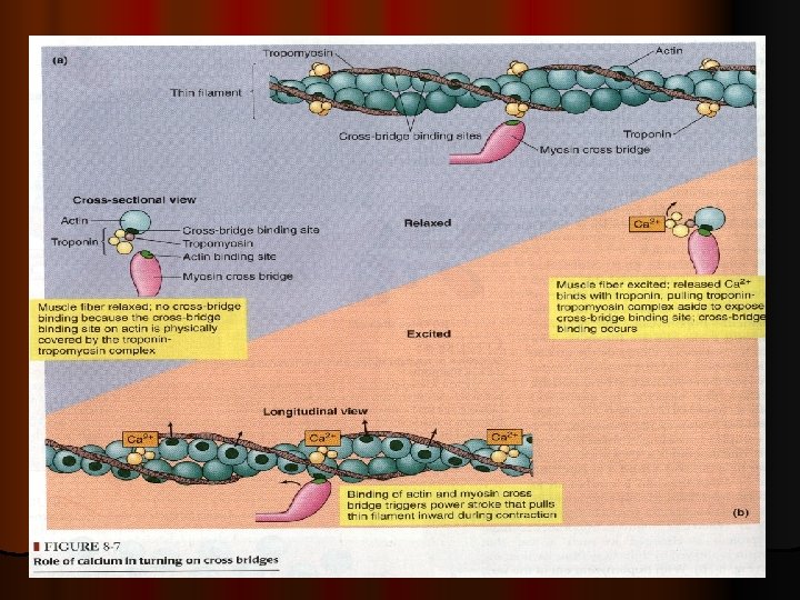
Events of muscle contraction: *** l Acetylcholine released by motor nerve » » » EPP » » » depolarization of CM (muscle AP) » » » l Spread of AP into T tubule » » » release of Ca from sarcoplasmic reticulum into the cytoplasm l » » » Ca combines with troponin » » » troponin pull tropomyosin sideway » » » exposing the active site on actin » » » myosin heads with ATP on them, attached to actin active site l » » » myosin cross bridges bend pulling actin toward center of sarcomere (Power stroke) using energy of ATP» » » ADP & P released » » » Linkage between actin & myosin broken as new ATP binds to myosin cross bridge >>> ATP hydrolyzed and cross bridge go back to its original conformation.

Events of muscle contraction: l When a new ATP occupies the vacant site on the myosin head, this triggers detachment of myosin from actin l The free myosin swings back to its original position, & attached to another actin, & the cycle repeat its self

Events of muscle relaxation: l When ca is pumped back into sarcoplasmic reticulum l » » » ca detached from troponin » » » tropomyosin return to its original position l » » » covering active sit on actin » » » prevent formation of cross bridge » » » relaxation
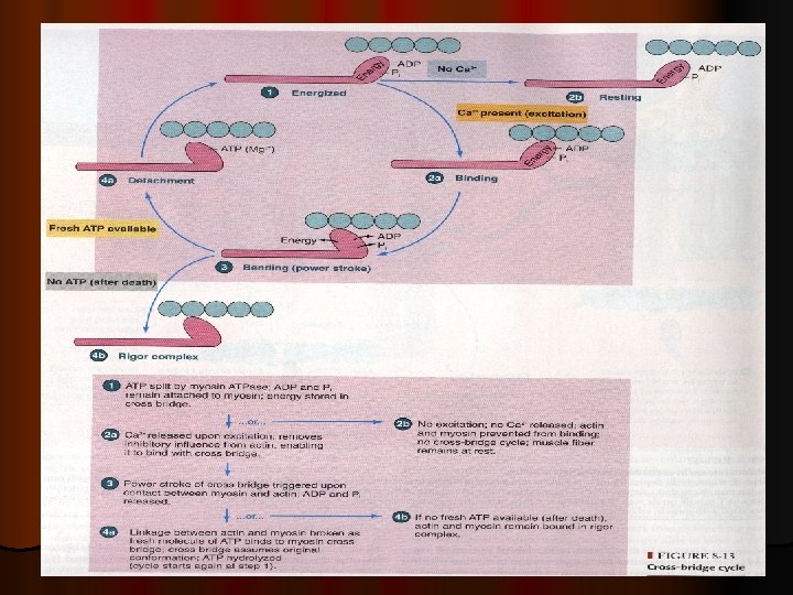

Therefore , on order to release the head of Myosin from Actin , a new ATP is needed to come and combine with the head of Myosin. l Q: What is Rigor Mortis ? l Q: ATP is needed for 3 things : what are they ? l l l l ATP is needed for 3 things : (1) Power stroke. (2) Detachment of myosin from actin active sites (3) Pumping C++ back into the Sarcoplasmic reticulum. Q: Is muscle relaxation a passive or active process ? A : it is active ; Why ? Because it needs ATP.

Q: What happens to A-band I-band during contraction ? l Q: Ca++ is needed in nerve & muscle : when and where ? l A : In nerve needed for exocytosis ( & release of Ach) l In Muscle needed for contraction. l

l. Thanks
- Slides: 49