RENAL ANATOMY DR AKANKSHA SINGH SR JNMC AMU

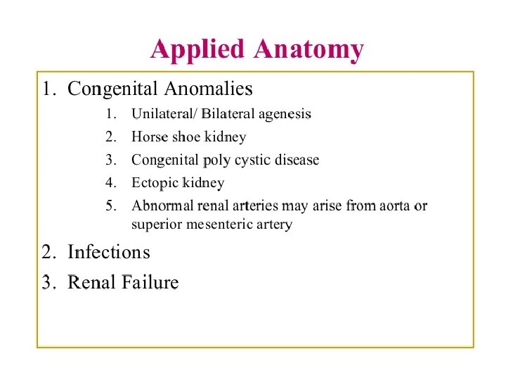
")


 – increased rate of")


- Slides: 61
RENAL ANATOMY DR. AKANKSHA SINGH SR, JNMC, AMU ALIGARH
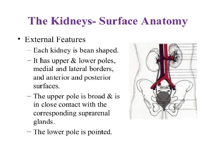
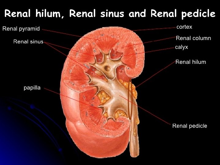
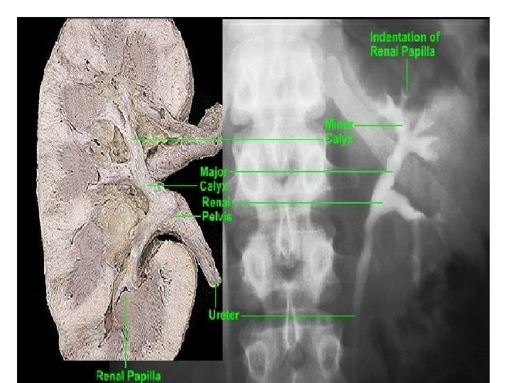
STRUCTURE OF HILUM OF KIDENEY(V. IMP)
CASE PRESENTATION • A 2 -year-old boy is referred by his pediatrician to a nephrologist for recurrent urinary tract infections. He was born at 39 weeks and appeared healthy at birth; however, he has developed significantly more urinary tract infections than expected for a child of his age. Physical exam reveals increased fullness in the abdomen and an MRI scan is obtained. Urine cultures are obtained and empirical antibiotics are started pending culture results.
Clinical definition congenital malformation of the renal system resulting in fused kidneys Epidemiology common malformation found in 1/400 live births twice as common in males Pathogenesis fusion of the lower poles of the kidneys (most common) trapping of the fused kidneys under the inferior mesenteric artery arrest of the normal retroperitoneal ascent of the kidneys Associated conditions Turner syndrome trisomy 18 (Edwards syndrome) Prognosis typically very good most patients remain asymptomatic
• • • Presentation Symptoms – asymptomatic (most common) – increased rate of urinary tract infections – hydronephrosis due to ureteropelvic junction obstruction – renal stones Physical exam – abdominal fullness upon palpation Imaging Kidney ultrasound – indications • initial evaluation of symptomatic patients Voiding cysturethrogram – indications • further evaluation of abnormal anatomy – findings • fusion of kidney poles trapped under the inferior mesenteric artery Intravenous pyelogram – indications • for evaluation of symptomatic kidney stones – findings • filling defect at the level of the obstruction
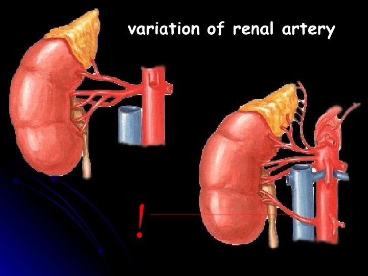
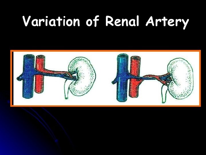
• • • Differential Unilateral renal agenesis Posterior urethral valves Multicystic dysplastic kidney Duplex collecting system • • Treatment Conservative – no treatment for asymptomatic patients • Medical – antibiotics • indication – urinary tract infections • outcomes – • very good Complications Postrenal kidney failure due to kidney stones – rare since most cases are asymptomatic – treatment • removal of kidney stones • • Hydronephrosis if obstruction is left untreated Anomalous original of multiple renal arteries may complicate surgery
• 1 IN 800 • It occurs due to fusion of the lower poles of the kidneys. • The ureter passes anterior to the isthmus. • The inferior mesentric artery also passes anterior to isthmus which limits the ascent of horse shoe shaped kidney.
MCQ--- CASE PRESENTATION • A 10 -year-old Caucasian female with Turner's syndrome underwent an abdominal imaging study and was discovered that the poles of her kidneys were fused inferiorly. Normal ascension of kidney during embryological development would be prevented by which of the following anatomical structures?
Correct answer is ----- • • • Inferior vena cava Superior mesenteric artery Inferior mesenteric artery Celiac artery Splenic artery
• Hydronephrosis is a condition that typically occurs when a kidney swells due to urine failing to properly drain from the kidney to the bladder. This swelling most commonly affects only one kidney, but it can involve both kidneys. Hydronephrosis isn’t a primary disease.
• Nephrolithiasis is characterized by the formation of crystalline material in the kidney and the urinary tract. Nephrolithiasis is also known as kidney stones and is formed as a result of a decrease in the volume of urine or an increase in the substances in urine that can form stones in the kidney or in the urinary tract. Nephrolithiasis is also called renal calculi and the stone formations often results in blood in the urine. Severe pain in the abdomen, flank or groin is also included in the manifestation of renal calculi.
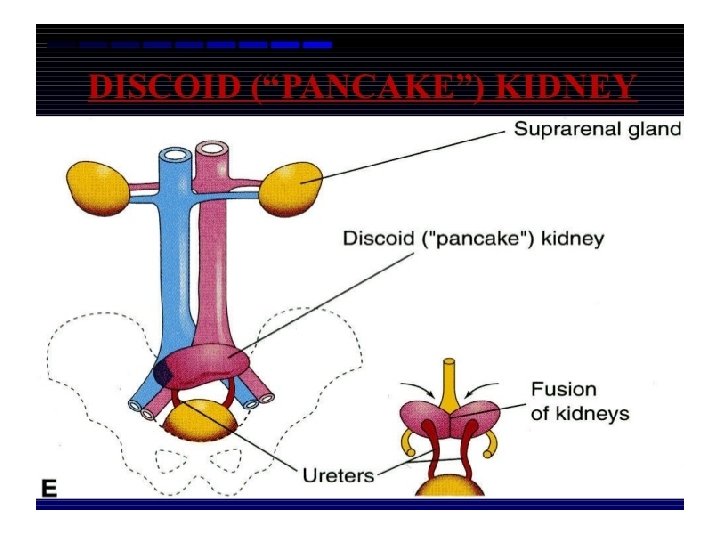
Pan cake kidney
• When luminal continuity between the nephrons and collecting tubules fail to establish. • The glomeruli continue to excrete urine which accumulates in the tubules due to lack of outlet. As a result tubules undergo cystic enlargements.
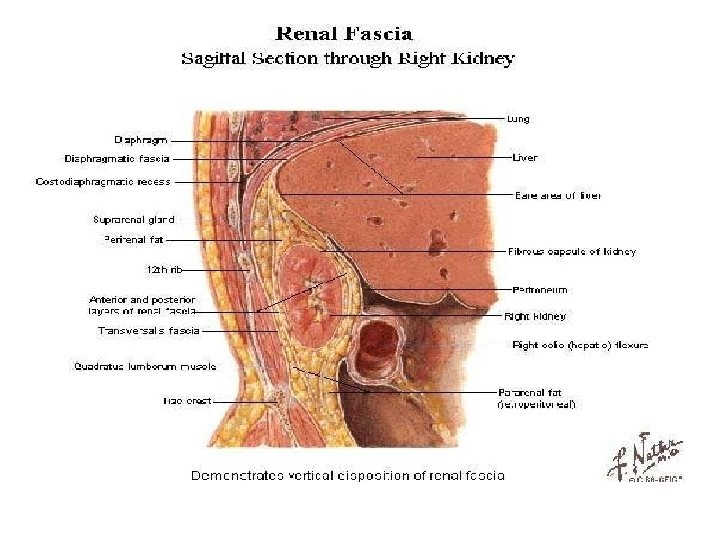
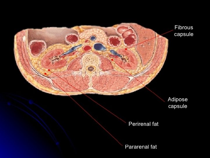
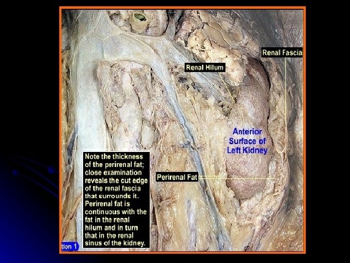
Floating kidney • The kidney is kept in position by the perirenal fat and renal fascia. However , each kidney moves up and down with respiration. If the amount of perinephric fat is reduced , the mobility of the kidney becomes excessive and may reduce the symptoms of the renal colic caused by the kinking of the ureter. A floating kidney can move up and down but not from the side to side within the renal fascia.
Transplantation of kidney • Renal transplantation is now a common procedure. It is done in patients with end stage renal failure/disease. The donor kidney is placed retroperitoneally in the iliac fossa with hilum parallel to the external iliac vessels. • The renal artery of donor kidney is anastomosed end to the internal iliac artey of recepient. Similarly the renal vein of donor kidney is anastomosed end to side to the external iliac vein of the recipient. The ureter is implanted into the urinary bladder. (ureterocystosto my)
Murphy’s punch sign • Medical test in which • Renal angle –it lies tenderness is elicited by between the lower tapping in the renal border of 12 th rib and angle during inspiration. outer border of erector This test is positive in spinae. perinephric abscess, renal stone etc.
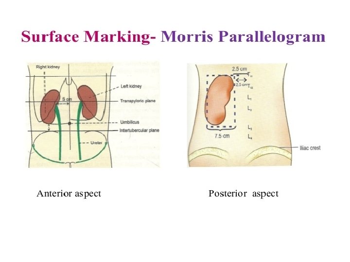
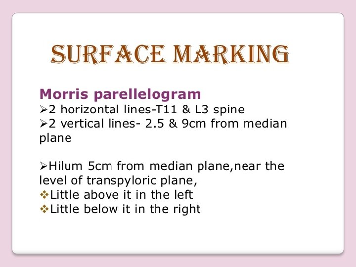
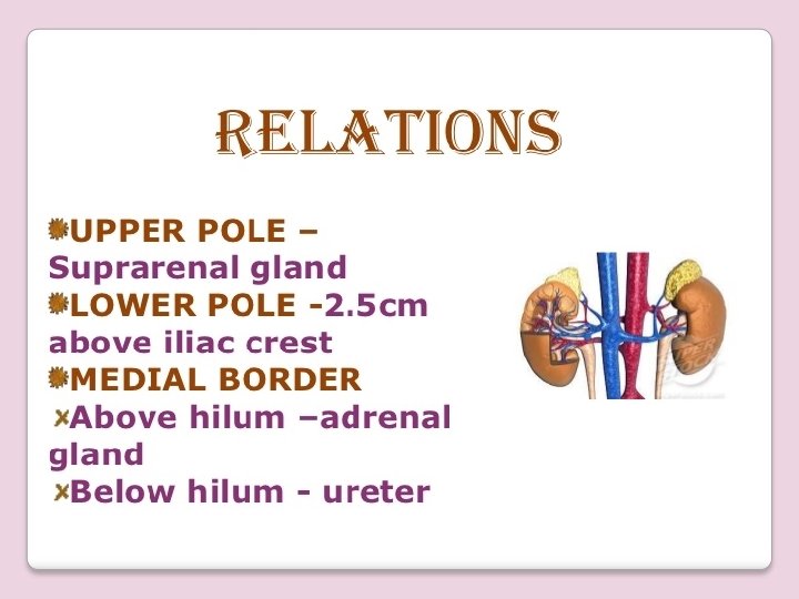
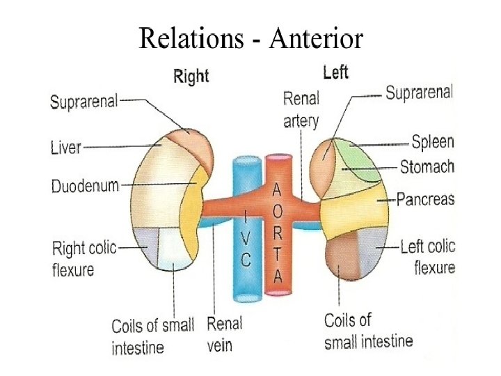
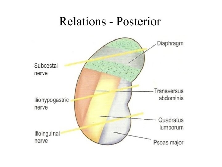
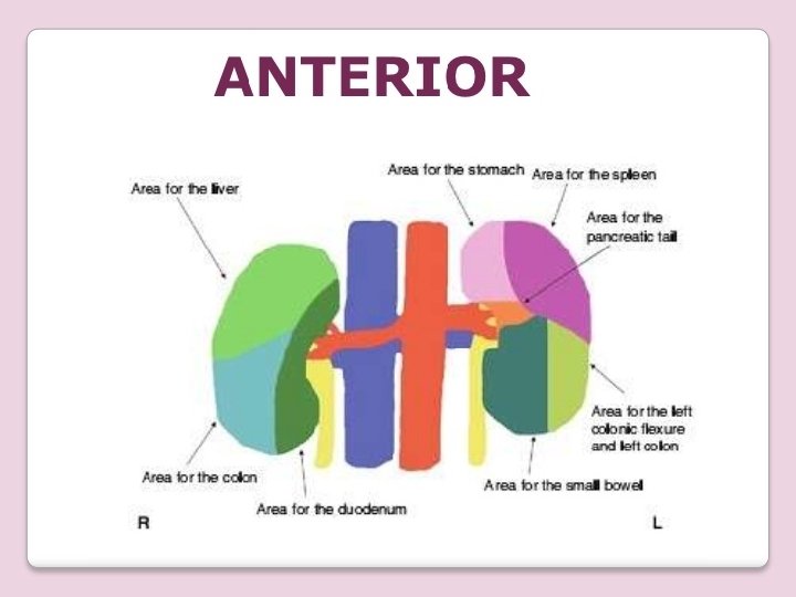
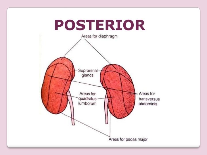
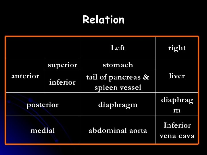
Imp question • Anterior relations of the kidney. • Posterior relation of the kidney. • Horseshoe shaped kidney. • Relations of renal artery, vein and pelvis in the hilum anterior to post and above downwards. • • Polycystic kidney disease Renal angle Morries parrallelogram Which artery prevents the ascent of horseshoe kidney.
Thank you