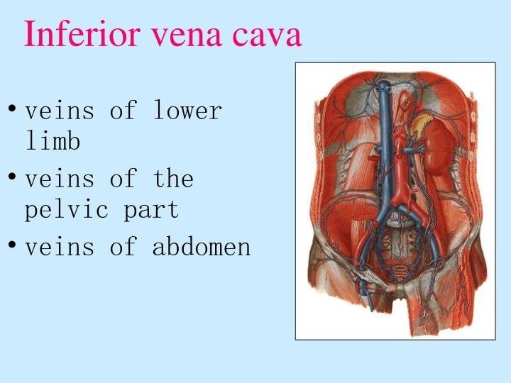
INFERIOR VENA CAVA DR AKANKSHA SINGH SR JNMC

 is the largest vein of the human body. It")


- Slides: 28
INFERIOR VENA CAVA DR. AKANKSHA SINGH SR, JNMC AMU ALIGARH
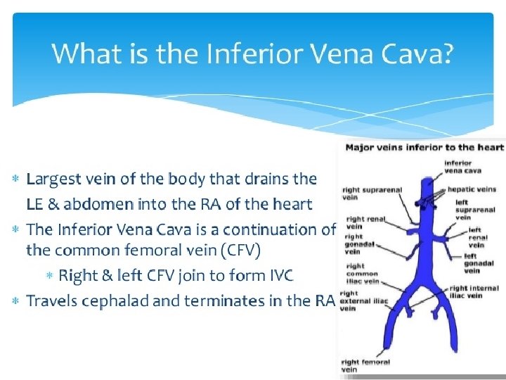
�The inferior vena cava (IVC) is the largest vein of the human body. It is located at the posterior abdominal wall on the right side of the aorta. The IVC’s function is to carry the venous blood from the lower limbs and abdominopelvic region to the heart.
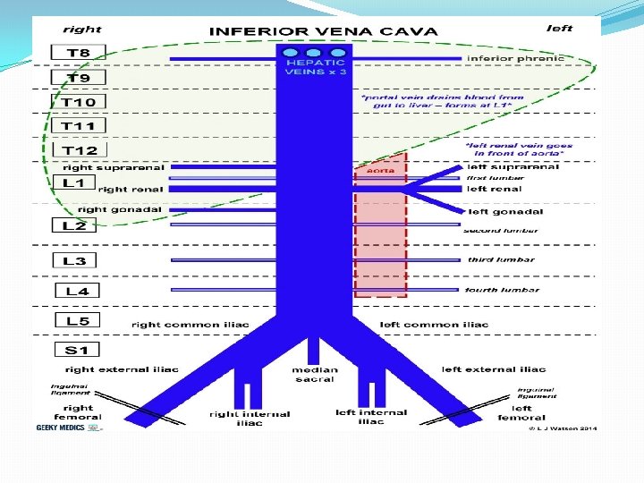
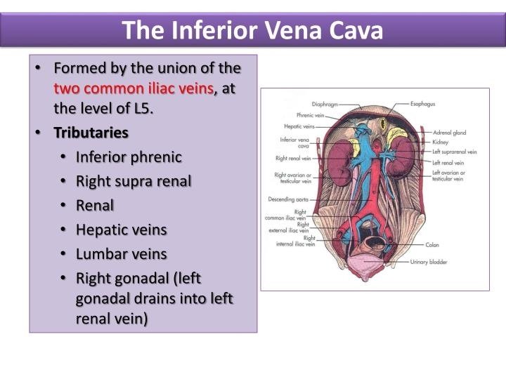
Key facts Definition and function The vein that collects deoxygenated blood from the abdomen, pelvis and lower limbs and carries it to the right atrium of the heart �Key facts. Definition and function. The vein that collects deoxygenated blood from the abdomen, pelvis and Common iliac veins (L 5) Source lower limbs and carries it to the right atrium of the heart. Source. Common iliac veins Tributaries Inferior Phrenic, right Suprarenal, Renal, (L 5)Tributaries. Inferior Phrenic, right Testicular (gonadal), Lumbar, right Suprarenal, Renal, right Testicular common Iliac and Hepatic veins (gonadal), Lumbar, common Iliac and Hepatic veins Mnemonic: Portal System Returns To Liv er In Humans �Mnemonic: Portal System Returns To Liver In Human s �Clinical relations. Inferior vena cava thrombosis Clinical relations
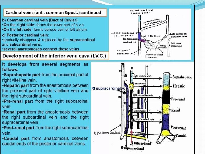
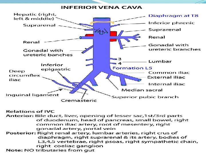
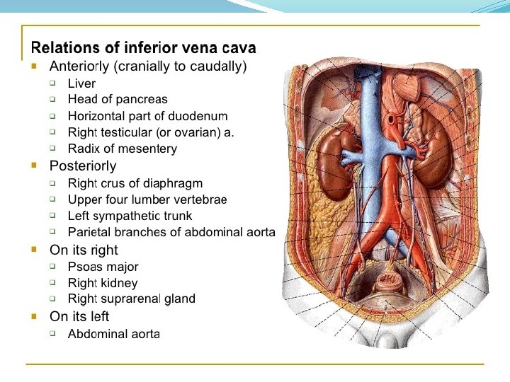
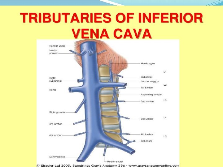
�The tributaries of the IVC correspond to the branches of the abdominal aorta. Note that some professors will want you to know at which vertebral level the IVC gets its direct tributaries, so they are as follows: �The direct tributaries are �the inferior phrenic veins (T 8), right suprarenal (L 1), right testicular (gonadal) (L 2), lumbar (L 1 -L 5), common iliac (L 5) and hepatic (T 8). �If you want an easy way to remember them just memorise the mnemonic 'Portal System Returns To Liver In Humans'. �Left gonadal and left suprarenal veins drain first into the left renal vein �The veins of the stomach, spleen, pancreas, small and large intestines first empty into the hepatic portal vein. The hepatic portal vein carries this blood to the liver to be processed and detoxified. Then, the blood reaches the IVC through the hepatic veins. �The inferior vena cava communicates with the superior vena cava through the collateral vessels, which include the azygos vein, lumbar veins, and vertebral venous plexuses.
Inferior vena cava in a cadaver. Notice how the largest tributaries are the left and right renal veins.
The IVC’s function is to convey the blood from the abdomen, pelvis, and lower limbs to the right atrium of the heart. Additional IVC functions are noticeable during some health disturbances, such as hepatic portal vein obstruction or the obstruction of the IVC itself. Specialized vessels called the portocaval (portosystemic) anastomoses open if the hepatic portal vein is obstructed. The intestinal blood then bypasses the liver and empties into the IVC directly. In cases where the IVC is occluded, the collateral vessels to the superior vena cava open.
STRUCTURE � What makes the IVC different from other veins is that there are no valves within the vein to keep blood moving forward instead of backward, which is how the typical anatomy of a vein works. To prevent the blood from moving back into the body, valves made up of tissue in the vein close as the blood through it. � But the anatomy of the IVC vein is slightly different. Instead of valves, the pressure from breathing and the contraction of the diaphragm as the lungs fill with air helps to pull the blood forward from the IVC all the way up to the heart. The IVC goes from the diaphragm into the right side of the heart, beneath the entrance of the superior vena cava. � A few veins merge and drain into the IVC before it makes its way up to the heart, including the left renal vein. The left adrenal and left gonadal veins go into the renal vein before all shifting to the IVC. � On the right side, the right adrenal and right gonadal veins go directly into the IVC without merging into the right renal vein first. This makes the IVC almost symmetrical. � Other veins that enter into the IVC through the spinal cord include the hepatic veins, inferior phrenic veins, and lumbar vertebral veins. � The IVC’s job is to drain all the blood from the lower half of the body including the feet, legs, thighs, pelvis, and abdomen.
LOCATION �The IVC starts in the lower back where the right and left common iliac veins (two major leg veins) have joined together. Once the IVC is formed it runs under the abdominal cavity along the right side of the spinal column. It goes into the right atrium of the heart, in through the back side. �From here, blood transported by the IVC and superior vena cava will pump out to the lungs for oxygen before traveling to the left side of the heart to be carried out to the body once again.
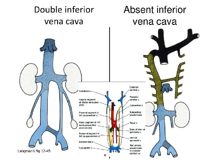
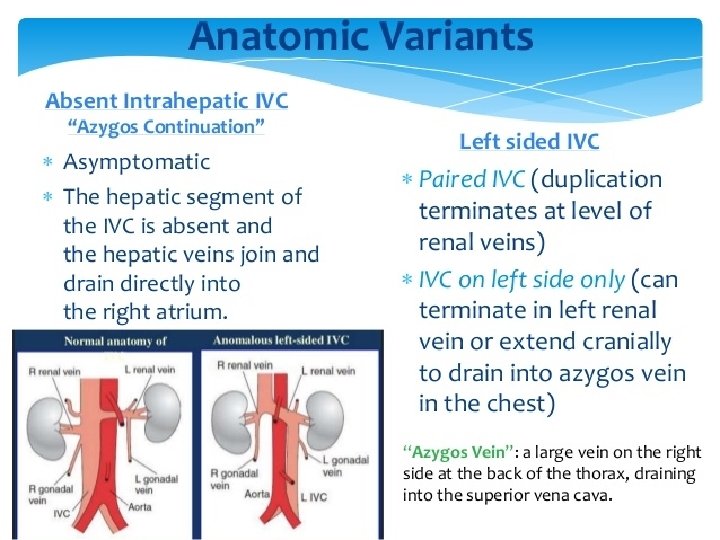
ANATOMICAL VARIATIONS � It’s possible for the IVC to have a congenital difference, and these are difficult to detect. 1� Often a person won’t have any symptoms to signal a defect in the IVC. Symptoms, when they do occur, include vague low back or abdominal pain. 2� � Some variations of the IVC are the left IVC, which happens when the left renal vein joins the left IVC but then crosses in front of the aorta before going into the right atrium if the heart. Left IVC has a prevalence rate of 0. 4% to 0. 5%. 3� � Another common variation is a duplicate or double IVC. In this case, a double IVC is just that: two IVC veins instead of one. Its prevalence rate is typically 0. 2% to 0. 3%. 4� � Other variations may include azygous continuation of the IVC, where blood coming from the lower body drains into a different venous system called the azygous system. This system drains the thoracic wall and upper lumbar area of blood. � The last, extremely rare, 2 �� variation is called absent infrarenal IVC. This results in the partial or complete absence of the IVC, likely due to another variation of the veins which merge into the IVC.
CLINICAL SIGNIFICANCE � The IVC is most commonly used for IVC filter placement, which can help reduce the risk of pulmonary embolisms (a blockage in the lung which can prevent blood flow). An IVC filter stops blood clots that form in the veins of the lower half of the body, or someone who suffers from deep vein thrombosis, from having those clots reach the lungs. 5 �� � An IVC filter is commonly used in patients that aren’t responding to medication for blood clots such as blood thinners. Depending on the severity and frequency of the blood clots, IVC filters can be left in permanently or removed once the risk of clots forming and traveling to the lungs has passed. � In some cases, an IVC filter that has not been removed may cause IVC thrombosis, creating blood clots in the IVC itself. 6� This is why if needed your physician will monitor the IVC filter and determine the best time to remove it to prevent blood clots from forming.
THANK YOU