Angiology is the branch of the anatomical sciences


Angiology: is the branch of the anatomical sciences which study and describe of the organs of circulation system of the blood and lymph. It is consisting of two systems (cardiovascular system and lymphatic system). Circulatory or cardiovascular system: group of organs 1 transports to the tissues and organs, the oxygen, nutritive &, immune substances, hormones and chemicals necessary for normal function and activities. 2 It also carries away waste products and carbon dioxide 3 helps to regulate body temperature 4 helps to maintain normal water and electrolyte balance

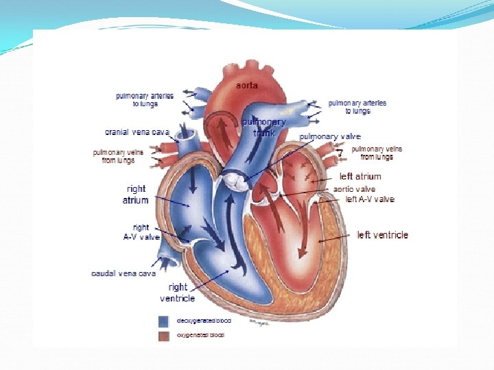
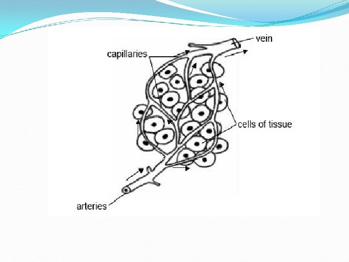
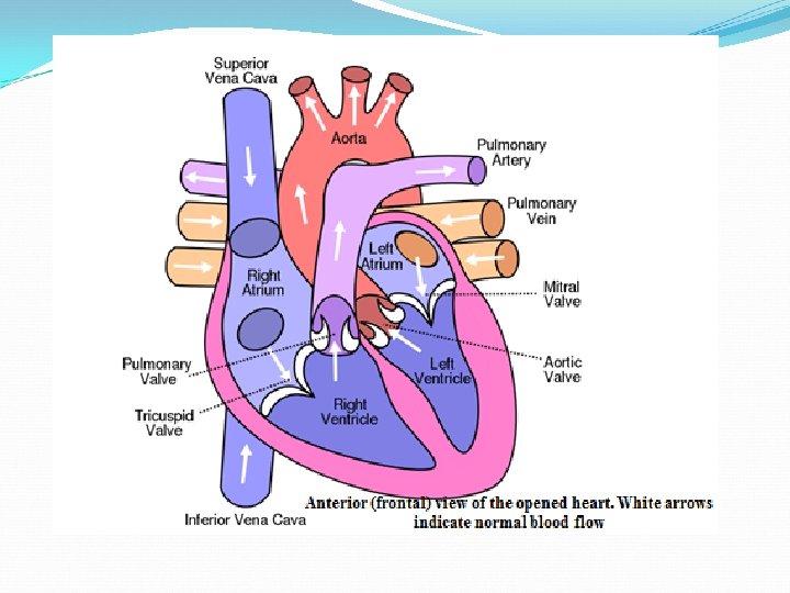
It is can be considered as composed of two parts: ASystemic circulation, which serves the body as a whole except for the lungs BPulmonary circulation, which carries the blood to and from the lungs. The organs of circulatory system consist of several types of vessels : A- Arteries which convey the blood from heart to the tissue. B- veins which convey the blood back from tissue to heart. C- capillaries are microscopic tubes permit to necessary gas exchange between the tissue and blood. C- A muscular pump , the heart that drives the blood. Note: 1 All arteries carry oxygenated blood except for the pulmonary artery that carries deoxygenated blood to the lungs 2 while all veins carry deoxygenated blood except for the pulmonary vein that carries oxygenated blood from the lungs to heart.


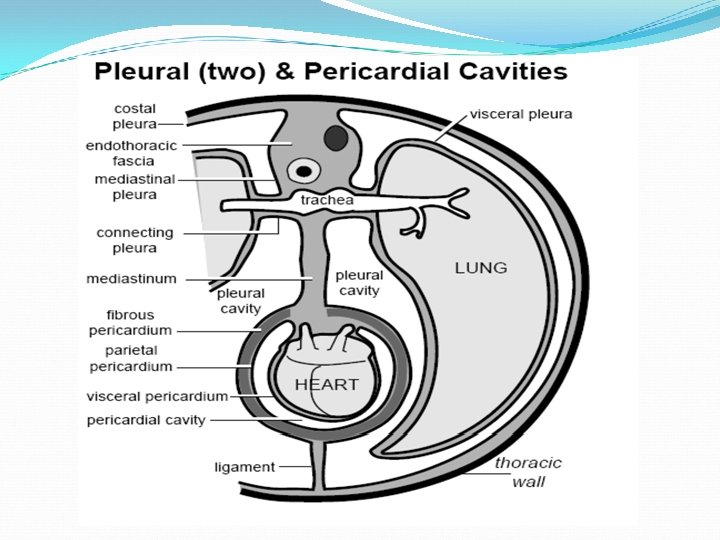
The Pericardium: 1 is a double-walled sac that contains the heart and the roots of the great vessels, 2 the base of it found dorsally toward the vertebrae, the greater vessels enter the base 3 the apex of it found ventrally toward the sternum. 4 Pericardium consists of two layers: AFibrous pericardium: is thin, strong, inelastic layer separated the pericardial cavities from pleural cavities. Its attached dorsally with great vessels at the base of the heart and ventrally with sternum by sternopericardial ligament which is found in horse, cattle and swine, but in carnivores it is attached with diaphragm by phernopericardial ligament. B-A serous layer : The serous pericardium, in turn, is divided into two layers 1 - The parietal pericardium, which is fused to and inseparable from the fibrous pericardium 2 -The visceral pericardium, which is a layer deep to the fibrous pericardium which comes into contact with the heart (not the great vessels), it is known as the epicardium. (The epicardium is the layer immediately outside of the myocardium). The pericardial cavity is space between the parietal and visceral pericardial layers which contains of serous fluid, ( the pericardial fluid). The pericardial cavity is found within the mediastinum of the thoracic cavity

dal cau ial cran dle mid um n i t ias d e m Phrenic nerve

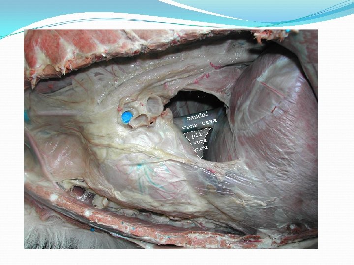
descending aorta parietal pleura viceral pleura caudal vena cava plica vena cava pericardial mediastinal pleura fibrous pericardium parietal serous pericardium visceral serous pericardium myocardium endocardium

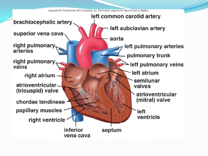
The heart: is conical, hollow, muscular organ constructed of a special kind of muscle called myocardium or cardiac muscle. it is enclosed in the pericardium and located in thoracic cavity between right and left lung in the middle of the mediastinum, opposite to the lateral wall of the thoracic wall from the 2 nd or 3 rd intercostal space to the 6 th rib or to 5 th rib in the (horse and dog) and ruminant respectively. it is attached at the base of the heart with large vessels, but in internally free in pericardium The wall of the heart consists of three layers from external to internal: 1 -the epicardium 2 -the myocardium: it is peculiar striated muscles. 3 -the endocardium: it is thin layer which lines the heart cavity. Morphology of the heart : Athe heart divided into two cavities: 1 left cavity pumps blood throughout the body. 2 right cavity pumps blood only through the lungs. Each cavity is in turn divided into two chambers, the upper ones called atria, the lower ones ventricles

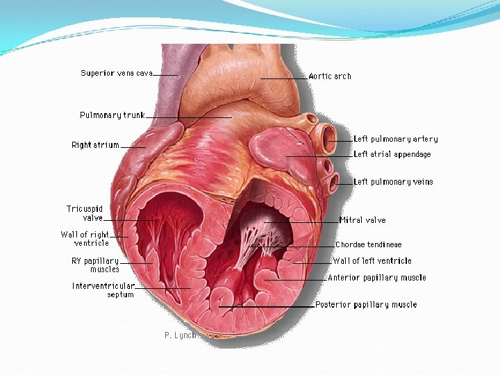
B- The heart consists of base, apex, and two surface and two borders: 1 -Base: it is directed dorsally, it is formed by the right and left atria, from which the cranial and caudal vena cava and pulmonary veins enter the base 2 -Apex: It is directed ventrally , caudally, and to the left, placed centrally dorsally to the sternum and is overlapped by the left lung and pleura 3 -The right or cranial border is convex, curved ventrally, the grated part parallel with the sternum, 4 -the left or caudal border is shorter and nearly vertical. 5 -The left and right surface , divided by four grooves (two coronary grooves and two longitudinally grooves)

The coronary grooves: it is called atrioventricular grooves, the indicate the division between the atria and ventricles, it is completely encircle the heart but interrupted at the origin of pulmonary trunk, these grooves occupied by coronary vessels and variable quantity of fat. The two ventricles of the heart are separated by two longitudinal grooves(interventricular sulcus), which are: 1 - The left longitudinal sulcus , is situated on the left surface. It began at the coronary groove , caudal to the origin of the pulmonary trunk and descends almost parallel to the caudal border and don’t reach the apex. 2 -The right longitudinal sulcus is situated on the right surface , it begin at the coronary groove ventral to the termination of the caudal vena cava and passes toward the apex.


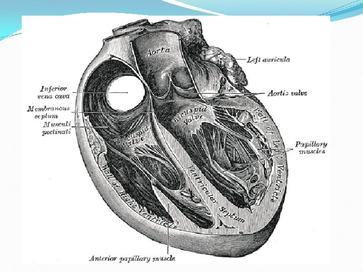
The four chambers of the heart are : The right atrium: A-it is form the right cranial part of the base of the heart, placed dorsally to right ventricle. B-it is separated from the left atrium by interatrial septum which contain diverticulum called oval fossa at the point of the entrance of the caudal vena cava, this fossa is remanent of the opening in the septum called oval foramen, through which the two atria communicate in fetal life. C-its consist of two parts a-the sinus venarum cavarum: into which the veins open b-Auricle: it is end ear shaped conical pouch, also termed auricular appendix appearing on the left side cranially at the pulmonary artery. It is curved around right and cranial surface of the atrium

. Right auricle; B. Left auricle; C. Right ventricle; D. Pulmonary trunk (gives rise to the two pulmonary arteries); E. Left atrium; F. Left ventricle; G 1. Aortic arch; G 2 Descending aorta ; H. Brachiocephalic trunk (gives rise to the carotid arteries and right subclavian artery); I. Left subclavian artery; J. Vertebral artery; K. Costocervical trunk; L. Superficial cervical artery; M. Internal thoracic artery; N. Axillary artery; O. Cranial vena cava; P. Caudal vena cava and p. Plica vena cava

D-In the right atrium, the internal surface of the atrium -the atrium is lined in all walls except the auricle by glistening membrane (endocardium ) which is smooth, while in front of it there are muscular fibers of the wall raised into parallel ridges resembling the teeth of a comb, and hence named the pectinate muscles , it is ended by curved crest known as the crista terminalis of His, On the external aspect of the right atrium, corresponding to the crista terminalis is the sulcus terminalis. The musculi pectinati are useful in increasing the power of contraction without increasing heart mass substantially

E- It receives deoxygenated blood from the body by five chief openings and pumps it into the right ventricle through the right atrioventricular orifice. The opening are: 1 -The cranial (superior) vena cava: is bringing de-oxygenated blood from the veins from the head and cranial body and empties into the right atrium of the heart cranially. 2 -The caudal (inferior) vena cava: is bringing de-oxygenated blood from veins from the legs and caudal body and empties into the right atrium of the heart caudally Inter-venous crest : project ventral and cranial from the dorsal wall of opening of the caudal vena cava. Function: It tends to direct the flow of blood from the cranial vena cava to the right atrioventricular opening.
 is just superior to the septal leaflet")
3 - The coronary sinus orifice (opening) is just superior to the septal leaflet of the tricuspid valve, and is guarded by the (Thebesian valve) which is a semicircular fold of the lining membrane of the atrium. The coronary sinus is a collection of veins joined together to form a large vessel that collects blood from the myocardium. It's located ventral to the caudal vena cava opening in the groove between the left atrium and ventricle on the caudal surface of the heart Thebesian valve: It may prevent the regurgitation of blood into the sinus during the contraction of the atrium. This valve may be double or it may be cribriform. 4 -Some small veins drain into any of the four chambers of the heart : is drains into the right atrium on the caudal surface, medial to the caudal vena cava opening. 5 - Right atrioventricular opening is separates the right atrium from the right ventricle. It’s guarded by Tricuspid Valve. It is triangular in shape

The left atrium : . a- It receives oxygenated blood from the pulmonary veins, and pumps it into the left ventricle, through the left atrioventricular orifice, which guarded by bicuspid or mitral valve. b-Attached to the left atrium is the left auricular appendix (auricle) extend lateral and cranial on the left side. It is consistently narrow and long. c-In the left atrium, the musculi pectinati, fewer and smaller than that in the right auricula, unlike the right atrium, the left atrium has no crista terminalis. This is due to the embryological origin of the auricles, which are the true atria. d-There are two opening in left atrium : 1 -Pulmonary Vein opening The pulmonary vein is the vessel transporting oxygen-rich blood from the lungs to the left atrium (seven to eight in number in horse and five in sheep) found caudal to auricle on the right side of atrium
, which contains the bicuspid or mitral valve. is")
2 - left atrioventricular orifice(mitral orifice), which contains the bicuspid or mitral valve. is situated ventral and cranially , it appear oval and smaller than that right one because the contraction of the left ventricle in the dead subject

The right atrium has been opened A. Cranial vena cava; B. Caudal vena cava; C. Pectinate muscles of the auricle; D. Intravenous tubercle; E. Fossa ovale; F. Coronary sinus


The right ventricle: . It form the cranial border of the heart. 1 -It receives deoxygenated blood from the right atrium by right atrioventricular orifice that guarded by the tricuspid valve, and pumps it into the pulmonary trunk by the pulmonary opening that guarded by the pulmonary valve. Pulmonary trunk : is branches into the pulmonary arterial system which transporting deoxygenated blood from the right ventricle to the lungs 2 -It is triangular in form, and extends from the right atrium to near the apex of the heart. 3 -The wall of the right ventricle is thinner than that of the left, the proportion between them being as 1 to 3; it is thickest at the base, and gradually becomes thinner toward the apex. The of right ventricle cavity equals in size that of the left ventricle. 4 -The upper left corner of the right ventricle, is called the infundibulum or conus arteriosus, is a conical pouch at the entrance of pulmonary trunk. 5 - all wall of the ventricular bear muscular ridge and bands termed the trabeculae carneae, except in the conus arteriosus is smooth

? ? left pulmonary auricle trunk ? aorta left ventrical ? right auricle ? right ventrical ?


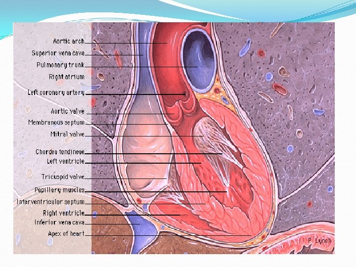
D-The left ventricle 1 -is longer and more conical in shape than the right, and on transverse section its concavity presents an oval or nearly circular outline. It form the left caudal part of ventricular mass. 2 - It receives oxygenated blood from the left atrium by left atrioventricular orifice that guarded by the bicuspid valve, and pumps it into the aorta by the aorta opening that guarded by the aorta valve 3 -it also forms the apex of the heart. 4 -The left ventricle is thicker and more muscular than the right ventricle because it pumps blood at a higher pressure. Interventricular septum (IVS), is separating between the two ventricles of the heart , and is directed obliquely backward, and is curved with the convexity toward the right ventricle. Tts margins correspond with the left and right longitudinal sulci. Disorders A hole in the interventricular septum is termed a ventricular septal defect (VSD).
 septum, located behind the right atrium and left ventricle,")
Note : The atrioventricular (AV) septum, located behind the right atrium and left ventricle, is divided into two portions: a superior portion (membranous) and an inferior portion (muscular). The atrioventricular (AV) node lies in the atrial septum, juxtaposed to the membranous and muscular portions of the AV septum Valves define as fold of the endocardium strengthened by fibrous tissue and at the periphery muscular fibres
")
1 -Tricuspid Valve a. The tricuspid valve (also known as the right atrioventricular valve) separates the right atrium from the right ventricle. it has, three leaflet(cusps ) : 1 -The anterior leaflet (infundibular in human been or angularis ), largest of the 3 leaflets, found between the atrioventricular opening and conus arteriosus. 2 - The posterior leaflet ( parietalis or marginal), smallest of the 3 leaflets, is usually scalloped found in the right margin. 3 -The septal leaflet (medial or septalis) usually attaches to the membranous and muscular portions of the inter ventricular septum. b-The valve is supported by a large anterior and several small posterior papillary muscle. The papillary muscles connect to valve by the chordae tendineae, which are more fragile in the right ventricle than those of the left ventricles.

c-The valve opens to allow the de-oxygenated blood collected in the right atrium( when right atrium contracts ) to flow into the right ventricle(relaxed ) and closes when the right ventricle contracts, preventing blood from returning to the right atrium; thereby, forcing it to exit through the pulmonary valve into the pulmonary trunk.
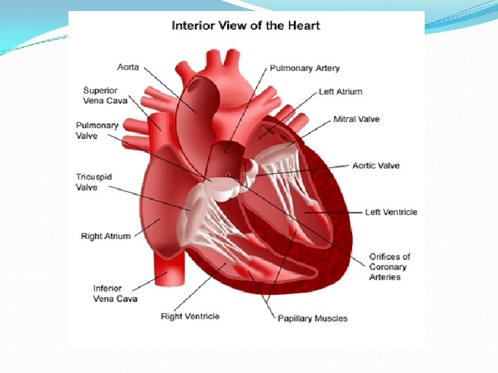

papillary muscle : a-serve to limit the movements of the mitral and tricuspid valves. The papillary muscles attach to the lower portion of the interior wall of the ventricles. b-These muscles contract to tighten the chordae tendineae, which in turn prevent inversion of the valve. This occurs in response to pressure gradients. c-The papillary muscles in that their contraction pulls on the chordae tendinae, closing the mitral (bicuspid) and tricuspid valves. This prevents backflow of blood from the ventricles into the atriums but when the papillary muscles relax, the valves open The septomarginal trabecula (or moderator band) : a-is a muscular band of heart tissue found in the ventricles and extends from the base of the papillary muscle to the ventricular septum. b-Its prevent over distension of the ventricle, and was named the "moderator band". c-it is considered part of the electrical conduction system of heart.

The chordae tendineae, or heart strings: a-They are cord-like tendons that connect the papillary muscles to the tricuspid valve and the mitral valve in the heart. b-The chordae tendineae prevents the flaps invert into the atria. c-As the papillary muscles contract and relax, the chordae tendineae transmit the resulting increase and decrease in tension. The trabeculae carneae (columnae carneae): a-They are rounded or irregular muscular columns which project from the whole of the inner surface of the ventricle, with the exception of the conus arteriosus. b-The purpose of the trabeculae carnae is most likely to prevent suction that would occur with a flat surfaced membrane and thus impair the heart's ability to pump efficiently , also serve a similar function to papillary muscles

2 -Mitral Value 1 -The mitral valve separates the left atrium from the left ventricle. It is bicuspid have two cusps (thicker than tricuspid vale ) ) : a-large anterior leaflet (septal or aortic) is triangular with a smooth texture. It found between the left atrioventricular opening and aortic opening. b- The posterior leaflet a smaller leaflet (ventricular) has a scalloped appearance. 2 -The chordae tendineae to the mitral valve originate from the 2 large papillary muscles of the left ventricle (anterolateral, posteromedial). 3 -It opens to allow the oxygenated blood collected in the left atrium to flow into the left ventricle and closes as the left ventricle contracts, preventing blood from returning to the left atrium; thereby, forcing it to exit through the aortic valve into the aorta


3 -The pulmonary valve: a-is the semilunar valve that lies between the right ventricle and the pulmonary trunk and has three cusps (right , left , and anterior ) ; a sinus is located behind each cusp. b-the pulmonic valve opens in ventricular systole as the ventricles contract, when the pressure in the right ventricle rises above the pressure in the pulmonary trunk; it opens to allow the de-oxygenated blood collected in the right ventricle to flow to the lungs. At the end of clos the pulmonic valve is close. c-Compared with the aortic valve, the pulmonary valve has thinner cusps, no associated with coronary arteries, and no continuity with the corresponding (anterior) tricuspid valve leaflet

The aorta: a-is originates at the left ventricle of the heart. b-After supplying the coronary arteries that nourish the heart itself, the aorta extends slightly toward the neck to feed branches serving the head and arms. c-It then arches caudally toward the thoracic, directing blood into the arterial system of the chest. d-Entering the abdomen through the aortic hiatus, an opening in the diaphragm, the aorta branches off to supply the stomach, kidneys, intestines, gonads, and other organs through extensive arterial networks. e-It finally divides into the two iliac arteries carrying blood to the legs. f-The elasticity of the aorta wall permits it to pulse in rhythm with the heartbeat, thus helping to propel blood through the body

4 -Aortic Valve a-The aortic valve separates the left ventricle from the aorta. It has 3 leaflets composed of fragile cusps (right, left, and posterior (or "noncoronary cusp")) and the sinuses of Valsalva( the aortic sinuses of Valsalva are 3 dilations of the aortic root that arise from the 3 closing cusps of the aortic valve ). The right and left sinuses give rise to the right and left coronary arteries; the non coronary sinus has no coronary artery. The sinus of Valsalva walls is much thinner than the aortic wall b-As the ventricles contract; it opens to allow the oxygenated blood collected in the left ventricle to flow through aorta to the body. It closes as the ventricles relax, preventing returning the blood through aorta

 that")
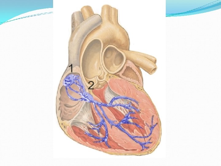
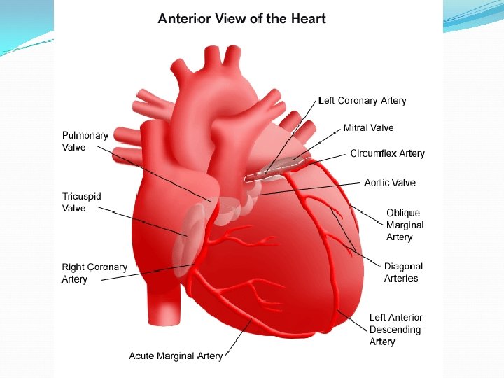
Coronary Arteries. : are two coronary arteries, ( "left" and "right" coronary arteries) that carry oxygen- and nutrient-rich blood to the cardiac muscle tissue, emerge from the beginning of the aorta, near the top of the heart. Left coronary artery : The initial segment of the left coronary artery is called the left main coronary (LCA) which arises from the left aortic sinus and courses between the left auricle and the pulmonary trunk to reach the coronary groove. When it reaches the left AV groove, the LCA bifurcates into the left anterior descending (LAD) and the Left circumflex artery (LCX) branches. The LCA supplies most of the left atrium, left ventricle, interventricular septum, and AV bundles.
 : runs along the right interventricular sulcus toward the apex,")
a--Left anterior descending artery(LAD) : runs along the right interventricular sulcus toward the apex, it turns sharply to anastomose with the posterior interventricular branch of the RCA. As the LAD artery courses anteriorly along the ventricular septum, it sends off diagonal branches to the lateral wall of the left ventricle. and supplies the apical portion of both ventricles b-Left circumflex artery (LCX) : 1 - arise at a right angle near the base of the left atrial appendage(auricle). 2 -The LCX artery courses in the coronary groove around the left border of the heart to the caudal surface of the heart to anastomose to the end of the Left coronary artery(RCA). In the atrioventricle (AV) groove, the LCX artery lies close to the mitral valve. 3 - The first branch off the LCX artery, supplies the left atrium, and gives off an oblique marginal (OM) branch at the left border of the heart near the base of the left atrial appendage to supply the posterolateral surface of the left ventricle
 1 -The RCA is a single large artery arises from")
Right coronary artery (RCA) 1 -The RCA is a single large artery arises from the right aortic sinus that courses along the right AV groove. 2 -The RCA supplies the right atrium, right ventricle, interventricular septum, and the sinuoatrial (SA) and atrioventricul (AV) nodes. 3 -As the RCA passes toward the caudal border of the heart, it gives off : a-A right marginal branch that supplies the apex of the heart. After this branching, the RCA turns left to enter the right interventricular groove to give off b-the right posterior descending artery PDA, which supplies both ventricles. c-The AV node artery. d-Terminal branches of the RCA supply the posteromedial papillary muscle of the left ventricle. (The LAD artery supplies the anterolateral papillary muscle of the right ventricle. ) e-Near the apex, the PDA anastomoses with the anterior interventricular branch of the LCA.

Cardiac veins : 1 -The great cardiac vein is the main tributary of the coronary sinus and drains areas of the heart supplied by the left main coronary artery (LCA). It begins at the apex of the heart, ascends in the left interventricular groove with the left anterior descending (LAD) artery, and enters the left end of the coronary sinus. 2 -The middle and small cardiac veins drain most of the heart supplied by the right coronary artery (RCA). The middle cardiac vein begins at the apex, ascends in the right interventricular groove with the right posterior interventricular artery, and empties into the right side of the coronary sinus. The small cardiac vein runs in the coronary groove along with the marginal branch of the RCA; this vein usually empties into the coronary sinus but may empty directly into the right atrium. 3 -Coronary veins of the right ventricle drain directly into the right atrium 4 -Thebesian veins drain into the right ventricle. 5 -The left ventricle venous return drains into the coronary sinus located next to the septal portion of the tricuspid valve.

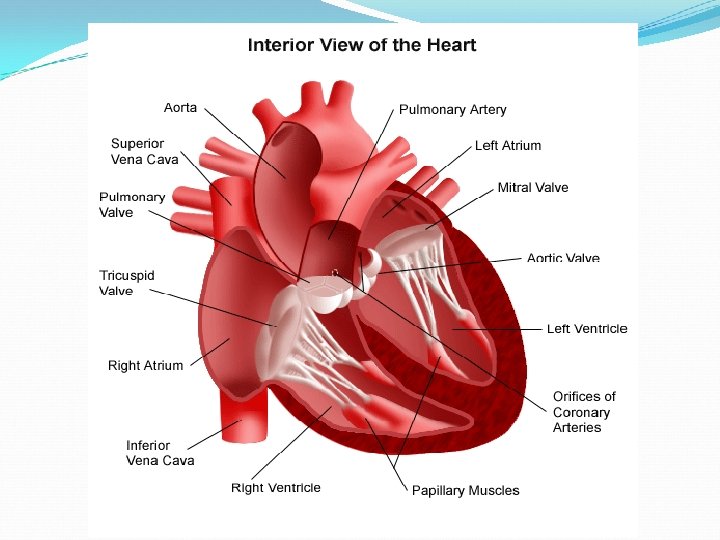
1. Right Coronary 2. Left Anterior Descending 3. Left Circumflex 4. Superior Vena Cava 5. Inferior Vena Cava 6. Aorta 7. Pulmonary Artery 8. Pulmonary Vein 1. Right Atrium 2. Right Ventricle 3. Left Atrium 4. Left Ventricle 5. Papillary Muscles 6. Chordae Tendineae 7. Tricuspid Valve 8. Mitral Valve 9. Pulmonary Valve Aortic Valve (Not pictured)

- Slides: 55