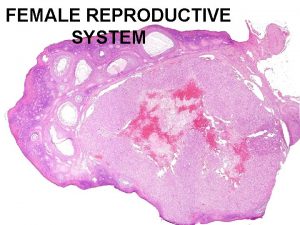
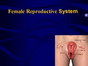
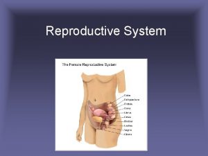
FEMALE REPRODUCTIVE SYSTEM The female reproductive system is


- Slides: 64

FEMALE REPRODUCTIVE SYSTEM • The female reproductive system is more complex than the male system because it serves more purposes – – Produces and delivers gametes Provides nutrition and safe harbor fetal development Gives birth Nourishes infant • Female system is more cyclic, and hormones are secreted in a more complex sequence 28 -1

Sexual Differentiation • The two sexes indistinguishable for first 8 to 10 weeks of development • Female reproductive tract develops from the paramesonephric ducts – Not because of the positive action of any hormone – Because of the absence of testosterone and müllerian-inhibiting factor (MIF) 28 -2

Sexual Differentiation • Without testosterone: – Causes mesonephric ducts to degenerate – Genital tubercle becomes the glans clitoris – Urogenital folds become the labia minora – Labioscrotal folds develop into the labia majora • Without MIF: – Paramesonephric ducts develop into the uterine tubes, uterus, and vagina 28 -3

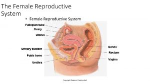
The Genitalia Copyright © The Mc. Graw-Hill Companies, Inc. Permission required for reproduction or display. • Internal genitalia – Ovaries, uterine tubes, uterus, and vagina Uterine tube Fimbriae Ovary Vesicouterine pouch Rectouterine pouch Posterior fornix Cervix of uterus Anterior fornix Round ligament Uterus Peritoneum Urinary bladder Pubic symphysis Mons pubis Urethra Clitoris Prepuce Labium minus Labium majus Rectum Anus Vaginal rugae Vaginal orifice • External genitalia – Clitoris, labia minora, and labia majora • Occupy the perineum • Primary sex organs – Ovaries • Secondary sex organs Figure 28. 1 – Other internal and external genitalia 28 -4

The Ovaries • Ovaries—female gonads that produce egg cells (ova) and sex hormones – Almond-shaped and nestled in the ovarian fossa of posterior pelvic wall – Tunica albuginea capsule, like on testes – Outer cortex where germ cells develop – Inner medulla occupied by major arteries and veins – Each egg develops in its own fluid-filled follicle – Ovulation: bursting of the follicle and releasing of the egg 28 -5

The Ovaries • Ligaments of ovary – Attached to uterus by ovarian ligament – Attached to pelvic wall by suspensory ligament • Contains ovarian artery, vein, and nerves – Anchored to broad ligament by mesovarium • Ovary receives blood from two arteries – Ovarian branch of the uterine artery – Ovarian artery • Equivalent to testicular artery in male 28 -6

The Ovaries • Ovarian and uterine arteries anastomose along margin of ovary – Give off multiple small arteries that enter the ovary • Ovarian veins, lymphatics, and nerves also travel through the suspensory ligaments 28 -7

Structure of the Ovary Figure 28. 2 28 -8

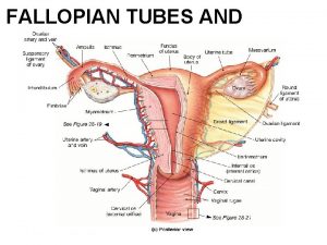
The Uterine Tubes • Uterine tube (oviduct) or (fallopian tube) • Canal about 10 cm long from ovary to uterus • Muscular tube lined with ciliated cells – Highly folded into longitudinal ridges Figure 28. 3 a 28 -9

The Uterine Tubes • Major portions – Infundibulum: flared, trumpet-shaped distal (ovarian) end – Fimbriae: feathery projections on infundibulum – Ampulla: middle and longest part – Isthmus: narrower end toward uterus • Mesosalpinx: superior portion of broad ligament that enfolds uterine tube Figure 28. 3 a 28 -10

The Uterus • Thick muscular chamber that opens into roof of the vagina – Usually tilts forward over urinary bladder – Harbors fetus, provides a source of nutrition, and expels the fetus at the end of its development – Pear-shaped organ • Fundus—broad superior curvature • Body (corpus)—middle portion • Cervix—cylindrical inferior end 28 -11

The Uterus (Continued) – Lumen is roughly triangular • Upper two corners are openings to uterine tubes • Not a hollow cavity, but a potential space in nonpregnant uterus – Cervical canal connects lumen of uterus to vagina • Internal os—superior opening of canal into body of uterus • External os—inferior opening of canal into vagina – Cervical glands: secrete mucus that prevents spread of microorganisms from vagina to uterus 28 -12

The Uterus Figure 28. 3 a 28 -13

Cervical Cancer • Cervical cancer common among women ages 30 to 50 – Smoking, early-age sexual activity, STDs, and human papillomavirus – Usually begins in epithelial cells of lower cervix • Best protection against cervical cancer is early detection by Pap smear – Cells removed from cervix and vagina and microscopically examined • Three grades of cervical intraepithelial neoplasia – Class I is mild dysplasia – Class II calls for a biopsy – Class III results may call for radiation therapy or hysterectomy 28 -14

Pap Smears and Cervical Cancer Figure 28. 5 a, b 28 -15

The Uterus • Uterine wall: perimetrium, myometrium and endometrium – Perimetrium—external serosa layer – Myometrium—middle muscular layer • Constitutes most of the uterine wall • Composed mainly of smooth muscle – Sweep downward from fundus; spiral around body – Less muscular and more fibrous near cervix – Produces labor contractions, expels fetus 28 -16

The Uterus (Continued) – Endometrium—inner mucosa • Simple columnar epithelium, compound tubular glands, and a stroma populated with leukocytes, macrophages, and other cells – Functional layer (stratum functionalis)—superficial half, shed each menstrual period – Basal layer (stratum basalis)—deep layer, stays behind and regenerates a new stratum functionalis with each menstrual cycle • During pregnancy, endometrium is the site of attachment of the embryo and forms the maternal part of the placenta from which the fetus is nourished 28 -17

The Uterus • Uterus is supported by ligaments – Broad ligament has two parts • Mesosalpinx • Mesometrium on each side of the uterus – Cardinal (lateral cervical) ligaments: support cervix and superior part of vagina extending to pelvic wall – Uterosacral ligaments: attach posterior side of uterus to the sacrum – Round ligaments: arise from anterior surface of uterus, pass through inguinal canals, and terminate in labia majora • Similar to gubernaculum terminating in male scrotum 28 -18

The Female Reproductive Tract Figure 28. 3 28 -19

The Uterus • Uterine blood supply is important to menstrual cycle and pregnancy • Uterine artery arises from internal iliac artery – Gives off several branches that penetrate myometrium and lead to arcuate arteries • Each travels in a circle around the uterus • Anastomose with arcuate artery on the other side • Spiral arteries penetrate through the myometrium into the endometrium 28 -20

Blood Supply to the Female Reproductive Tract Suspensory ligament Ovarian branch of uterine artery Mesosalpinx Aorta Common iliac artery Ovarian artery Arcuate artery Ovary Vaginal artery Uterine artery Internal iliac artery Spiral arteries Copyright © The Mc. Graw-Hill Companies, Inc. Permission required for reproduction or display. Figure 28. 7 28 -21

The Vagina • Vagina (birth canal)— 8 to 10 cm distensible muscular tube – Allows for discharge of menstrual fluid, receipt of penis and semen, and birth of baby – Outer adventitia, middle muscularis, and inner mucosa – Tilted posteriorly between rectum and urethra – Vagina has no glands • Transudation lubricates vagina—“vaginal sweating” – Serous fluid through its walls and by mucus from the cervical gland above it 28 -22

The Vagina (Continued) – Fornices: blind-ended spaces at top of vagina that extend slightly beyond the cervix – Transverse friction ridges (vaginal rugae) at lower end – Mucosal folds form hymen across vaginal opening 28 -23

The Vagina • Vaginal epithelium – Undergoes metaplasia: transformation from one tissue type to another • Childhood: simple cuboidal • Puberty: estrogens transform it to stratified squamous – Bacteria ferment glycogen producing acidic p. H in vagina to inhibit growth of pathogens – Has antigen-presenting dendritic cells: route by which HIV from infected semen invades the female body 28 -24

Vaginal canal Photos © Mc. Graw-Hill Education Posterior vaginal fornix Anterior vaginal fornix

The External Genitalia • External genitalia are collectively called the vulva or pudendum – Mons pubis: mound of fat over pubic symphysis bearing most of the pubic hair – Labia majora: pair of thick folds of skin and adipose tissue inferior to the mons • Pudendal cleft—fissure between labia majora – Labia minora: thin, hairless folds medial to labia majora • Space between forms vestibule which contains urethral and vaginal openings • Anterior margins of labia minora join to form hood-like prepuce over clitoris 28 -26

The External Genitalia Vulva (Continued) – Clitoris: erectile, sensory organ • Primary center for sexual stimulation • Glans, body, and crura – Vestibular bulbs: erectile tissue deep to labia majora • Bracket the vagina – Greater and lesser vestibular and paraurethral glands open into vestibule for lubrication 28 -27

The Female Perineum Copyright © The Mc. Graw-Hill Companies, Inc. Permission required for reproduction or display. Mons pubis Labium majus Prepuce Clitoris Vaginal orifice Urethral orifice Hymen Vestibule Labium minus Figure 28. 8 a Perineal raphe (a) Anus 28 -28

External Genitalia (Vulva) (Pudendum) Mons pubis Vaginal orifice Perineum Labium majus Vestibule Labium minus Photos © Mc. Graw-Hill Education

External Genitalia (Vulva) (Pudendum) Clitoris Vestibular bulbs Photos © Mc. Graw-Hill Education Body of clitoris Greater vestibular glands Prepuce of clitoris Labium majus Crus of clitoris Mons pubis

The Female Perineum Copyright © The Mc. Graw-Hill Companies, Inc. Permission required for reproduction or display. Clitoris Glans Crus Paraurethral gland Greater vestibular gland Pubic symphysis Ramus of pubis Urethral orifice Vestibular bulb Vaginal orifice Ischial tuberosity Anus Figure 28. 8 b (b) 28 -31

The Breasts and Mammary Glands • Breast—mound of tissue overlying pectoralis major – Enlarges at puberty and remains so for life – Most of the time it contains very little mammary gland • Mammary gland—develops within the breast during pregnancy – Remains active in the lactating breast – Atrophies when a woman ceases to nurse 28 -32

The Breasts and Mammary Glands • Two principal regions of the breast – Body: conical to pendulous, with nipple at its apex – Axillary tail: extension toward armpit • Lymphatics in axillary tail are important as a route for breast cancer metastasis • Nipple surrounded by circular colored zone, called the areola – Capillaries and nerves close to skin surface: more sensitive – Sensory nerve fibers of areola trigger a milk ejection reflex when an infant nurses 28 -33

The Breasts and Mammary Glands (Continued) – Areolar glands: intermediate between sweat glands and mammary glands • Secretions protect the nipple from chapping and cracking during nursing – Smooth muscle fibers in dermis of areola that contract in response to cold, touch, and sexual arousal, wrinkling the skin and erecting the nipple 28 -34

The Breasts and Mammary Glands • The nonlactating breast consists mostly of adipose and collagenous tissue – Breast size determined by amount of adipose tissue • Suspensory ligaments attach breast to dermis of overlying skin and fascia of the pectoralis major 28 -35

The Breasts and Mammary Glands • Ducts branch through fibrous stroma and converge on the nipple – Mammary gland develops during pregnancy – 15 to 20 lobes around the nipple – Lactiferous duct drains each lobe • Dilates to form lactiferous sinus which opens into nipple – Ducts ends in acini (sacs) • Acini are surrounded by myoepithelial cells 28 -36

The Breast in Lactating State Copyright © The Mc. Graw-Hill Companies, Inc. Permission required for reproduction or display. Adipose tissue Suspensory ligaments Lobe Lobules Areolar glands Areola Nipple Lactiferous sinus Lactiferous ducts (a) Anterior view Figure 28. 9 a 28 -37

Breast Anatomy Nipple Photos © Mc. Graw-Hill Education Lactiferous sinus Areola Retromammillary space Suspensory ligaments

The Breast Figure 28. 9 c, d 28 -39

Breast Cancer • Breast cancer occurs in 1 out of 8 or 9 American women – A leading cause of female mortality • Tumors begin with cells from mammary ducts – May metastasize by mammary and axillary lymphatics • Signs may include lump, skin puckering, changes in skin texture, and drainage from nipple 28 -40

Breast Cancer • Contibuting causes – BRCA 1 and BRCA 2: two breast cancer genes • But most breast cancer is nonhereditary – Some breast cancers are stimulated by estrogen • Risk factors – Aging, exposure to ionizing radiation, carcinogenic chemicals, excessive alcohol and fat intake, and smoking – Over 70% of cases lack identifiable risk factors 28 -41

Breast Cancer • Tumor discovery usually during breast selfexamination (BSE)—monthly for all women • Mammograms (breast X-rays) – Late 30 s: baseline mammogram – Ages 40 to 49: every 2 years – Over age 50: yearly 28 -42

Breast Cancer • Treatment of breast cancer – Lumpectomy: removal of tumor only – Simple mastectomy: removal of breast tissue only or breast tissue and some axillary lymph nodes – Radical mastectomy: removal of breast, underlying muscle, fascia, and lymph nodes • Rarely done as it generally does not improve outcome – Surgery is followed by radiation or chemotherapy – Breast reconstruction from skin, fat, and muscle from other parts of the body 28 -43

Puberty • Puberty begins at age 8 to 10 for most girls in United States • Triggered by rising levels of Gn. RH which stimulates anterior pituitary to secrete FSH and LH • FSH stimulates ovarian follicles and they begin to secrete estrogen, progesterone, inhibin, and a small amount of androgen • Estrogens are feminizing hormones with widespread effects on the body – Estradiol (most abundant), estriol, and estrone 28 -44

Puberty • Thelarche—onset of breast development, the earliest noticeable sign of puberty – Estrogen, progesterone, and prolactin trigger development of ducts and lobules – Completion of duct and lobules is governed by glucocorticoids and growth hormone – Adipose and fibrous tissue enlarge breast • Pubarche—appearance of pubic and axillary hair, sebaceous glands, and axillary glands – Androgens from ovaries and adrenal cortex stimulate pubarche and libido 28 -45

Puberty • Menarche—first menstrual period – Requires at least 17% body fat in teenager • Improved nutrition has lowered age of onset to age 12 • Leptin stimulates gonadotropin secretion • If body fat and leptin levels drop too low, gonadotropin secretion declines and menstrual cycle might cease – Menstruation ceases when body fat drops below 22% in adult woman • First few menstrual cycles are anovulatory (no egg ovulated) • Girls begin ovulating regularly about a year after they begin menstruating 28 -46

Puberty • Estradiol stimulates many changes in puberty – Stimulates vaginal metaplasia – Stimulates growth of ovaries and secondary sex organs – Stimulates growth hormone secretion • Increase in height and widening of pelvis – Stimulates fat deposition (breast, hips, etc…) – Thickens skin • But girls’ skin is still thinner, softer, and warmer than boys • Progesterone – Primarily acts on the uterus preparing it for possible pregnancy in the second half of the menstrual cycle 28 -47

Puberty • Estrogens and progesterone suppress FSH and LH secretion through negative feedback • Inhibin selectively suppresses FSH secretion • Hormone secretion is distinctly cyclic and the hormones are secreted in sequence 28 -48

Climacteric and Menopause • Climacteric—midlife change in hormone secretion – Accompanied by menopause: cessation of menstruation • Female born with about 2 million eggs, climacteric begins when there about 1, 000 follicles left – Follicles less responsive to gonadotropins – secrete less estrogen and progesterone – Uterus, vagina, and breast atrophy – Intercourse may become uncomfortable – Vaginal infections more common – Skin becomes thinner 28 -49

Climacteric and Menopause (Continued) – Cholesterol levels rise, increasing risk of cardiovascular disease – Bone mass declines, increasing risk of osteoporosis – Blood vessels constrict and dilate in response to shifting hormone balances • Dilations may produce hot flashes: spreading sense of heat from abdomen to thorax, neck, and face • Hormone replacement therapy (HRT)—low doses of estrogen and progesterone to relieve some of these symptoms – Risks and benefits still being debated 28 -50

Climacteric and Menopause • Menopause—cessation of menstrual cycles – Usually occurs between ages of 45 and 55 – Age of menopause has increased in last century – Menopause considered complete when there has been no menstruation for a year 28 -51

Oogenesis and the Sexual Cycle • Reproductive cycle—sequence of events from fertilization to giving birth and returning to fertility • Sexual cycle—events that recur every month when pregnancy does not intervene – Consists of two interrelated cycles controlled by shifting patterns of hormone secretion • Ovarian cycle—events in ovaries • Menstrual cycle—parallel changes in uterus 28 -52

Oogenesis • Oogenesis—egg production – Produces haploid gametes by means of meiosis – Distinctly cyclic event that normally releases one egg each month – Accompanied by cyclic changes in hormone secretion – Cyclic changes in histological structure of the ovaries and uterus • Uterine changes result in monthly menstrual flow 28 -53

Oogenesis • Embryonic development of ovary – Female germ cells arise from yolk sac – Colonize gonadal ridges the first 5 to 6 weeks of development – Differentiate into oogonia and multiply until the fifth month • 6 to 7 million in number 28 -54

Oogenesis (Continued) – Transform into primary oocytes: early meiosis I – Most degenerate (atresia) by the time the girl is born – Egg, or ovum: any stage from the primary oocyte to the time of fertilization – By puberty, 200, 000 oocytes remain • Lifetime supply—probably will ovulate only 480 times 28 -55

Oogenesis • Egg development resumes in adolescence – FSH stimulates monthly cohorts of about 24 oocytes to complete meiosis I – Each oocyte divides into two haploid daughter cells of unequal size and different destinies • Important to produce an egg with as much cytoplasm as possible • If fertilized, it must divide repeatedly and produce numerous daughter cells – Secondary oocyte: large daughter cell that is the product of meiosis I – First polar body: smaller one that ultimately disintegrates • A means of discarding the extra set of haploid chromosomes 28 -56

The Menstrual Cycle • Menstrual cycle—consists of a buildup of endometrium during most of the sexual cycle, followed by its breakdown and vaginal discharge – Divided into four phases: proliferative phase, secretory phase, premenstrual phase, and menstrual phase – First day of noticeable vaginal discharge is defined as day 1 of the sexual cycle • Menstrual phase average: 5 days 28 -57

The Menstrual Cycle • Proliferative phase—rebuilding of functional layer of endometrium that was lost in last menstruation – At day 5 of menstruation, the endometrium is about 0. 5 mm thick and consists only of basal layer – As new cohort of follicles develops, they secrete more estrogen – Estrogen stimulates mitosis in basal layer and regrowth of blood vessels to regenerate the functional layer – By day 14, endometrium is 2 to 3 mm thick – Estrogen also stimulates endometrial cells to produce progesterone receptors 28 -58

The Menstrual Cycle • Secretory phase—endometrium thickens more in response to progesterone from corpus luteum – Day 15 to day 26 – Thickening due to secretion and fluid accumulation rather than mitosis – Endometrial glands secrete glycogen – Glands grow wider, longer, and more coiled – Endometrium 5 to 6 mm thick – Soft, wet, nutritious bed available for embryonic development 28 -59

The Menstrual Cycle • Premenstrual phase: period of endometrial degeneration – – – – Last 2 days of the cycle Corpus luteum atrophies and progesterone levels fall Triggers spasmodic contractions of spiral arteries Causes endometrial ischemia (interrupted blood flow) Brings about tissue necrosis and menstrual cramps Pools of blood accumulate in the functional layer Necrotic endometrium mixes with blood and serous fluid: menstrual fluid 28 -60

The Menstrual Cycle • Menstrual phase (menses)—discharge of menstrual fluid from the vagina • First day of discharge is day 1 of the new cycle • Average woman expels about 40 m. L of blood and 35 m. L of serous fluid over a 5 -day period • Contains fibrinolysin so it does not clot 28 -61

The Female Sexual Cycle (b) Menstrual cycle Progesterone Estradiol Menstrual fluid Thickness of endometrium Ovarian hormone secretion Copyright © The Mc. Graw-Hill Companies, Inc. Permission required for reproduction or display. Days 1 3 Menstrual phase 5 7 9 11 13 15 Proliferative phase 17 19 21 Secretory phase 23 25 27 1 Premenstrual phase Figure 28. 14 b 28 -62

Endometrial Changes Figure 28. 16 28 -63

Endometriosis • Endometriosis—growth of endometrial tissue outside of the uterus – Growth is often on peritoneum of pelvic cavity or on surface of ovary – Prevalence: 6% - 10% of women – Causes pain, sometimes infertility – Believed to result from retrograde menstruation— backward flow of menstrual fluid that exits uterine tube 28 -64
 Duct system female reproductive
 Seminal tubules
 Fetus reproductive system
 Ovarian ligament
 Epilization
 Parts of male and female reproductive system
 Unit 5 lesson 3 the female reproductive system
 Ovary diagram
 Female reproductive system
 In pila fertilization is...
 Fetal pig masseter muscle
 Figure 28-2 the female reproductive system
 Female reproductive organs sagittal section
 Chapter 16 lesson 3 the female reproductive system
 Female cow reproductive system
 Rabbit reproductive organs
 Similarities between male and female reproductive system
 Oviduct funnel
 The reproductive system chapter 16
 Oogenesis
 Fsh and lh in female reproductive system
 Oogonium
 Lesson 3 the female reproductive system
 Female reproductive system color
 Female reproductive system bones
 Chapter 8 female reproductive system
 Drawing of the male and female reproductive system
 Conclusion of female reproductive system
 Prostatic urethra male
 Female reproductive system pathology
 Male reproductive system labeled
 Cow reproductive anatomy
 Female reproductive system pathology
 Female reproductive system pathology
 Female reproductive system
 Hysterosalpingoophorectomy
 Uterine seal
 Bovine female reproductive system
 Oviduct
 Female reproductive system pathology
 Figure 28-2 the female reproductive system
 Pearson
 Female reproductive system labeled
 Female reproductive
 Fertilization
 Female anatomy pictures images photos
 Female anatomy
 Female reproductive system
 Female reproductive system
 External genitalia of female
 Similarity between male and female reproductive system
 Animal female reproductive system diagram
 Chapter 16 lesson 1 the endocrine system
 Poem about female reproductive system
 Female reproductive system diseases
 Ovari
Ovari Oogenesis diagram
 Hyogonadism
 Fish reproduction
 Distinguish between ova ovaries and ovulation
 Main part of seed
 Inguinal
 Mucous membrane reproductive system
 Gynoecium female reproductive part
 Cervix function
