Histology of Reproductive system Female Reproductive The female


- Slides: 24

Histology of Reproductive system

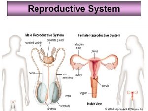
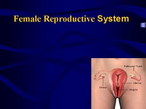
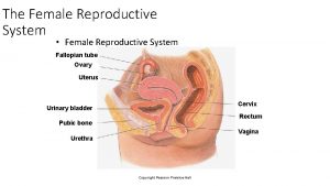
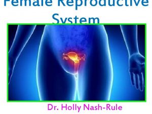
Female Reproductive The female reproductive system consists of the ovary, oviduct, uterus and vagina. The oviduct, uterus and vagina have a common structure which is adapted for their particular functions. The breast or mammary glands are also considered here, as they are important during pregnancy.

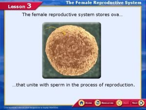
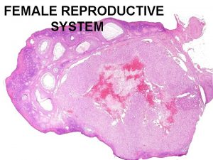
The Ovary The ovary is where oogenesis occurs Ovaries are stimulated by gonadotrophin from the anterior pituitary. Ovaries also have an endocrine function - they release oestrogen and progesterone. The genital tract makes up the rest of the female reproductive system: fallopian tubestake the ova to the uterus. The uterus is a muscular organ, and its mucosal lining undergoes hormone dependent changes. The vagina is a muscular tube that leads to the outside. The ovaries are small almond shaped structures, covered by a thick connective tissue capsule - the tunica albuginea. This is covered by a simple squamous mesothelium called the germinal epithelium. The ovary has a cortex, which is where the ovarian follicles can be found, and a highly vascular medulla, with coiled arteries called arteries. The oocytes are surrounded by epithelial cells and form follicles. The ovary contains many primordial follicles, which are mostly found around the edges of the cortex. There are fewer follicles in different stages of development.


Formation of Ova Follicular development. Primordial germ cells multiply during fetal development. At birth, the ovary contains around 400 000 primordial follicles which contain primary oocytes. These primary oocytes do not undergo further mitotic division, and they remain arrested in the prophase stage of meiotic division I, until sexual maturity -At sexual maturity, two hormones, produced by the pituitary gland: follicle stimulating hormone (FSH) and lutenising hormone (LH) cause these primordial follicles to develop. In each ovarian cycle, about 20 primordial follicles are activated to begin maturation. however, normally one follicle fully matures, and the rest contribute to the endocrine function of the ovary. -When activated, the first meiotic division is completed. When this happens, the primary follicle has matured into a secondary follicle. The second division then starts, and a Graafian follicle is formed. This contains a secondary oocyte. This second division is not completed, unless the ovum is fertilised.

Uterus -The uterus is made up of an external layer of smooth muscle called the myometrium, and an internal layer called the endometrium. -The endometrium has three layers: stratum compactum, stratum spongiosum(which make up the stratum functionalis) and stratum basalis. -The Stratum compactum and stratum spongialis develop into the stratum functionalis during the first half of the menstrual cycle (proliferative phase) The wall of the uterus changes during the menstrual cycle, as shown diagramatically here.


Proliferative Phase In the proliferative phase, facilitated by FSH, the endometrium thickens, connective tissue is renewed, along with glandular structures and ehlicrine arteries. Oestrogen causes the endometrial stroma to become deep and richly vascularised. Simple tubular glands in the stratum functionalis open out onto the surface, and the endometrium thickens. �

Vagina -The vagina is a muscular tube. The lining epithelium is stratified squamous -Underneath the epithelium is a layer of lamina propria, which is rich in elastic fibres, and does not have any glands. -Under the lamina propria layer is a layer � of smooth muscle, which has an inner circular and outer longitudinal layer. -Finally, there is an adventitial layer, which � merges with that of the bladder (anteriorly) and rectum (posteriorly).

The vagina is lubricated by cervical mucus, � which is derived from the rich vascular network, and mucus from glands in the labia minora. The smooth muscle contracts during/after � coitus to keep the pool of semen close to the cervix.


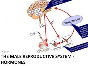
Male Reproductive The main functions of the male reproductive system, are to produce spermatozoa, androgens (sex hormones - principally testosterone) and to facilitate fertilisation, by introducing spermatozoa into the femal genital tract (copulation). The male reproductive system includes the testis, genital ducts, accessory sex glands and penis.

Testis The pair of testes produces spermatozoa androgens. Several accessory glands produce the fluid constituents of semen. Long ducts store the sperm and transport them to the penis. The male reproductive system consists of paired testes and genital ducts, accessory sex glands and the penis. The testes and ducts are shown in this diagram.


Epidymis The genital ducts -The looped seminiferous tubules in � the testes are connected to the genital duct system which transports the spermatozoa and fluid component of the semen to the outside. -This duct system is made up of the tubuli recti (short straight tubules connected to the seminiferous tubules), the rete testis - which is found in the mediastinum testis. The rete testis empties into the ductuli efferentes that lead into the ductus epididymus. -The ductus epididymus empties into the vas � deferens, which empties into the ejaculatory duct, which empties into the urethra and passes to the outside.

The epididymis � -The ductulis efferentes and the ductus � epididymus make up the epididymis. These ducts are highly coiled, and make up a single tube in the epididymis that can be up to 6 metres long. This means that several sections of it can be found on a single slide. -The ductus epidymis is a storage reservoir for � spermatozoa that become matured here, and competent to fertilise ova. Fluid is absorbed here, and the epithlium is also secretory. -The epithelium is pseudostratified, with long � immotile 'sterocilia' which are in fact very long microvilli. These are thought to be involved in absorption of fluid. The cells are also thought to be secretory, but the nature of the secretions is unknown.


-Spermatogenesis -The production of sperm and eggs/ova (gametes) is a procedure called gametogenesis (spermatogenesis and oogenesis). Gametogenesis involves two rounds of meiotic cell division. -The germinal (seminiferous epithelium) of the seminiferous tubules contains spermatogenic cells and Sertoli cells. -The spermatogenic cells divide by mitosis, then meiosis to form gametes, which mature into sperm by the process of spermiogenesis.
 Uocyhtuzwza -site:youtube.com
 Female testes
 Female reproductive system with baby
 Female external reproductive system
 Female reproductive system pregnancy
 What is reproductive system
 Lesson 3 the female reproductive system
 Ovary diagram
 Renal
Renal Female reproductive system of pila
 Mammary papilla pig
 Figure 28-2 the female reproductive system
 Sagittal female reproductive system
 Chapter 16 the reproductive system
 Female cow reproductive system
 Cow reproduction system
 Similarities between male and female reproductive system
 Parts of and functions of female reproductive system
 Pearson education
 Female anatomy
 Fsh and lh in female reproductive system
 Corpus luteum in female reproductive system
 Lesson 3 the female reproductive system
 Female reproductive system colour
 Female reproductive system bones
