Reproductive System Reproductive System Male and female system


- Slides: 46
Reproductive System
Reproductive System • Male and female system are a connected series of organs and glands • Produce and nurture sex cells – Transport the sex cells to fertilization site • Secrete hormones vital for development and maintenance of secondary sex characteristics • Regulate reproductive physiology
Male Reproductive Organs • Produce and maintain male sex cells, sperm cells • Transport these cells and supporting fluid to outside • Secrete male sex hormones
Male Reproductive Organs • Primary sex organ: 2 testes – Make sperm cells and male sex hormones • Accessory sex organs: internal and external reproductive organs
Testes • Ovoid structures about 5 cm in length and 3 cm in diameter • Both testes are within the saclike cavity of the scrotum
Testes Structure • Composed of lobules separated by connective tissue and filled with seminiferous tubules – Epithelium lining the seminiferous tubules produces sperm cells – Interstitial cells produce male sex hormones
Sperm Formation • Epithelium lining the seminiferous tubules include: – Supporting cells: support and nourish spermatogenic cells – Spermatogenic cells: give rise to sperm cells • Sperm cells consists of a head, midpiece, and tail
Spermatogenesis • Give rise to sperm • Meiosis reduces the number of chromosomes in sperm cells by ½ – 46 23 chromosomes • Produces 4 sperm cells for each primary spermatocyte
Spermatogenesis
Male Internal Accessory Organs • Epididymis: a tightly coiled tube that leads into the vas deferens – Stores and nourishes immature sperm cells and promotes their maturation
Male Internal Accessory Organs • Vas deferens: muscular tube – Passes through the inguinal canal and enters the abdominal cavity – Courses medially into the pelvic cavity and end behind the urinary bladder – Fuses with the duct from the seminal vesicle to form the ejaculatory duct
Male Internal Accessory Organs • Seminal Vesicle: a saclike structure attached to the vas deferens – Secretes an alkaline fluid that contain nutrients, such as fructose, prostaglandins
Male Internal Accessory Organs • Prostate Gland: surrounds the urethra just inferior to the urinary bladder – Secretes a thin, milky fluid that neutralizes the p. H of semen and acidic secretions of the vagina
Male Internal Accessory Organs • Bulbourethral glands: two small structures inferior to the prostate gland – Secrete a fluid that lubricates the penis in preparation for sexual intercourse • Semen: consists of sperm cells and secretions of the seminal vesicles, prostate gland, and bulbourethral glands – This fluid is slightly alkaline and contains nutrients and prostaglandins
Male Internal Accessory Organs • Sperm Cell: located in semen – Swim and have the ability to fertilize egg cells once in female reproductive tract
Male External Reproductive Organs • Scrotum: a pouch of skin and subcutaneous tissue – Encloses the testes • Penis: specialized to become erect for insertion into the vagina during sexual intercourse – During erection, the vascular spaces within the tissue become engorged with blood – Semen moves along the reproductive tract as smooth muscle in the walls of the tubular structures contracts by reflex
Male Hormones • Hypothalamic and Pituitary Hormones – Male body remains reproductively immature until the hypothalamus releases gonadotropin-releasing hormone (Gn. RH) – Stimulates the anterior pituitary gland to release gonadotropin – Follicle-stimulating hormone (FSH) stimulates spermatogenesis – Luteinizing hormone (LH) stimulates interstitial cells to produce male sex hormones
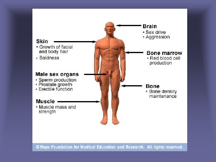
Male Sex Hormones • Androgens – Production increases rapidly at puberty – Testosterone: most important • Stimulates development of male reproductive organs • Male sexual characteristics
Regulation of Male Sex Hormones • A negative feedback mechanism regulates testosterone concentration – Rising testosterone concentration inhibits the hypothalamus and reduces the anterior pituitary’s secretion of gonadotropins – As testosterone concentrations falls, the hypothalamus signals the anterior pituitary to secrete gonadotropins – Testosterone concentrations remain stable from day to day
Organs of the Female Reproductive System
Female Reproductive System • Produce and maintain the female sex cells – Egg cells or ova • Transport these cells to the site of fertilization • Provide a favorable environment for a developing offspring • Move offspring to the outside • Produce female sex hormones • Primary Female Sex Organs: two ovaries
Ovaries • Two ovaries: solid, ovoid structures – 3. 5 cm long, 2 cm wide, and 1 cm thick – Lie in the shallow depressions in the lateral wall of the pelvic cavity
Ovary Structure • Subdivided into a medulla and a cortex – Medulla: connective tissue, blood vessels, lymphatic vessels, and nerves – Cortex: ovarian follicles and is covered by cuboidal epithelium
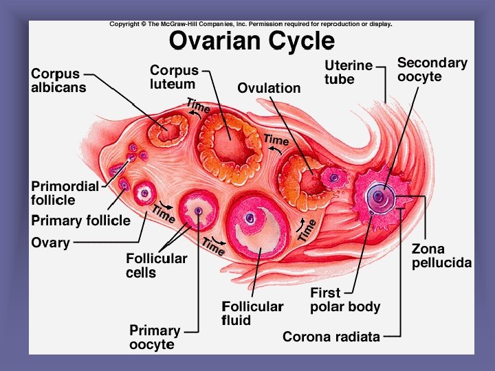
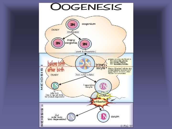
Preimordial Follicles • During prenatal development, groups of cells in the ovarian cortex form millions of preimordial follicles – Each contains a primary oocyte and a layer of follicular cells – Primary oocyte begins meiosis • Process stalls until puberty – The number of oocytes steadily declines throughout a female’s life
Oogenesis • At puberty, some oocytes are stimulated to continue meiosis • Primary oocyte secondary oocyte – Chromosome number is cut in half • 46 23 • Fertilization of a secondary oocyte produces a zygote
Follicle Maturation • At puberty, initiated by FSH • Generally, one follicle matures at a time • Maturation: – oocyte enlarges – Follicular cells multiply – Fluid filled cavity forms
Ovulation • The release of an oocyte from an ovary – A rupturing follicle releases a oocyte • After ovulation, the oocyte is drawn into the opening of the uterine tube
Internal Accessory Organs • Uterine tubes – the end of each tube expands – Margins bears irregular extensions – Ciliated cells that line the tube and peristaltic contractions in the wall of the tube help transport the egg cell down the uterine tube
Internal Accessory Organs • Uterus: receives the embryo &sustains it – Wall includes the endometrium, myometrium, and perimetrium • Vagina: receives the erect penis, coveys uterine secretions to the outside, and provides an open channel for the fetus during birth
External Accessory Organs • Labia Majora: rounded folds of adipose tissue and skin – Upper ends form a rounded elevation over the symphysis pubis • Labia minora: are flatted, longitudinal folds between the libia majora – Well supplied with blood vessels
External Accessory Organs • Clitoris: a small projection at the anterior end of the vulva – Corresponds to the penis – Composed of two columns of erectile tissue • Vestibule: space between the labia minora – Vestibule glands secrete mucus
Female Hormones and Mammary Glands
Female Hormones • Sex cell maturation • Development & maintained of female secondary sexual characteristics • Glands – Hypothalamus – Anterior pituitary gland – Ovaries
Female Hormones • Most important: estrogen & progesterone – Estrogen: develops and maintains most female secondary sex characteristics – Progesterone: changes in the uterus
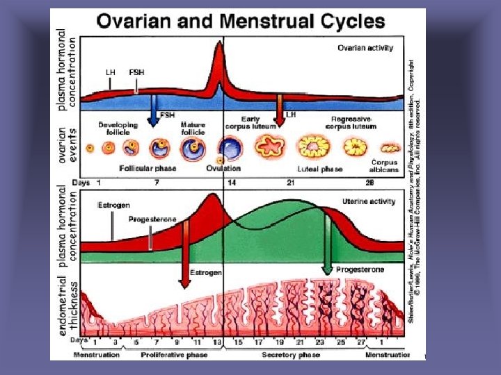
Female Reproductive Cycle • FSH initiates a menstrual cycle by stimulating follicle maturation – Maturing follicular cells secrete estrogen • Maintains secondary sexual traits • Thickening of the uterine lining • Anterior Pituitary: secretes large amount of LH – Triggers ovulation
Female Reproductive Cycle • Following ovulation, follicular cells give rise to the corpus letuem – Secretes progesterone • Causes the uterine lining to become more vascular and glandular • If an oocyte is not fertilized, the corpus luteum begins to degenerate • As estrogen and progesterone concentrations decrease, the uterine lining disintegrates – Menstrual flow
Ovulation
Female Reproductive Cycle • During the cycle, estrogen and progesterone inhibit the release of LH and FSH • As estrogen and progesterone decline, anterior pituitary secretes FSH and LH again – Stimulate a new menstrual cycle
Menopause • Termination of menstrual cycle due to the aging of the ovaries • Reduced estrogen concentrations and lack of progesterone may cause regressive changes in female secondary sex characteristics
Menopause
Mammary Glands • Located in the subcutaneous tissue of the anterior thorax – Composed of lobes that contain ducts and glands • Dense connective and adipose tissue separates the lobes
Mammary Glands • Ovarian hormones stimulates female breast development – Alveolar glands and ducts enlarge – Fat deposits around and within the breasts