
FALLOPIAN TUBES AND OVARIES FEMALE REPRODUCTIVE SYSTEM OVIDUCT
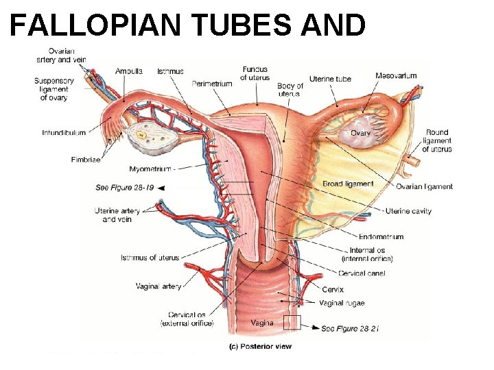
FALLOPIAN TUBES AND OVARIES
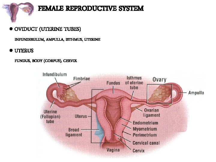
FEMALE REPRODUCTIVE SYSTEM OVIDUCT (UTERINE TUBES) INFUNDIBULUM, AMPULLA, ISTHMUS, UTERINE UTERUS FUNDUS, BODY (CORPUS), CERVIX
FEMALE REPRODUCTIVE SYSTEM OVARIES OVIDUCT (UTERINE TUBES) UTERUS VAGINA
FALLOPIAN TUBE • Paired hollow muscular tubes one on either sides of uterus • 10 cm long and 1 cm broad at ampullary part.
• Each from medial to lateral divided into 4 parts • Interstitial or intramural • Isthumus • Ampulla • Infundibulum
• Intramural part: narrowest. 1. 25 cms in length and 1 mm in breadth Isthumus : thick 2. 5 cms length and 1 -2 mm in diameter. tubal sterlization operations don here
• Ampulla : tortous. 5 cms length with its lumen measuring 1 -2 mm medially and 6 mm at outer end • Infundibulum: funnel shaped opening of distal end of fallopian tube being 1. 25 cms in length 3 mm wide. Leading to processes called fimbriae which touch ovary to pick up ovum during ovulation.

The Female Reproductive System in Midsagital View Figure 28. 13
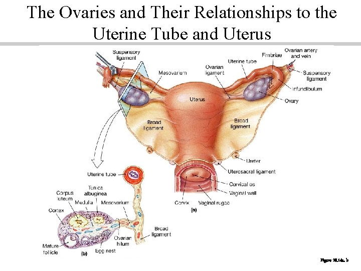
The Ovaries and Their Relationships to the Uterine Tube and Uterus Figure 28. 14 a, b
• 3 layers: serous • Muscular • Mucus membrane thrown into longitudnal folds • Lined by tall columnar non cilliated secretory cells with intervening cilliated columnar cells.
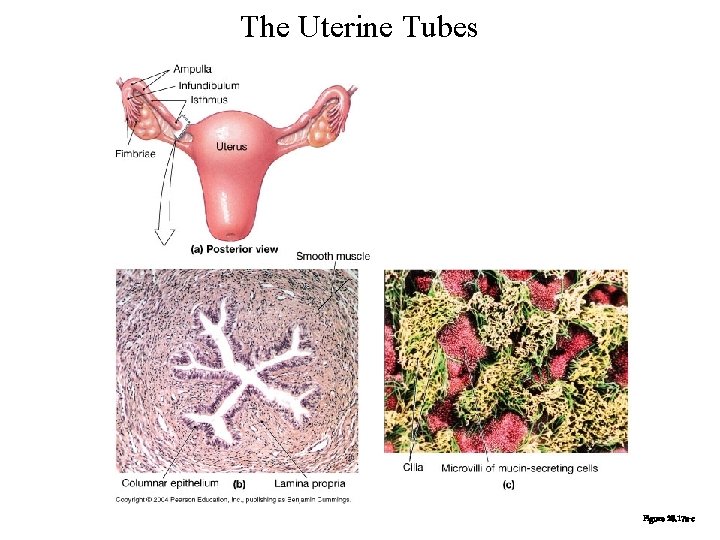
The Uterine Tubes Figure 28. 17 a-c
• Functions ; • Site for fertilization at the ampulla. • Transportation of gametes. tubal peristalysis and mucosal cilliary function. • Tubal secetion contains pyruvate for nuturing the zygote
• BLOOD SUPPLY AND NERVE SUPPLY • Ovarian and uterine arterial anastomosis in the mesosalpinx. • Venous drainage through pampiniform plexus into ovarian veins. • Nerves derived from ovarian and uterine sympathetic nerves from T 11 and T 12.
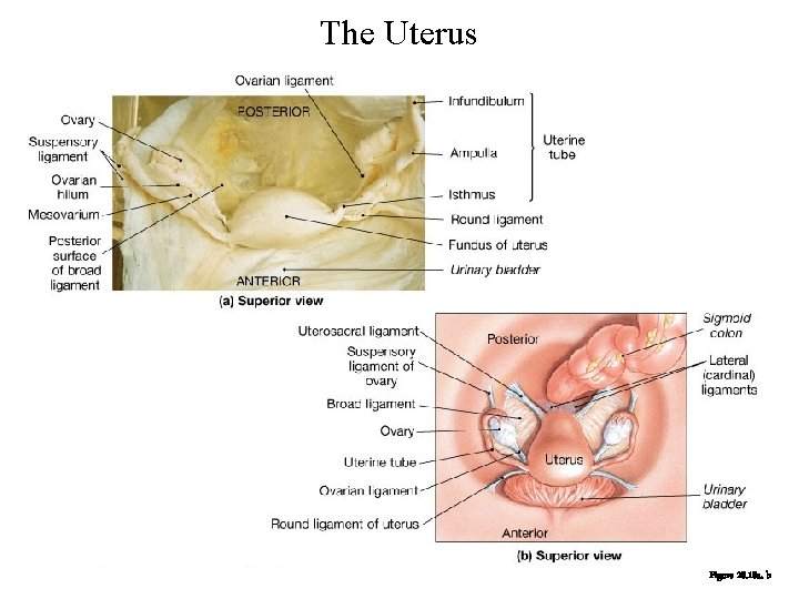
The Uterus Figure 28. 18 a, b
• OVARY: • One on either side is a solid flat ovoid sex gland or gonad. • Responsible for maturation , storage and release of germ cells and production of sex hormones
5 -10 gms weight. POSITION ovaries situated in upper part of pelvic cavity. in a slight depression on lateral wall of pelvis between internal and extrnal illia vessels. called ovarian fossa of waldeyer.
• STRUCTURE • Surface epithelium ; single layer of cubical cells. • Cortex comprised of highly cellular spindle shaped stromal cells and ovarian follicles which are functional units scattered in cortex. Medulla : loose connective tissue , blood vessels and nerves.
• BLOOD SUPPLY • Ovarian artery , a branch from abdominal aorta. • Venous drainage through pampiniform plexus to form ovarian veins which drain into inferiior venecava.
Functions of the Ovary • Production of a mature oocyte, capable of fertilization and embryonic development. • Production of ovarian steroids (estradiol, progesterone). • Production of gonadal peptides (inhibin, activin).
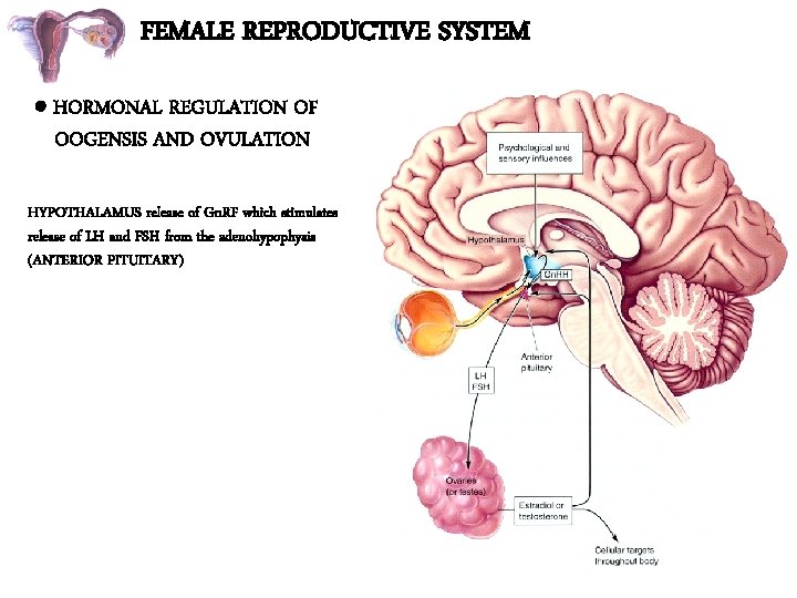
FEMALE REPRODUCTIVE SYSTEM HORMONAL REGULATION OF OOGENSIS AND OVULATION HYPOTHALAMUS release of Gn. RF which stimulates release of LH and FSH from the adenohypophysis (ANTERIOR PITUITARY)
The Menstrual Cycle: The Ovary • Follicular phase: small antral follicles develop, a dominant follicle is selected and grows to the preovulatory stage. • Midcycle: the gonadotropin surges cause ovulation of the dominant follicle. • Luteal phase: the corpus luteum forms and becomes functional, secreting large amounts of progesterone, followed by estradiol (results in negative feedback, not positive feedback, because P increases before E 2). • If pregnancy does not take place, the corpus luteum regresses, and P and E 2 levels decrease.
The Menstrual Cycle: The Uterus • Proliferative stage: increasing estradiol levels stimulate proliferation of the functional layer of the uterine endometrium. - Results in increased thickness of the endometrium. - Increased growth of uterine glands (secrete mucus) and uterine arteries. • Secretory stage: progesterone acts on the endometrium. - uterine glands become coiled and secrete more mucus - uterine arteries become coiled (spiral arteries)

THANK YOU. • THANK YOU
- Slides: 23