DR VEERENDRA V KERUDI Growth sites in maxilla


 b) The vertical lengthening of the nasomaxillary complex involves: remodeling")


 is")


 direction and that it lies towards the")

 b) The bony arch becomes increased & the corpus lengthens by: Deposits")


 n")


 b) c) deposition on the alveolar surface of the labial cortex resorption on")


 & n deposition")
- Slides: 96
DR. VEERENDRA V KERUDI
Growth sites in maxilla n n Maxillary tuberosity Sutures Alveolar border Nasal septum
Maxillary tuberosity n Horizontal lengthening of the bony maxillary arch is produced by growth at the maxillary tuberosity. n Represented by a backward movement of PTM from the vertical reference line
n It is a depository field & receives continued deposits of new bone as long as growth in the face continues. n Because the posterior surface of the maxillary tuberosity points toward the direction of arch elongation, this is the particular surface that is depository
n Endosteal side of the cortex within the interior of the tuberosity is resorptive. n Cortex thus drifts progressively posteriorly and also, to a lesser extent, in a lateral direction. n Deep to the tuberosity is the maxillary sinus
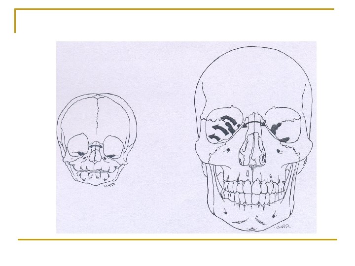
n In newborn, the sinus is quite small, but it becomes greatly expanded as growth continues and occupies greater part of the large suborbital compartment.
n The maxillary tuberosity is a major "site" of maxillary growth. n Provides for the growth of the whole maxilla associated only with the posterior part of the lengthening arch
Nasal septum n a) b) The vertical lengthening of the nasomaxillary complex involves: remodeling growth (deposition and resorption on the various bony cortices) and displacement.
n Bony portion of the internasal septum (the vomer and the perpendicular plate of the ethmoid) lengthens vertically at the various sutural junctions n The bony septum also drifts laterally in relation to variable amounts and directions of septal deviation
n The remodeling patterns involved are individually variable, and the thin plate of bone typically shows alternate fields of deposition and resorption on the right and left sides, which produce a drift and buckling to one side or the other.
n Breadth of the nasal bridge in the region just below the frontonasal sutures does not markedly increase from early childhood to adult n Inferiorly in the interorbital area, the medial wall of each orbit expands and balloons out considerably in a lateral direction in conjunction with the considerable extent of lateral enlargement of the nasal chambers. n The ethmoidal sinuses are thereby enlarged greatly
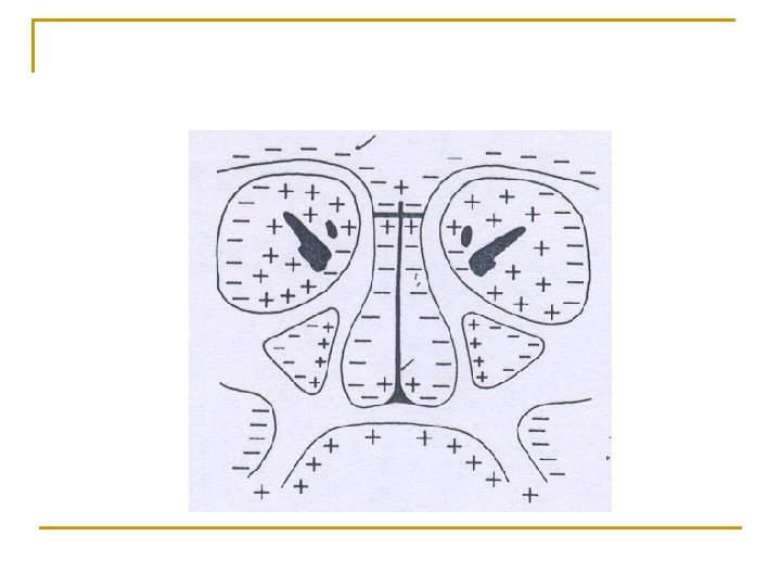
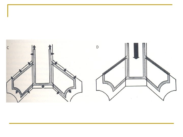
n Ø Ø In the growth of the bony maxillary arch, bone deposition occurs in 3 directions: it lengthens posteriorly by deposition on the posteriorfacing maxillary tuberosity it grows laterally by deposits on the buccal surface (widens the posterior part of the arch); and
Ø it grows downward by deposition of bone along the alveolar ridges and also on the lateral side, because this outer surface slopes (in the child) so that it faces slightly downward
Vertical drift of teeth a. Resorptive b. Depository c. Resorptive periosteal surface As the maxilla and mandible d. Depository enlarge and develop the dentition e. Resorptive drifts both vertically and horizontally f. Depository to keep pace in respective anatomic positions
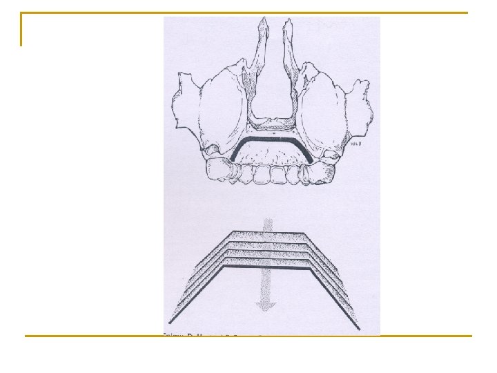
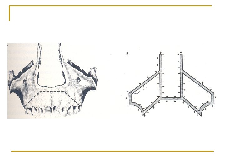
Palatal remodelling n Though anterior part of max. arch is resorptive, bone is added onto the inside of the arch; arch thus increases in width and palate becomes wider n Growth along mid palatal suture is known to participate to a greater or lesser extent in the progressive widening of the palate and alveolar arch. n As the palate grows inferiorly by the remodeling process a nearly complete exchange of the old for new hard and soft tissue occurs
n At each succeeding descending level the palate becomes a different palate n It occupies different position and is composed of diff bone conn. tissue, epithelia, bld vessels, nerve extentions and so on. n Natural increase in palatal width is the result of vertical drift of post teeth with expansion laterally occuring according to V principle of growth.
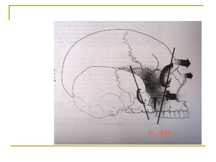
Maxillary sutures n Primary displacement of the whole ethmomaxillary complex in an inferior direction
n Its displacement movement accompanies simultaneous enlargement in all areas throughout the entire nasomaxillary region. n The system of sutures that unites the bones of upper part of face to cranium include the following:
n n n Fronto-nasal Fronto-maxillary Zygomatico-temporal Zyomatico-maxillary Pterygo-palatine
n These sutures are oblique and more or less parallell to each other. n Thus growth in these areas would serve to move the maxilla downwards and forwards and increases the ht of the orbit. n However it does not contribute to the infranasal ht. n By 7 yrs the orbit attain almost their full adult size
n After these sutures play a little role in vertical growth of face. n The sutures of midline are probably of little significance after 12 yrs. n The surface apposition accounts for lateral growth which takes place later on.
n Maxillary width follows neural growth curve which will be completed earlier than the downward and forward growth which follows the general body curve and continues to parallel pubertal changes else where
Alveolar process n Developes in response to the presence of tooth buds n As the teeth erupts the alveolar process developes and increases in height by bone deposition at the margins.
n The alveolar bone adds to height and thickness of the body and is particularly manifested as a ledge extending lingual to ramus to accommodate third molars n In absence of teeth it fails to develop; and it resorbs after tooth extraction
Cheek bone and zygomatic arch n The posterior side of the malar protuberance within the temporal fossa is depository; n Together with a resorptive anterior surface, the cheek bone relocates posteriorly as it enlarges
n The zygomatic process move posteriorly as the max and mand arches develop posteriorly to complement each other. n The inferior edge of the zygoma is heavily depository n The ant part of the zygomatic arch and malar region thereby become greatly enlarged vertically as the face developes in depth.
n The zygomatic arch moves laterally by resorption on the medial side within the temporal fossa and by depostion on the lateral side n This enlarges the temporal fossa and keeps the cheekbone proportionately broad in relation to face and jaw size and the masticatory musculature
n The zygoma and the cheekbone becomes displaced anteriorly and inferiorly in the same directions and amount as the primary displacement of maxilla
n Malar region grows and becomes relocated posteriorly; nasal region grows and becomes relocated anteriorly; this results in more protrusive appearing nose and anteroposteriorly much deeper face.
Orbital growth n The remodelling changes of the orbit are complex because many separate bones comprise its enclosing walls n Most of the lining roof and floor are depository n As the frontal lobe of the cerebrum expands forwards and downwards the orbital roof remodels anteriorly and inferiorly by resorption on the endocranial side and deposition on the orbital side.
n The cone shaped cavity moves in a direction towards its wide opening; deposits on the inside thus enlarge rather than reduce the volume.
n The lateral wall of the orbital rim remodels by resorption on the medial side and deposition on the lateral side n The cutaneous side of the supraorbital ridge is depository, and this combination causes the superior orbital rim to become protrusive n The upper rim extends beyond the lower rim
Maxillary height: n Sutural lowering of the maxilla was found on average to be 11. 2 mm (range 9 -13. 5 mm). n The height of the nasal cavity increased up through puberty as result of resorptive lowering of nasal floor. n Appositional growth in the height of the alveolar process.
Maxillary width: n n Bjork showed that growth in the suture continues until puberty. By measuring the distance of separation between the lateral implants on the frontal cephalogram over time, it was shown that sutural growth was the most important factor in the development of the width of the maxilla.
Bimolar width: n n n Bjork’s implant studies show that though growth in the median suture from eruption of 1 st permanent molars to adulthood is 4. 8 mm, increase in arch width during this period was on average, 3. 1 mm. The reason for this difference in growth in width is related to transverse rotation of the maxillae.
Bicanine width: n n n The development in width of the dental arches between the canines was different from that of molar region. Though there was an overall increase of 3. 1 mm in intercanine width from 4 years to adulthood, most of this increase occurred early, with only 1. 1 mm increase from age 6 years to adulthood.
n Mutual transverse rotation of maxillae also results in greater separation of lateral segments of dental arch posteriorly, than anteriorly. n There is thus, greater increase in intermolar width than intercanine width, and also a corresponding decrease in arch length. n Thus, shortening of arch length is related to transverse growth of the maxilla.
n At birth, mandible is little more than a curved bar of bone. n The coronoid , angular and alveolar process are not formed.
Growth sites in mandible n n n Ramus Lingual tuberosity Mandibular condyle Alveolar process Chin Suture
Macerated mandibles of a neonate, a 5 year old an adult. Growth is considerably faster during the early postnatal period than during adolescence.
n Mandible is an “expanding V” additive growth at the ends of “V” naturally increases the distance between the terminal points.
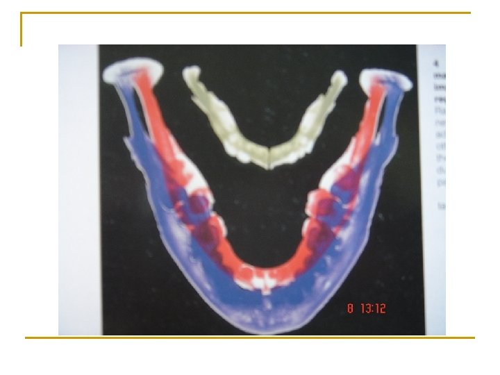
n Scott divides the mandible into 3 basic types of bone basal, muscular and alveolar. 1. The basal portion is a tube-like central foundation running from the condyle to the symphysis.
2. n 3. n n The musculature (the gonial angle and coronoid process) is under the influence of the massetter, internal pterygoid and temporal muscles. Muscles function determines the ultimate form of the mandible in these areas. The alveolar bone, exists to hold the teeth. When the teeth are lost there is gradual resorption of the alveolar bone. Reduced muscle activity accounts for the flattening of the gonial angle and reduced coronoid process.
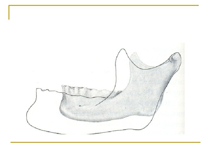
Ramus : n Mandibular corpus should extent so that it matches its counterpart, the maxillary arch n In this process, one notable structural difference between mandible & maxilla exists: the mandibular ramus. n The posterior growth of mandibular bony arch undergoes remodeling conversion from ramus to corpus
n Whole ramus relocates posteriorly and the former anterior part of the ramus is structurally altered into an addition to the corpus, which thereby becomes lengthened by remodeling
n Some of the key anatomic parts that participate in the relocation and remodeling processes of the ramus and corpus cannot be represented in conventional twodimensional headfilms and tracings n Eg : lingual tuberosity n As important as maxillary tuberosity
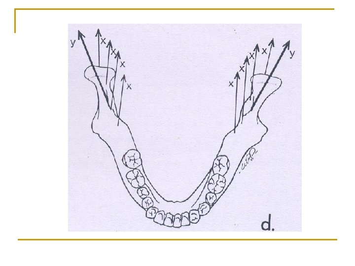
Lingual tuberosity n Lingual tuberosity is a boundary between two basic parts of the mandible: the ramus & corpus n Lingual tuberosity grows posteriorly by deposition on its posterior facing surface
n Lingual tuberosity protrudes in lingual (medial) direction and that it lies towards the midline from the ramus n The prominence of the tuberosity is increased by presence of large resorptive field just below it. n This resorptive field produces lingual fossa.
n Tuberosity grows in almost posterior direction n Part of the ramus just behind the tuberosity grows medially
n a) b) The bony arch becomes increased & the corpus lengthens by: Deposits on posterior surface of lingual tuberosity & on lingual side of ramus Resultant lingual shift of the part of the ramus becoming added to the corpus
n Resorption of the anterior border of the ramus is described as “making room for the last molar” n Growth takes place as follows
Coronoid process n The coronoid process has a propeller-like twist, so that its lingual side faces three general directions all at once posteriorly, superiorly medially
n When bone is added onto the lingual side of coronoid process, its growth proceeds superiorly and the ramus increases in vertical dimension n Each coronoid process lengthens vertically, even though additions are made on the medial (lingual) surfaces of the right and left coronoid processes. n Eg: of enlarging V principle, with the V oriented vertically.
n Same deposits of bone on the lingual side also bring about a posterior direction of growth movement, because this surface also faces posteriorly n This produces a backward movement of the two coronoid processes, even though deposits are added on the inside (lingual) surface. Eg: of the expanding V principle, with the V oriented horizontally n
n Same deposits of bone on the lingual side also function to carry the base of the coronoid process and the anterior part of the ramus in medial direction in order to add to the lengthening corpus n Eg: of the V principle
n Buccal side of coronoid has resorptive type of periosteal tissue n It faces away from the combined superior, posterior, & medial directions of growth
n Lower part of the ramus below the coronoid process also has a twisted contour. n Its buccal side faces posteriorly toward the direction of backward growth and thus, characteristically, has a depository type of surface n The opposite lingual side, facing away from the direction of growth, is resorptive,
n Single field of surface resorption is present on the inferior edge of the mandible at the ramuscorpus junction. n This forms the antegonial notch by remodeling from the ramus just behind it as the ramus relocates posteriorly
Mandibular foramenn As the whole ramus grows posteriorly and superiorly the mandibular foramen also drifts backward and upward to maintain a constant position i. e. midway between the anterior and posterior border of ramus.
Condyle : n Has an oblique upward & backward growth direction n Angle of growth – variable depending on whether horizontal or vertical growers with respect to mandible
n Condyle was & is still believed by some to be the ultimate determinant that, essentially, establishes the mandibular rate of growth, amount of growth, growth direction, overall mandibular size, and overall mandibular shape n But now it is no longer believed to represent a pace setting “master center”
n During development, the condyle functions as a regional field of growth that provides an adaptation for its own localized growth circumstances just as all the other regional fields accommodate their own particular (but different) localized growth circumstances. n The growth of the mandible is a product of all the different regional forces and regional functional agents
n Condylar growth mechanism is clear-cut n Endochondral growth mechanism is required, because condyle grows in a direction towards its articulation in face of direct pressure n Endochondral growth occurs only at articular contact part of condyle because this is where pressure exists at levels beyond tolerance of bone’s soft tissue membrane
n Condylar cartilage is a secondary type of cartilage n It is developed on dentary bone to provide for lower jaw articulation with the cranium n It is a believed unique connective tissue covering condylar cartilage is an original periosteum n Its undifferentiated connective tissue stem cells converted into chondroblasts instead of osteoblasts because of the compressive forces acting on the membrane
n Condyle have a special multidirectional capacity for growth and remodeling in selective response to varied mandibular displacement movements and rotations n Unique capsular layer of poorly vascularized connective tissue covers the articular surface of the condyle (a)
n Just deep to it is a special layer of prechondroblast cells (b) n This is a predominant site for cellular proliferation
n The proliferative process produces the "upward and backward" growth movement of the condyle n The condylar cartilage moves by prechondroblast cell divisions on the articular side
Neck of the condyle n Lingual and buccal sides of the neck characteristically have resorptive surfaces n This is because the condyle is quite broad and the neck is narrow. n The neck is progressively relocated into areas previously held by the much wider condyle, and it is sequentially derived from the condyle as the condyle moves in a superoposterior course.
n What used to be condyle in turn becomes the neck as one is remodeled into the other. n Occurs by periosteal resorption combined with endosteal deposition. n Another way, the endosteal surface of the neck actually faces the growth direction; the periosteal side points away from the course of growth n Eg: of enlow principle
n For many years it was presumed that primary displacement of mandible was due to the growth of condylar cartilage n As it is pressure adapted it provides thrust of the mandible against its articular bearing area
n Later observations explained that the mandible is displaced away from its cranial base articular contact by secondary displacement & that there is no space or gap created
n n n The rate & direction of condylar growth are subject to the influence of extracondylar agents, including intrinsic & extrinsic biomechanical forces & physiologic inductors amount of pressure - cell division & growth pressure – accelerate growth
n Recent research studies by Mc. Namara show that the nature of the condylar stimulus is more complex than simple forces acting directly on the condyle; nerve-muscle-connective tissue pathways are involved, and changes utilize a composite of such tissue responses and chain feedbacks with the condyle as well as the other parts of the mandible that also participate.
n The key point is that regional areas within the condyle can be stimulated or inhibited by resultant, localized forces to grow regionally either more or less. n This alters the amount of ramus growth in different directions, thereby continually adjusting both the alignment and the shape of the ramus to accommodate its multiple anatomic and functional relationships.
Ramus up righting n The ramus becomes more vertically aligned as its development occurs n As the ramus is growing in the posterior direction greater amount of deposition takes place on the inferior part than on the superior part and on the anterior surface greater resorption takes place inferiorly than superiorly.
n Thus the Condylar growth becomes directed more vertically along with rest of the mandible n The ramus continues to grow vertically even after its horizontal growth slows/stops. n This is to match the vertical growth of the mid face
n The Condylar growth becomes more vertically directed. n Different type of remodeling of the ramus takes place n The direction of deposition and resorption is reversed
n Resorption takes place on the upper portion of the posterior border and deposition takes place on the anterior border on the upper part of the Coronoid process
n The result is more upright alignment and a longer vertical dimension of the ramus of the mandible with out the increase in the breadth n There is an increase in the length of the corpus n The angle of the mandible gets changed in order to retain the positional relationship between the upper and lower arches
n During the descent of the maxilla and the vertical drift of the mandibular teeth, the mandibular teeth drift lingually and superiorly. n This produces a greater or lesser over jet & over bite n This involves the Periosteal resorption at the labial cortex :
a) b) c) deposition on the alveolar surface of the labial cortex resorption on the alveolar surface o the lingual cortex deposition on the lingual side of the lingual cortex
Human chin n In infancy chin is under developed As age advances growth of chin becomes significant It is influenced by sexual and specific genetic factors; males –more prominent than females
n During the descent of the maxillary arch & the vertical drift of the mandibular teeth, ant mandibular teeth simultaneously drift lingually & superiorly. this produces a greater amount of ant overjet &overbite
n The remodelling process that brings this about involves n periosteal resorption on the labial bony cortex (a), n deposition on the alveolar surface of the labial cortex (b),
n resorption on the alveolar surface of the lingual cortex (c) & n deposition on the lingual side of the lingual cortex (d).