Maxilla maxillary sinus mandible Dr Gallatz Katalin Parts

Maxilla, maxillary sinus, mandible Dr Gallatz Katalin

Parts of the maxilla: 1. body 2. processes frontal process zygomatic process alveolar process palatine process Surfaces of the body of the maxilla anterior infratemporal nasal orbital

Anterior, orbital and infratemporal surface frontal process orbital surface infraorbital foramen post. alveolar foramina alveolar process maxillary tuber zygomatic process canine fossa

ANTERIOR AND INFRAORBITAL FORAMEN CANINE FOSSA INFRATEMPORAL SURFACE MAXILLARY HIATUS NASAL SURFACE

NASAL SURFACE maxillary hiatus lacrimal sulcus → nasolacrimal canal conchal crest → inf. nasal concha. INCISIVE CANAL PALATINE PROCESS

ANTERIOR, INFRATEMPORAL AND ORBITAL SURFACE forms the anterior wall of the pterygopalatine fossa forms the inferior surface of the orbit

HARD PALATE PALATINE PROCESS OF THE MAXILLA HORIZONTAL PLATE OF PALATINE BONE

Orbital surface Inferior orbital fissure infraorbital sulcus infraorbital canal infraorbital foramen

PTERYGOPALATINE FOSSA Infratemporal surface of the MAXILLA, maxillary surface of the GREATER WING and PTERYGOID PROCESS perpendicular plate of PALATINE BONE

MAXILLARY SINUS 1. The maxillary sinus is the largest paranasal sinus. 2. It is intimately related to the upper teeth, tear duct, and the floor of the orbit.

Recesses of the maxillary sinus 1. infraorbital recess 2. zygomatic recess 3. alveolar recess frontal recess basal recess tuberal recess

Maxillary sinus has infraorbital, zygomatic and alveolar recesses.

Maxillary sinus opens at its top into the middle nasal meatus through the posterior one-third of the semilunar hiatus. SEMILUNAR HIATUS bordered by the uncinate process and the ethmoidal bulla.
a large, irregular")
MAXILLARY SINUS Nasal wall/base On the nasal wall, ( or base )a large, irregular aperture the maxillary hiatus can be seen The maxillary hiatus is reduced in size by the following bones: - ethmoidal bulla and uncinate process of the ethmoid above, - the ethmoidal process of the inferior nasal concha below, - inferior nasal concha below - the perpendicular plate of the palatine bone behind, a small part of the lacrimal bone above and in front. The sinus communicates through an opening into the semilunar hiatus Maxillary sinusitis is imflammation of the maxillary sinuses.

Ethmoidal bulla Uncinate process

coronal section

1. Frontal sinus 2. Orbit 3. Maxillary sinus 4. Nasal septum 5. Nasal cavity 6. Hard palate

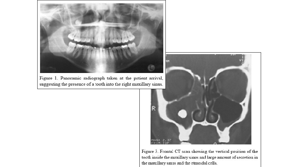
Maxillary sinusitis

Maxillary sinusitis

Frontal and maxillary sinuses are almost completly missing at the time of birth but it grows continuosly. 1 year 4 years 8 years 12 years 20 years 60 years

MANDIBLE

Parts of the mandible Corpus or body base of the mandible alveolar process Ramus coronoid process condylar process

Mandible – external surface masseteric tuberosity, oblique line mental foramen, mental protuberance mental tubercle alveolar juga mental protuberance masseteric tuberosity

Mandible - inner surface pterygoid tuberosity, pterygoid fovea mandibular foramen, lingula, mylohyoid sulcus mylohyoid line, submandibular fossa, sublingual fossa, digastric fossa, mental spines,

INSERTION SURFACES, LINES OF THE MUSCLES

TRAJECTORIAL STRUCTURE OF THE MANDIBLE MANDIBULAR CANAL

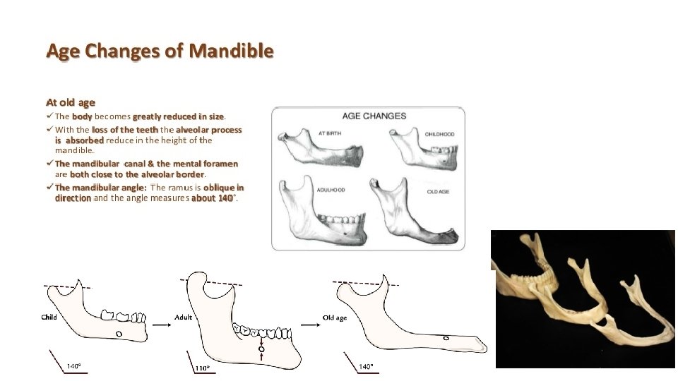
Neonatal mandible Mental symphysis Angle of the mandible: ~150° No alveolar process

Thank you for your attention!
- Slides: 30