NOSE AND NASAL SINUSES Nose Is the part


 of the Nasal Cavity The nasal cavity has a roof, floor, medial")


 is also supplied by")


- Slides: 37
NOSE AND NASAL SINUSES
Nose Is the part of the respiratory tract Superior to the hard palate contains the peripheral organ of smell. Parts: External nose Nasal cavity, divided into right and left cavities by the nasal septum. The functions of the nose are olfaction (smelling), respiration (breathing), filtration of dust, humidification of inspired air, and reception and elimination of secretions from the paranasal sinuses and nasolacrimal ducts.
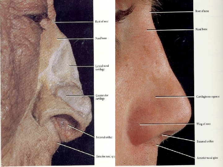
External Nose Is the part seen in face ; vary in shape and size Its skeleton is mainly cartilaginous Dorsum extends from the root to the apex (tip) of the nose. Inferior surface of the nose is pierced by two piriform openings, the nares (nostrils, anterior nasal apertures), bound laterally by the alae (wings) Superior bony part of the nose, including its root, is covered by thin skin. The skin over the cartilaginous part of the nose is covered with thicker skin, which contains many sebaceous glands. The skin extends into the vestibule of the nose , where it has a variable number of stiff hairs (vibrissae). The junction of the skin and mucous membrane is beyond the hairbearing area.
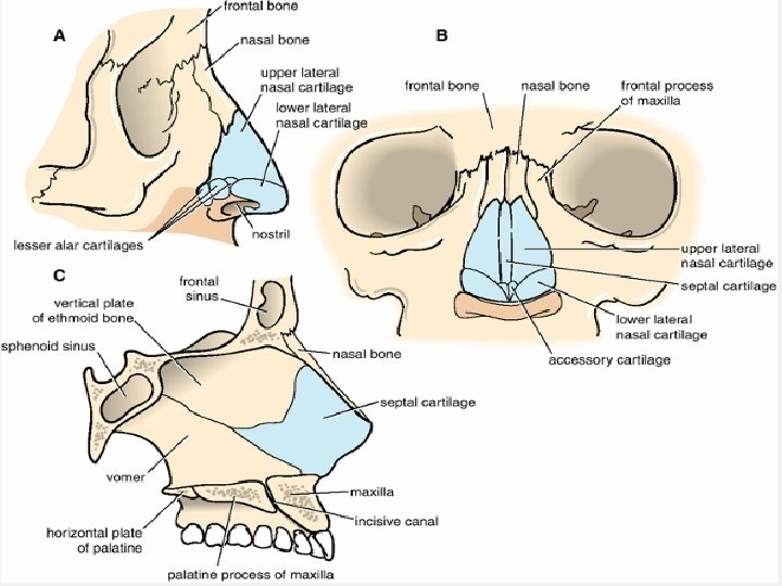
Skeleton of the External Nose Is composed of bone and hyaline cartilage. 1. The bony part consists of: Nasal bones Fontal processes of the maxillae Nasal part of the frontal bone and its nasal spine Bony parts of the nasal septum. 2. The cartilaginous consists of five main cartilages: Two lateral cartilages Two alar cartilages One septal cartilage. �The U-shaped alar cartilages are free and movable; they dilate or constrict the nares when the muscles acting on the nose contract.
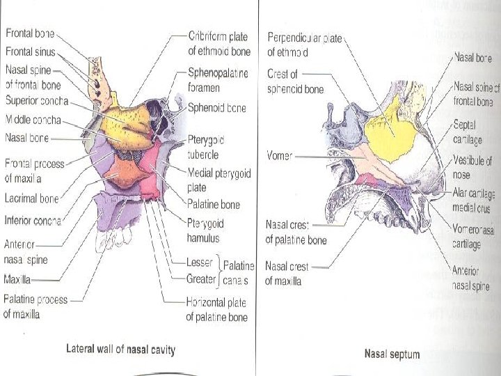
External Nose- Nasal Septum Divides the chamber of the nose into two nasal cavities Has a bony part and a soft mobile cartilaginous part. The main components of the nasal septum are: The thin perpendicular plate of the ethmoid �Forms the superior part of the nasal septum, descends from the cribriform plate and is continued superior to this plate as the crista galli The vomer (a thin flat bone) �Forms the posteroinferior part of the nasal septum, with some contribution from: � the nasal crests of the maxillary and palatine bones. . The septal cartilage. �Has a tongue-and-groove articulation with the edges of the bony septum
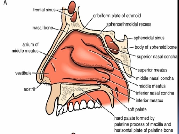
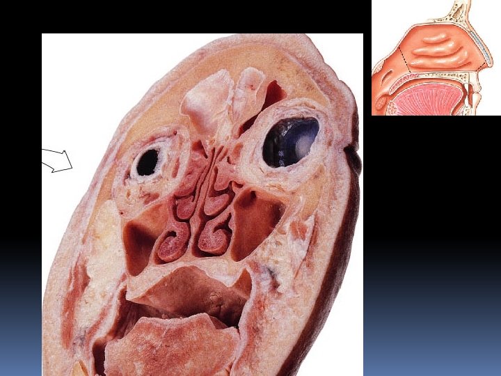
Nasal Cavity Refers to either the entire cavity or one of its halves Is entered anteriorly through the nares. It opens posteriorly into the nasopharynx through the choanae. Mucosa lines the nasal cavity, except for the nasal vestibule, which is lined with skin Nasal mucosa is firmly bound to the periosteum and perichondrium of the supporting bones and cartilages of the nose. The mucosa is continuous with the lining of all the chambers with which the nasal cavities communicate: the nasopharynx posteriorly, the paranasal sinuses superiorly and laterally, and the lacrimal sac and conjunctiva superiorly. The inferior two thirds of the nasal mucosa is the respiratory area (moisture) and the superior one third is the olfactory area(smell)
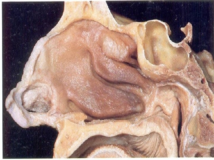
Boundaries (walls) of the Nasal Cavity The nasal cavity has a roof, floor, medial and lateral walls. The roof of the nasal cavity: is curved and narrow, except at its posterior end is formed anteriorly beneath the bridge of the nose by the nasal and frontal bones, in the middle by the cribriform plate of the ethmoid, located beneath the anterior cranial fossa, and posteriorly by the downward sloping body of the sphenoid The floor of the nasal cavity is wider than the roof is formed by the palatine processes of the maxilla and the horizontal plates of the palatine bone. The medial wall of the nasal cavity is formed by the nasal septum. The lateral wall has three projections of bone called the superior, middle, and inferior nasal conchae. The space below each concha is called a meatus
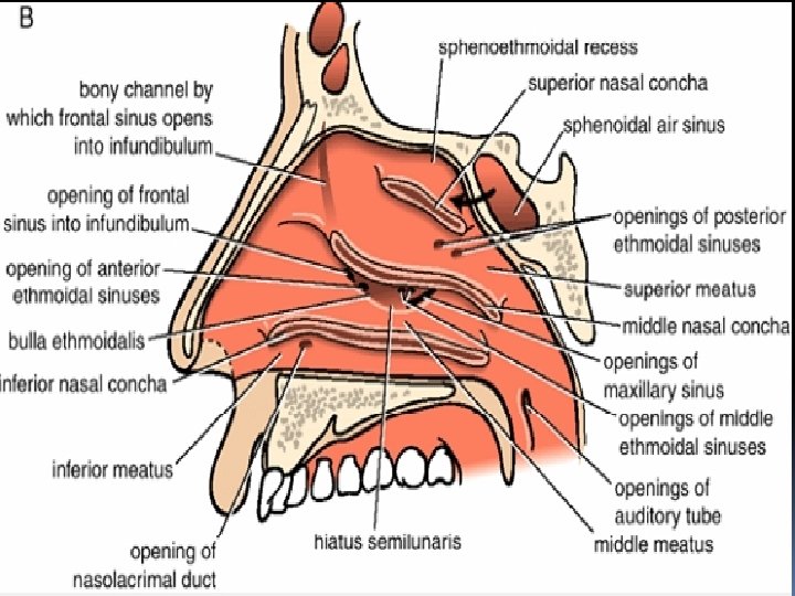
Features of the Nasal Cavity The nasal cavity is thus divided into five passages: a posterosuperiorly placed sphenoethmoidal recess three laterally located nasal meatus (superior, middle, and inferior) a medially placed common nasal meatus into which the four lateral passages open. The inferior concha is the longest and broadest and is formed by an independent bone (of the same name, inferior concha) covered by a mucous membrane that contains large vascular spaces that can enlarge to control the caliber of the nasal cavity. When infected or irritated, the mucosa may swell rapidly, blocking the nasal passage(s) on that side.
Features of the Nasal Cavity The sphenoethmoidal recess: lying superoposterior to the superior concha, receives the opening of the sphenoidal sinus. The superior nasal meatus is a narrow passage between the superior and the middle nasal conchae into which the posterior ethmoidal sinuses open by one or more orifices.
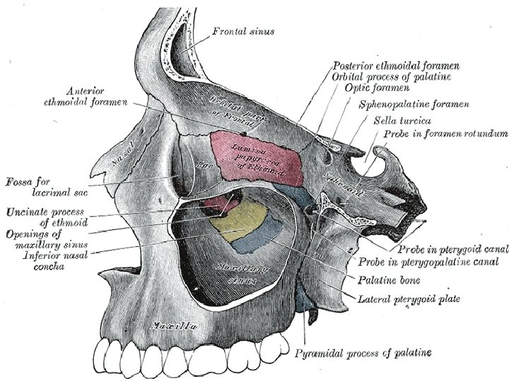
Features of the Nasal Cavity The middle nasal meatus; is longer and deeper than the superior one. It has a rounded swelling called the bulla ethmoidalis that is formed by the middle ethmoidal air sinuses, which open on its upper border A curved opening, the hiatus semilunaris, lies just below the bulla. The anterior end of the hiatus leads into a funnel-shaped channel called the infundibulum, which is continuous with the frontal sinus. The maxillary sinus opens into the middle meatus through the hiatus semilunaris.
Features of the Nasal Cavity The inferior nasal meatus: is a horizontal passage inferolateral to the inferior nasal concha. The nasolacrimal duct, which drains tears from the lacrimal sac, opens into the anterior part of this meatus. The common nasal meatus is the medial part of the nasal cavity between the conchae and the nasal septum, into which the lateral recesses and meatus open.
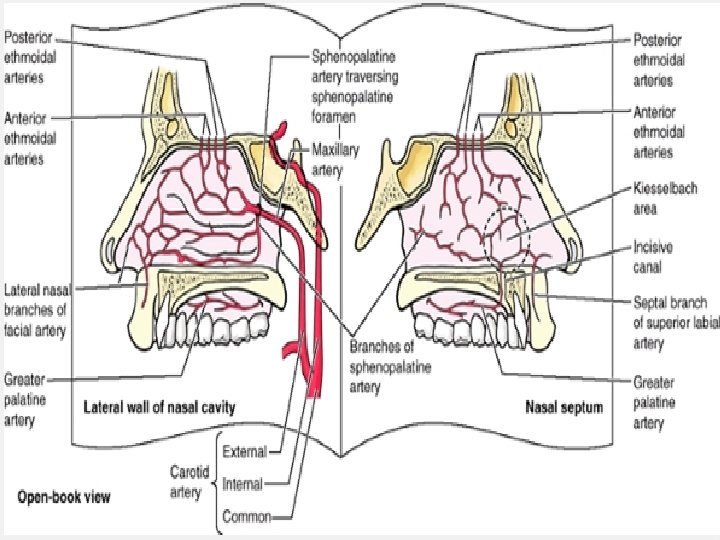
Blood Supply of Nose- Arterial The arterial supply of the medial and lateral walls of the nasal cavity is from five sources: Anterior ethmoidal artery (from the ophthalmic artery). Posterior ethmoidal artery (from the ophthalmic artery). Sphenopalatine artery (from the maxillary artery). Greater palatine artery (from the maxillary artery). Septal branch of the superior labial artery (from the facial artery). �The first three arteries divide into lateral and medial (septal) branches. �The greater palatine artery reaches the septum via the incisive canal through the anterior hard palate. The anterior part of the nasal septum is the site (Kiesselbach area) of an anastomotic arterial plexus involving all five arteries supplying the septum. The external nose also receives blood from first and fifth arteries listed above plus nasal branches of the infraorbital artery and the lateral nasal branches of the facial artery.
Blood Supply of Nose Venous: A rich submucosal venous plexus deep to the nasal mucosa drains into the sphenopalatine, facial, and ophthalmic veins. This venous plexus is an important part of the body's thermoregulatory system, exchanging heat and warming air before it enters the lungs. Venous blood from the external nose drains mostly into the facial vein via the angular and lateral nasal veins. However, recall that it lies within the danger area of the face because of communications with the cavernous (dural venous) sinus of the Facial Vein Lymph: The lymph vessels draining the vestibule end in the submandibular nodes. The remainder of the nasal cavity is drained by vessels that pass to the upper deep cervical nodes.
Nerve supply The nasal mucosa can be divided into posteroinferior and anterosuperior portions by an oblique line passing approximately through the apex of the nose and the sphenoethmoidal recess. The nerve supply of the posteroinferior portion of the nasal mucosa is chiefly from the maxillary nerve, by way of the nasopalatine nerve to the nasal septum, and posterior superior lateral nasal and inferior lateral nasal branches of the greater palatine nerve to the lateral wall. The nerve supply of the anterosuperior portion is from the ophthalmic nerve (CN V 1) by way of the anterior and posterior ethmoidal nerves, branches of the nasociliary nerve.
Nerve supply Most of the external nose (dorsum and apex) is also supplied by CN V 1 (via the infratrochlear nerve and the external nasal branch of the anterior ethmoidal nerve), but the alae are supplied by the nasal branches of the infraorbital nerve (CN V 2). The olfactory nerves, concerned with smell, arise from cells in the olfactory epithelium in the superior part of the lateral and septal walls of the nasal cavity. The central processes of these cells (forming the olfactory nerve) pass through the cribriform plate and end in the olfactory bulb, the rostral expansion of the olfactory tract.
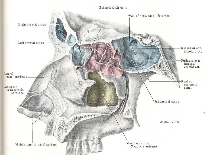
Paranasal Sinuses Are air-filled extensions of the respiratory part of the nasal cavity into the following cranial bones: frontal, ethmoid, sphenoid, and maxilla. They are named according to the bones in which they are located. The sinuses continue to invade the surrounding bone, and marked extensions are common in the crania of older individuals.
Frontal sinus Situated behind the superciliary arches and the root of the nose , are rarely symmetrical, and the septum between them frequently deviates to one or other side of the middle line. Their average measurements are as follows: height, 3 cm. ; breadth, 2. 5 cm. ; depth from before backward, 2. 5 cm. Each sinus drains through a frontonasal duct into the ethmoidal infundibulum, which opens into the semilunar hiatus of the middle nasal meatus. Absent at birth, they are generally fairly well developed between the seventh and eighth years, but only reach their full size after puberty The frontal sinuses are innervated by branches of the supraorbital nerves (CN V 1).
Ethmoid sinus Air-space enclosed within the ethmoid bone Anatomically, ethmoidal sinuses can be classified as the anterior, middle, and posterior ethmoid sinuses and each contain air cells Ethmoidal Air Cells (cellulae ethmoidales) Consist of numerous thin-walled cavities situated in the ethmoidal labyrinth They lie between the upper parts of the nasal cavities and the orbits, and are separated from these cavities by thin bony laminæ. The anterior ethmoidal sinus( up to 11 air cells) drain directly or indirectly into the middle nasal meatus through the ethmoidal infundibulum The middle ethmoidal sinus (about 3 air cells) open directly into the middle meatus and are sometimes called bullar cells because they form the ethmoidal bulla, a swelling on the superior border of the semilunar hiatus The posterior sinus (1 -7 air cells) open directly into the superior meatus; sometimes one or more opens into the sphenoidal sinus The ethmoidal cells are supplied by the anterior and posterior ethmoidal branches of the nasociliary nerves (CN V 1) The ethmoidal cells usually are not visible in plain radiographs before 2 years of age but are recognizable in CT scans
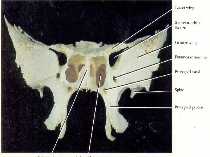
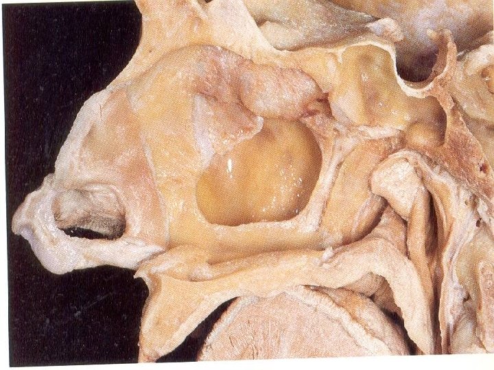
Sphenoidal Sinuses Contained within the body of the sphenoid vary in size and shape; owing to the lateral displacement of the intervening septum they are rarely symmetrical. Average dimensions: vertical height, 2. 2 cm. ; transverse breadth, 2 cm. ; anteroposterior depth, 2. 2 cm. When exceptionally large they may extend into the roots of the pterygoid processes or great wings, and may invade the basilar part of the occipital bone Drains anteriorly to the sphenoethmoidal recess. Minute cavities at birth, but their main development takes place after puberty Derived from a posterior ethmoidal cell that begins to invade the sphenoid at approximately 2 years of age. In some people, several posterior ethmoidal cells invade the sphenoid, giving rise to multiple sphenoidal sinuses that open separately into the sphenoethmoidal recess. Only thin plates of bone separate the sinuses from several important structures: the optic nerves and optic chiasm, the pituitary gland, the internal carotid arteries, and the cavernous sinuses. The posterior ethmoidal arteries and posterior ethmoidal nerve supply the sphenoidal sinuses
MAXILLARY AIR SINUS The maxillary sinuses are the largest of the paranasal sinuses. They occupy the bodies of the maxillae and communicate with the middle nasal meatus Is pyramidal in shape. The apex extends toward and often into the zygomatic bone. The base forms the inferior part of the lateral wall of the nasal cavity. The roof is formed by the floor of the orbit. The floor is formed by the alveolar part of the maxilla. The roots of the maxillary teeth, particularly the first two molars, often produce conical elevations in the floor of the sinus. It is intimately related to the upper teeth, nasolacrimal duct, and the floor of the orbital cavity.
MAXILLARY AIR SINUS It is lined with respiratory epithelium, contains air, and is linked to the middle meatus by the hiatus semilunaris. Drains by one or more openings, the maxillary ostium (ostia), into the middle nasal meatus of the nasal cavity by way of the semilunar hiatus. The arterial supply is mainly from superior alveolar branches of the maxillary artery ; however, branches of the descending and greater palatine arteries supply the floor of the sinus. Innervation is from the anterior, middle, and posterior superior alveolar nerves, which are branches of the maxillary nerve
Facts about maxillary sinus Its nasal wall, or base, presents, in the disarticulated bone, a large, irregular aperture, communicating with the nasal cavity. In the articulated skull this aperture is much reduced in size by the following bones: the uncinate process of the ethmoid above the ethmoidal process of the inferior nasal concha below the vertical part of the palatine behind a small part of the lacrimal above and in front �the sinus communicates with the middle meatus of the nose, generally by two small apertures left between the above-mentioned bones. In the fresh state, usually one small opening exists, near the upper part of the cavity; the other is closed by mucous membrane. On the posterior wall are the alveolar canals, transmitting the posterior superior alveolar vessels and nerves to the molar teeth.
Facts about maxillary sinus The floor is formed by the alveolar process of the maxilla, and, if the sinus be of an average size, is on a level with the floor of the nose; if the sinus be large it reaches below this level. Projecting into the floor of the antrum are several conical processes, corresponding to the roots of the first and second molar teeth; in some cases the floor is perforated by the fangs of the teeth. The infraorbital canal usually projects into the cavity as a wellmarked ridge extending from the roof to the anterior wall; additional ridges are sometimes seen in the posterior wall of the cavity, and are caused by the alveolar canals. The size of the cavity varies in different skulls, and even on the two sides of the same skull.