UNITVII PULSE PRESENTED BY Mrs PRASHMA LEARNING OBJECTIVES

UNIT-VII PULSE PRESENTED BY: Mrs. PRASHMA

LEARNING OBJECTIVES • • Define pulse Explain the physiology and regulation of pulse Explain the characterestics of pulse List out the factors affecting pulse Explain the sites of pulse Explain the assessment of pulse Discuss the alterations in pulse

INTRODUCTION • A pulse represents the tactile arterial palpation of the heartbeat by trained fingertips. The pulse may be palpated in any place that allows an artery to be compressed near the surface of the body. • It is the palpable bounding of blood flow noted at various points on the body. • It is an indicator of circulatory status. As the heart pushes blood through the arteries, the arteries expand contract with the flow of the blood

PULSE DEFINITION : • • A Pulse is a wave of blood created by contraction of the left ventricle of the heart. OR A pulse is a series of pressure waves in an artery, caused by contraction of the left ventricle of the heart. The pulse rate is a measurement of the heart rate, or the number of times the heart beats per minute. •

PHYSIOLOGY AND REGULATION: • • • Electrical impulse originating from the sinoatrial node (SA node) travel through heart muscle to stimulate cardiac contraction. Approximately 60 -70 ml of blood enters the aorta with each ventricular contraction With each stroke volume ejection , the walls of the aorta distend , creating a pulse wave that travels rapidly toward the distal ends of the arteries

PHYSIOLOGY AND REGULATION: • when the pulse wave reaches a peripheral artery , it can be felt by palpating the artery lightly against underlying bone or muscle. • The number of pulsing sensations occurring in 1 minute is the pulse rate • The volume of blood pumped by the heart during 1 minute is called cardiac output , the product of heart rate and the ventricle’s stroke volume

PHYSIOLOGY AND REGULATION: • In an adult the heart normally pumps 5000 ml of blood per minute. • Mechanical, neural and chemical factors regulate the strength of heart contractions and its stroke volume • The cause of an abnormally slow, rapid or irregular pulse may alter cardiac output • The nurse assess the heart’s ability to meet the demands of the body’s tissue for nutrients by palpating a peripheral pulse or by using a stethoscope to listen to heart sounds.

• • A peripheral pulse is a pulse located away from the heart The apical pulse , in contrast, is a central pulse, that is located at the apex of the heart. Characteristics of pulse: • • Rate Rhythm Strength, and Equality

RATE : • The number of pulsing sensations occurring in 1 minute is the pulse rate. • In an adult Normal rate-60 -100 pulsations/minute • Before measuring a pulse , the nurse reviews the client’s baseline rate for comparison • Postural changes cause changes in pulse rate because of alterations in blood volume and sympathetic activity. • When assessing the pulse, the nurse must consider the variety of factors influencing the pulse rate • If the nurse detects any abnormal rate while palpating peripheral pulse next step is to assess the apical pulse.
")
• Identify the first and second heart sounds. Two common abnormalities are 1) Tachycardia 2) Bradycardia Rhythm : - • Normally cardiac contractions occur at evenly spaced intervals, resulting in a regular rhythm • Heart diseases, medications or electrolyte imbalances may alter the heart’s normal rhythmic beating, causing an irregular pulse. This may be , *regularly irregular *irregularly irregular • To document dysrhythmias, a physician may order *electrocardiogram(ECG), holter monitor, telemetry.

Strength: - • The strength or amplitude of a pulse reflects the volume of blood ejected against the arterial wall with each heart contraction and the condition of the arterial vascular system leading to the pulse site. • Normally the pulse strength remains the same with each heart beat • Pulse strength may be graded or described as strong, weak, thready or bounding Equality: - • Pulses on both sides of the peripheral vascular system should be assessed • All symmetrical pulses can be assessed simultaneously except carotid artery

Factors affecting the pulse: - • Age : - as age increases the pulse rate decreases specific variations in pulse rates from birth to adulthood Age heart rate infant --------120 -160 toddler-------90 -140 preschooler-------80 -110 school-age----75 -100 adolscent-----60 -90 adult--------60 -100 • Gender • Exercise • Fever

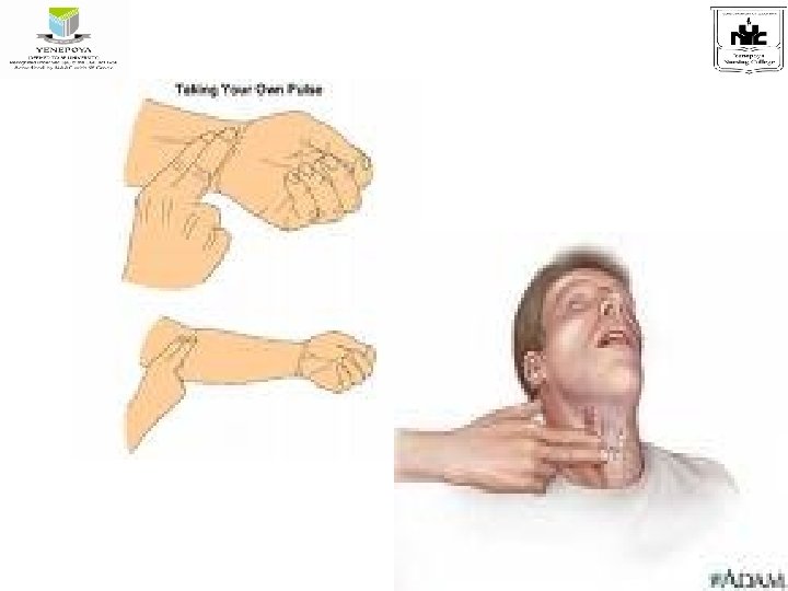
• • • Medications Hypovolemia Stress Position changes Pathology PULSE SITES: 1. where the temporal artery passes over the temporal bone of the head. The site is superior and lateral to the eye 2. A t the neck where the carotid artery runs between the trachea and the sternocleidomastoid muscle

3. Apical: -at the apex of the heart 4. Brachial: - at the inner aspect of the biceps muscle of the arm or medially in the antecubital space 5. Radial : - where the radial artery runs along the radial bone , on the thumb side of the inner aspect of the wrist 6. Femoral: - where the femoral artery passes alongside the inguinal ligament 8) Popliteal: - where the Popliteal artery passes behind the knee
 Posterior tibial: - on the medial surface of the ankle where the posterior")
8) Posterior tibial: - on the medial surface of the ankle where the posterior tibial artery passes behind the medial malleolus. 9) Pedal( dorsalis pedis): - where the dorsalis pedis artery passes over the bones of the foot , on the imaginary line drawn from the middle of the ankle to the space between the big and second toes.

• PULSE SITES

Reasons for use of each sites Pulse sites -Radial -Temporal -carotid -apical -brachial -femoral Rationale -Readily accessible -Used when radial pulse is not accessible -used in cases of cardiac arrest, used to determine the circulation to the brain -routinely used for infants and children up to 3 yrs -used to measure blood pressure, used during cardiac arrest for infants -used in cases of cardiac arrest, used for infants and children, used to determine circulation to leg -

Pulse sites Rationale popliteal - determine circulation to lower leg posterior tibial Pedal - determine circulation to foot

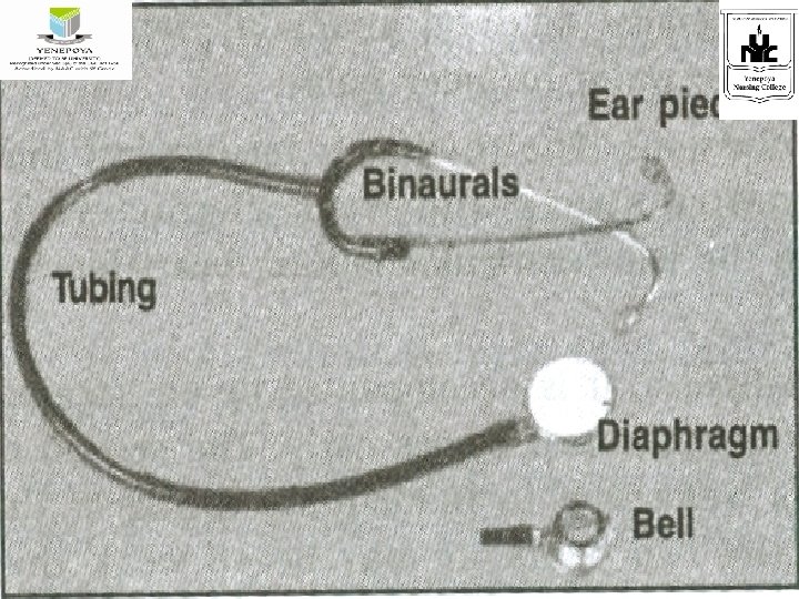
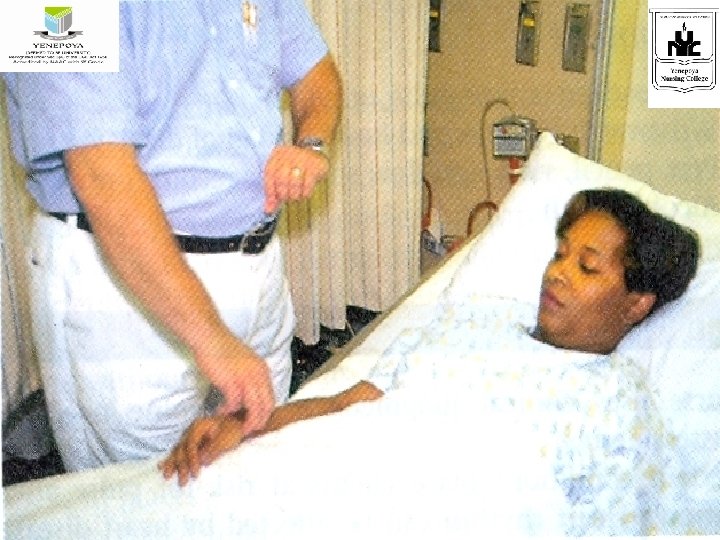
Assessment of the pulse: - • Commonly assessed by palpation or auscultation • The middle three fingers are used for palpating all the sites except the apex of the heart • A stethoscope is used for assessing apical pulse and fetal heart tones • A pulse is palpated by applying moderate pressure with the three middle fingers of the hand

Assessment of the pulse: • When checking the pulse the nurse should aware of: *any medication that could affect the heart rate *whether the client has been active *baseline data • When assessing the pulse , should collect rate, rhythm , volume and equality

DOPPLER: • Peripheral pulse that cannot be detected by palpation may be assessed with an ultrasonic Doppler device Assessment of peripheral pulse Purposes: • To establish baseline data for subsequent evaluation • To identify whether the pulse rate is with in the normal range • To determine whether the pulse rhythm is regular and the pulse volume is appropriate Assessment: • Clinical signs of cardiovascular alterations

Procedure: • • • Explain to the client Perform hand hygiene Provide privacy Select the pulse point, normally the radial pulse is taken Assist the client to a comfortable position, ( with forearm across chest or at side with wrist extended in lying down position and arm on the thigh when sits) Palpate and count the pulse, place two or three middle finger tips lightly over the pulse point. Count for 15 minutes and multiply by 4 or check for 1 minute when the pulse is irregular or checking for the first time Assess the pulse rhythm and volume Document the pulse rate, rhythm and volume on the client’s record

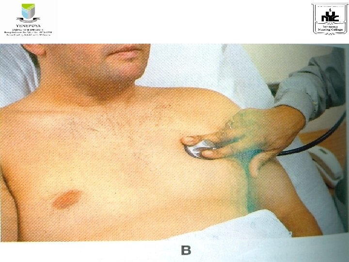
Assessment of apical pulse: Purposes; • To obtain the heart rate of newborns, infants and children and of an adult with irregular pulse rate • To identify whether the pulse rate is with in the normal range Procedure: • Explain to the client • Perform hand hygiene • Provide privacy • Position the client appropriately in a comfortable supine position or in a sitting position • Expose the area of the chest over the apex of the heart

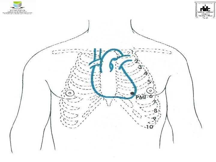
Assessment of apical pulse: - • Locate the apical impulse. This is the point over the apex of the heart where the apical pulse can be most clearly heard. It is also referred to as the point of maximal impulse (PMI) * palpate the angle of Louis just below suprasternal notch between the sternal body and manubrium, it is felt as prominence * slide your index finger just to the left of the client’s left of the sternum , and palpate the second intercostal space * place your middle or next finger in the third intercostal space and move fingers down left side of sternum to fifth intercostal space and laterally to the left midclavicular line. * Auscultate and count heart beats
 * warm the diaphragm of the stethoscope")
ASSESSMENT OF APICAL PULSE (CONT. . ) * warm the diaphragm of the stethoscope by holding it in the palm of the hand for a moment to avoid being from startled * place the diaphragm over point of maximal impulse (PMI) and auscultate heart sounds • Assess the rhythm and strength of the heart beat • If rhythm is clear check for 30 minutes and multiply by 2 if not check for 1 minute • Assist the client in returning to comfortable position • Wash hands • Compare readings with the previous baseline • Record the rate, rhythm and volume

. ALTERATIONS IN PULSE; Rate -tachycardia-rate over 100 beats per minute -bradycardia-rate below 60 beats per minute Rhythm: -Arrhythmias: -indicates any variation from normal rhythm -Intermittent pulse: -the beats are missed in regular intervals -Extra systoles: -when the cardiac contractions occur prematurely, i. e, before they are normally due in the cardiac cycle, it is called extra systolic pressure -Atrial fibrillation: -rapid contractions of the atrium causing irregular contractions of the ventricles in both rhythm and force -Ventricular fibrillation; -rapid twitching of the ventricles
; • Sinus arrhythmia: -pulse rate is rapid during inspiration and slow")
ALTERATIONS IN PULSE(cont…); • Sinus arrhythmia: -pulse rate is rapid during inspiration and slow during expiration • Dicrotic pulse: -there is one heart beat and two arterial pulsations giving the sensation of a double beat • Volume: It refers to the fullness of the artery. It is the force of the blood felt at each beat. Volume depends upon the amount of blood in the arteries
; Water hammer pulse or Corrigan's pulse or collapsing pulse: -it is")
ALTERATIONS IN PULSE(cont…); Water hammer pulse or Corrigan's pulse or collapsing pulse: -it is a full volume pulse but rapidly collapsing pulse occurring in aortic regurgitation or incompetence, where the blood having been forced into the artery by the ventricular contraction, regurgitates back into the ventricle, owing to the nonclosure of the aortic valve Bounding pulse: -signifies in increased stroke volume as seen in exercises, anxiety, anaemia, hepatic failure etc. .
; • Pulsus alternans : -the rhythm is regular but the volume")
ALTERATIONS IN PULSE(cont…); • Pulsus alternans : -the rhythm is regular but the volume has an alternative strong and weak character. This may be noticed in the left ventricular failure, heart block and digitalis toxicity • Bigeminal pulse: -it is accompanied by an irregular rhythm in which every other beat comes early. The second or premature beat feels weak due to inadequate filling of the ventricles between the two beats. It may be so weak that it fails to produce a palpable a peripheral pulse. It is seen in myocardial infarction and digitalis toxicity
; • Weak/wiry/thready pulse: - a small weak pulse that feels like")
ALTERATIONS IN PULSE(cont…); • Weak/wiry/thready pulse: - a small weak pulse that feels like a wire or thread on the palpation of arteries. . It signifies a decreased stroke volume and is seen in hemorrhagic shock or loss of fluid from the body • Paradoxical pulse: -in this case the force or strength of the pulse wave varies , feeling weaker when the client takes in a breath. During inspiration, less blood is returned to the left side of the heart, reduces the stroke volume and therefore decreases the strength of the pulse. This may occur normally , but if pronounced this may indicate cardiac damage

SUMMARY • Today we discussed about meaning of pulse, characteristics of pulse, factors affecting pulse, sites of pulse , assessment of the pulse and alterations in pulse.

CONCLUSION Pulse is the number of times heart beats per minute. Normal heart rate varies from person to person. As an age increases, changes in the rate and regularity of the pulse can change and may signify a heart condition or other condition that needs to be addressed.

EVALUATION MCQ’s 1. Abnormal increase in heart rate is termed as • Tachypnea • Bradycardia • Tachycardia 2. An heart rate of over 100 beats per minute is referred as • A. Tachpnea • B. Tachy cardia • C. Bradypnea • D. Brady cardia

THANK YOU
- Slides: 39