GROWTH OF MAXILLA The study of head form


 Growth at sutures: . - Sutural connective tissue, - Proliferation - Ossification -")
Surface Remodeling: Remodeling occurs by bone deosition & resorption to bring about: a) Increase")


- Slides: 31
GROWTH OF MAXILLA
• The study of head form in man has always been of considerable interest to anthropologist, anatomists, & other students of human growth. In fact, the wide array of students involved in solving the complex phenomenon of “GROWTH “have been aptly described by Krogman as early as 1943 in these golden words: “ Growth was conceived by an anatomist , born to a biologist , delivered by a phisician, left on a chemist’s doorstep & adopted by a physiologist. At an early age she eloped with a statistician, divorced him for a psychologist & is now being wooed , alternately & concurrently by an endocrinologist, a pediatrician , a physical anthropologist, an educationalist , a biochemist , a physicist , a mathematician , an orthodontist , a eugenicist & the children’s bureau”.
According to “ TODD” “Growth is an increase in size & , Development is progress towards maturity
Some definitions related to Growth As is the nature of growth where in the concepts keep changing with new research findings there has been no single definitions associated with it: Different researchers have defined growth in various ways. - The self multiplication of living substance – JX Huxely. - Increase in size, change in proportion & progressive complexity. - Krogman -Entire series of sequential anatomic & physiological changes taking place from the beginning of prenatal life to senility –Meredith. -Quantitative aspect of biologic development per unit of time -Mayers -Change in any morphological parameter, which is measurable. Moss.
i Terminology Related To Growth: Growth Fields : The outside & inside surfaces of a bone are blanketed by a mosaic-like, pattern of soft tissues, cartilage or osteogenic membrane called as Growth Fields. There when altered are capable of producing an alteration in the growth of the particular bone. Growth Sites : Growth sites are growth fields that have a special significance in the growth of a particular bone. Eg. Mandibular condyle in the mandible, Maxillary tuberosity in the maxilla. The growth sites may process some intrinsic potential to growth.
Remodeling : It is the differential growth activity involving simultaneous deposition & resorption on all the inner & outer surfaces of the bone. Eg. Ramus moves posteriorly by a combination of resorption & deposition. Growth Centers: Growth centers are special growth sites , which control the overall growth of the bone. Eg. Epiphyseal plates of long bone.
Mechanism Of Bone Growth • Bone is a specialized tissue of mesodermal origin. It forms the structural framework of the body. • Bone is calcified tissue that supports the body & gives points of attachment to the musculature. • Normal bone contains between 32 -36% of organic matter. -Bone deposition & deposition -Cortical drift -Displacement
Bone deposition & resorption: Bone changes in shape & size by two basic mechanisms, bone deposition & bone resorption. The bone deposition & resorption together is called “ BONE REMODELING”. The changes that bone deposition & resorption can produce are: A) Change in size B) Change in shape C) Change in proportion D) Change in relationship of the bone with adjacent structures.
Cortical Drift: - Most bones grow by interplay of bone deposition & resorption. - A combination of bone deposition & resorption resulting in a growth movement towards the deposition surface is called “Cortical Drift”. - If bone deposition & resorption on either side of a bone are equal the thickness of the bone remains constant. - If in case more bone is deposited on one side & less bone resorbed on the opposite side – The thickness of the bone increases.
Displacement: • It is the movement of the whole bone as a unit. • Displacement can be of two types. Primary displacement: If a bone gets displaced as a result of its own growth, it is called “Primary displacement”. eg. Growth of the maxilla at the tuberosity region results in pushing of the maxilla against the cranial base which results in the displacement of the maxilla in a forward & downward direction. Secondary displacement: If the bone gets displaced as a result of growth & enlargement of an adjacent bone, it is called Secondary displacement. eg. The growth of the cranial base causes the forward & downward displacement of the maxilla.
Characteristics of Bone Growth Bone formation occurs by 2 methods of differentiation of mesenchymal tissues that may be of mesodermal or ectomesenchymal origin. Accordingly 2 types of bone growth is normally seen. 1). Intra-membranous ossification : The transformation of mesenchymal connective tissue usually in membranous sheets, into osseous tissues.
Endochondral ossification: The conversion of hyaline cartilage prototype models into bone. The interstitial growth expansion capability of cartilage, even under weight pressure due to its avascularity precluding ischemia, (Cartilage nutrition is provited by perfusing tissue fluids that are not easily obstructed by load pressures) allows for directed prototype cartilage growth. The cartilage ‘template ‘ is then replaced by endochondral bone accounting for indirect bone growth.
Growth and development of an individual can be divided into PRENATAL & the POSTNATAL periods. The pre-natal period of development is a dynamic phase in the development of a human being. During this period, the height increases by almost 5000 times as compared to only a threefold increase during the post-natal period. The re-natal life can be arbitrarily divided into three periods. 1. Period of the Ovum 2. Period of the Embryo 3. Period of the Fetus
1. Period of the ovum: This period extends for a period of approximately two weeks from the time of fertilization. During this period the cleavage of the ovum and the attachment of the ovum to the intra -uterine wall occurs. 2. Period of the embryo: This period extends from the fourteenth day to the fifty sixth day of intra-uterine life. During this period the major part of the development of the facial & the cranial region occurs. 3. Period of the fetus: This phase extends between the fifty sixth day of intra-uterine life till birth. In this period , accelerated growth of the cranio-facial structures occurs resulting in an increase in their size. In addition, a change in proportion between the various structures also occurs.
Prenatal Growth Of Maxilla Around the fourth week of intra-uterine life, a prominent bulge appears on the ventral aspect of the embryo corresponding to the developing brain. Below the bulge a shallow depression which corresponds to the primitive mouth appears called “ STOMODEUM”. The floor of the stomodeum is formed by the buccopharyngeal membrane which separates the stomodeum from the foregut.
By around the 4 th week of intra-uterine life, five branchial arches form in the region of the future head & neck. Each of these arches gives rise to muscles, connective tissue, vasculature, skeletal components, & neural components of the future face.
The first branchial arch is called the mandibular arch & plays an important role in the development of the naso- maxillary region. The mesoderm covering the developing forebrain proliferates & forms a downward projection that overlaps the upper part of stomodeum. This downward projection is called “FRONTO-NASAL PROCESS”.
The stomodeum is thus overlapped superiorly by the fronto-nasal process. The mandibular arches of both The sides form the lateral walls of the stomodeum. The mandibular arch gives off a bud from its dorsal end called the “MAXILLARY PROCESS”.
The maxillary process grows ventro-medio-cranial to the main part of the mandibular arch which is now called the “MANDIBULAR PROCESS”. Thus at this stage the primitive mouth or stomodeum is overlapped from above by the frontal process, below by the mandibular process & on either side by the maxillary process.
The ectoderm overlying the fronto-nasal process shows bilateral localized thickenings above the stomodeum. These are called the “NASAL PLACODES”. These placodes soon sink and form the nasal pits. The formation of these nasal pits divides the fronto-nasal process into two parts: a)The medial nasal process & b)The lateral nasal process
The two mandibular processes grow medially & fuse to form the lower lip & lower jaw. As the maxillary processe become narrow so that the two nasal pits come closer. The line of fusion of the maxillary process & the medial nasal process corresponds to the naso-lacrimal duct.
POST-NATAL GROWTH Of MAXILLA Since, the maxillary complex is attached to the cranial base, there is a strong influence of the latter on the former. Although there is no sharp line of demarcation between the cranium & maxillary growth gradients, yet the position of the maxilla is dependent upon the growth at spheno-occipital & spheno-occipital synchondroses. Hence, while discussing the growth of nasomaxillary complex, we have to look into two aspects. 1)The displacement in the position of the maxillary complex, -Secondary displacement- Occurs in a downward & forward direction as the cranial base grows.
-Primary displacementoccurs in a forward direction. This occurs by gowth of the maxillary tuberosity in a posterior direction. This results in the whole maxilla being carried anteriorly.
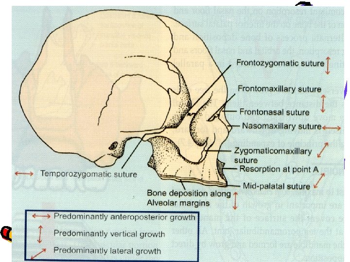
2) Growth at sutures: . - Sutural connective tissue, - Proliferation - Ossification - Surface apposition - Resorption - Translation are the mechanisms for maxillary growth. - Maxilla is related to cranium at least partially by the, - Frontomaxillary suture - Zygomaticotemporal suture - Pterygogopalatine suture These sutures are all oblique & more or les parallel with each other. The growth in these areas would serve to move the maxilla downward & forward.
3)Surface Remodeling: Remodeling occurs by bone deosition & resorption to bring about: a) Increase in size b) Change in shape c) Change functional relationship
Bone remodeling seen in the midfacial region
Bone remodeling of the palate resulting in its downward displacement
Growth of the palate exhibiting V pattern of growth
The naso-maxillary complex as it emerges from beneath the cranium
Moss Cites three types of bone growth changes to be observed in the maxilla 1) Those changes that are associated with compensations for the passive motions of the bone brought about by the primary expansions of the orofacial capsule. 2) There are changes in bone morphology associated with alterations in the absolute volume, size shape or spatial position of any or all the several relatively independent maxillary functional matrices, such as orbital mass. 3) There are bone changes associated with the maintenance of the form of the bone itself. All these changes do not occur simultaneously but rather differentially or squentially.