CERVICAL LESIONS DR LAMA ALMEHAISEN MD MRCOG CONSULTANT


 1. Undetermined Significance (ASC-US) Cytologic changes suggestive of a low")
 / CIN 1 CIN I being the morphologic manifestation")

/ CIN 2 -3 is a cervical cancer precursor")


 are")


 is central to cervical carcinogenesis Worldwide, the")


 ; results may suggest invasive cancer")


 staging system is")


 • Magnetic Resonance Imaging")
![Stage Determines Treatment Early Stage (I-IB 1 [IB 2 -IIa]) Primary Surgery Chemoradiation •](https://slidetodoc.com/presentation_image_h2/6e28e4a228613f8df0ab708d15fd3475/image-42.jpg "Stage Determines Treatment Early Stage (I-IB 1 [IB 2 -IIa]) Primary Surgery Chemoradiation •")
/ SURGERY • Pros 1. Preserves ovarian function 2. Opportunity")


 1")


 Cervical Cancer Radiation therapy Addition of chemotherapy can reduce the risk")

 Cervical Cancer Rarely- recurrent disease can be cured Surgery for")


- Slides: 55
CERVICAL LESIONS DR LAMA AL-MEHAISEN, MD, MRCOG CONSULTANT OBSTETRICIAN AND GYNAEDCOLOGY UROGYNAECOLOGY
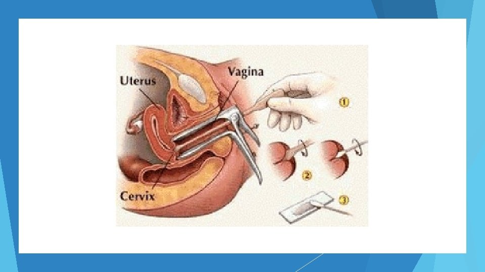
WHAT IS ABNORMAL PAP SMEAR? 1. Abnormal due to inadequacy 2. Abnormal due to inflammation 3. Abnormal due to infection 4. Abnormal due to dysplastic changes
PAP SMEAR UNSATISFACTORY • TX ANY INFECTION • GIVE A COURSE OF ESTROGEN IF POST MENOPAUSE WITH ATROPHY REPEAT 6/12 2 ND SMEAR UNSATISFACTORY REPEAT 6/12 3 RD SMEAR UNSATISFACTORY COLPOSCOPY
Inflammatory smear Inflammation on Pap smear results, does not indicate any particular pathology Therefore, does not necessitate routine treatment.
POSSIBLE CAUSES • Infection • Chronic cervicitis • Atrophic cervicitis • Chemical or mechanical irritation to cervix tampon, douching
Infection COMMON INFECTIONS…. • Tricomonas vaginalis • Fungal ie candidiasis • Bacterial Vaginosis • Actinomyces • Herpes Simplex ORGANISM TREATMENT
Dyspalsia DYSPLASTIC CHANGES SQUAMOUS CELL • ASCUS • ASC-H • LGSIL • HGSIL INVASIVE SQUAMOUS CELL CARCINOMA ABNORMALITY GLANDULAR ABNORMALITY • AGS • AIS • INVASIVE ADENOCARCINOMA
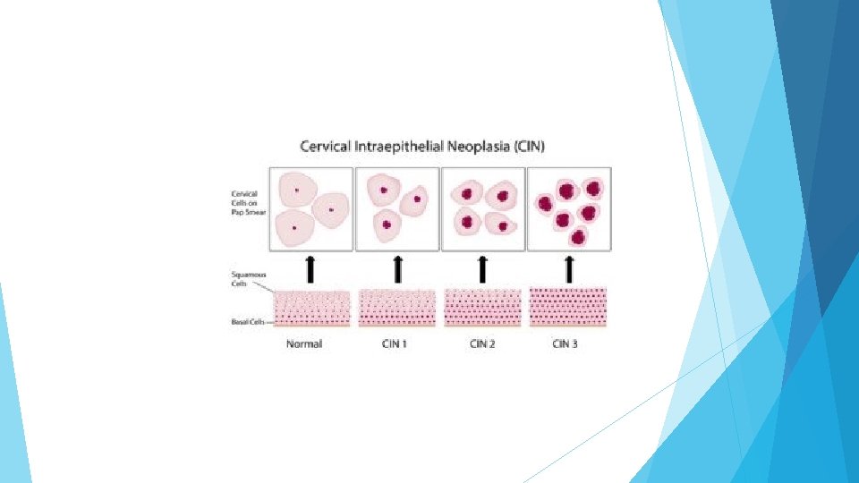
Squamous cell changes ABNORMAL PAP SMEAR DUE TO DYSPLASTIC CHANGES – SQUAMOUS CELL ABNORMALITIES 1 -Atypical Squamous Cells (ASC) Atypical Squamous Cells-Undetermined Significance (ASC-US) Atypical Squamous Cells, Cannot Exclude High Grade Lesion(ASC-H) 2 -Low-grade Squamous Intraepithelial Lesion (LSIL) (Mild Dyskaryosis / HPV/CIN 1) 3 -High-grade Squamous Intraepithelial Lesion (HSIL) (Mod or Severe Dyskaryosis / CIN 2, 3) 4 - Invasive Squamous Cell Carcinoma
ATYPICAL SQUAMOUS CELL (ACS) 1. Undetermined Significance (ASC-US) Cytologic changes suggestive of a low grade squamous lesion but lack criteria for definitive interpretation. 2. Cannot Exclude High Grade Lesion (ASC-H) Cytologic changes suggestive of a high grade squamous lesion but lack criteria for definitive interpretation.
LOW GRADE INTRAEPITHELIAL LESSION (LGSIL) / CIN 1 CIN I being the morphologic manifestation of a self-limited sexually transmitted HPV infection 60% of CIN I regress spontaneously 30% of CIN I persists. 10% of CIN I lesions progress to CIN III 1% may ultimately progress to invasive cancer.
MANAGEMENT APPROACH A lesion that persist after 1 -2 years or any progression during follow up suggest need of treatment If HPV testing is available, +ve HPV: indication for treatment. Treatment- local ablative/ excision Follow up after treatment for CIN 1 repeat smear in 6/12 repeat smear and colposcopy in 12/12 If normal, yearly pap smear x 2 years then back to normal routine
3. HIGH GRADE INTRAEPITHELIAL LESSION (HGSIL)/ CIN 2 -3 is a cervical cancer precursor 1. CIN 2 • 40% of CIN II regress • 30% of CIN II persist • 20% of CIN II progress to CIN III • 5% of CIN II progress to CIN III 2. CIN 3 • 33% of CIN III regress • 18% of CIN III progress to invasive disease over a 10 years • 36% of CIN III progress to invasive disease over a 20 years
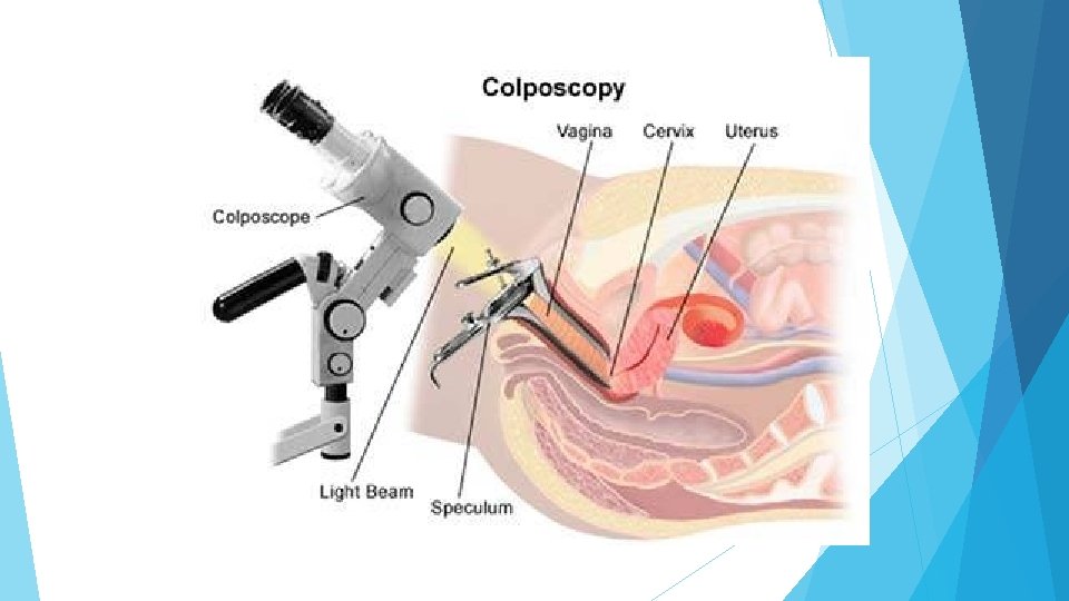
PAP SMEAR HGSIL COLPOSCOPY AND BIOPSY Subsequent management depends on: Whether lesion identified Whether colposcopy satisfactory Annual smear following treatment MANAGEMENT APPROACH EXCISION METHOD LLETZ Cold knife cone biopsy Hysterectomy
ABLATIVE METHODS Cryocautery Electrodiathermy Cold coagulation
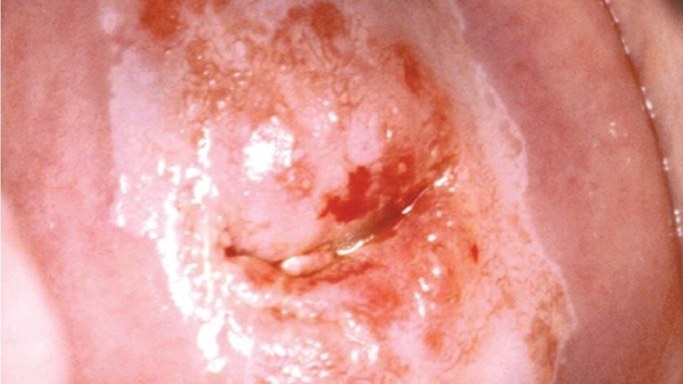
INVASIVE SQUAMOUS CELL CANCER PAP SMEAR COLPOSCOPY AND BIOPSY Subsequent management depends on Stage of the disease
glandular ABNORMAL PAP SMEAR DUE TO DYSPLASTIC CHANGES- GLANDULAR CELL ABNORMALITIES 1. Atypical Glandular Cells (AGS) (undetermined or favour neoplastic) 2. Adenocarcinoma in Situ (AIS) 3. Invasive Adenocarcinoma
GLANDULAR ABNORMALITIES The most common significant lesions associated with AGC (Atypical Glandular Cells) are actually squamous Management should include colposcopy and endocervical sampling ATYPICAL ENDOMETRIAL CELLS Always perform endometrial sampling If endometrial sampling is negative : colposcopy with endocervical sampling
ATROPHY SMEAR LOCAL ESTROGEN CREAM 1 G ON FOR 2 WEEKS THEN TWICE WEEKLY FOR 6 WEEKS ATROPHY SMEAR REPEAT IN 6 MONTHS
REACTIVE CELLULAR CHANGES DUE TO RADIATION REPAIR IUCD REACTIVE CELLULAR CHANGES REPEAT IN 1 YEAR
ABNORMAL PAP SMEAR IN PREGNANCY Reported abnormal smear during pregnancy 1%- 8% Follow-up should be similar to non pregnant state-every trimester regardless of gestation suspicious lesion should be biopsied. Cervical biopsy does not increase the risk of miscarriage If evidence of invasive cancer- require excision
Cervical cancer Epidemiology Worldwide, cervical cancer is the second leading cause of cancer mortality in women Among the ~500, 000 new cases each year, ~75% occur in developing countries Cervical cytologic testing has reduced the incidence of cervical cancer by 70% in countries where it is easily available
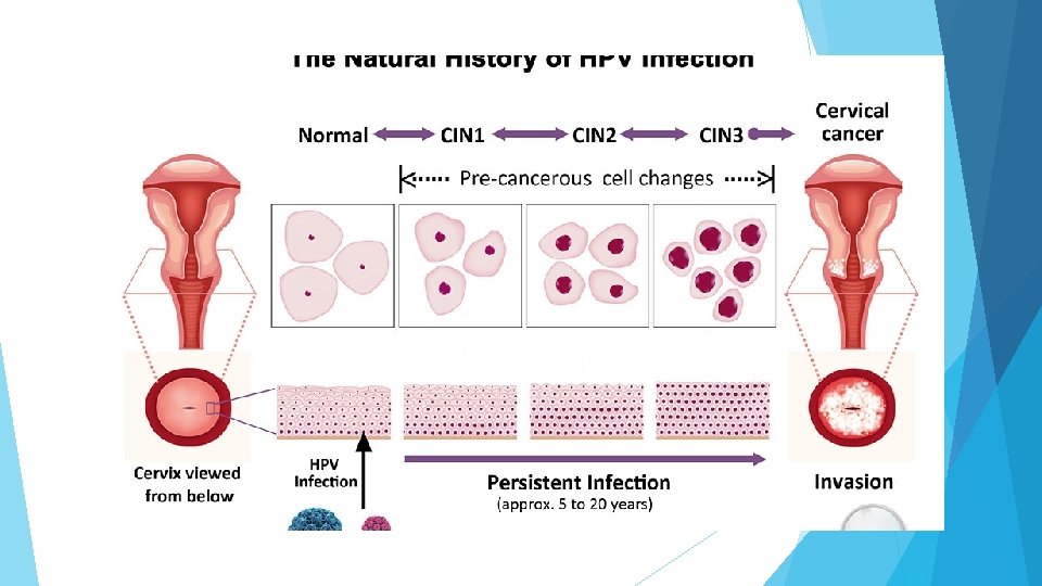
Cervical Cancer and HPV Human papillomavirus (HPV) is central to cervical carcinogenesis Worldwide, the prevalence of HPV in cervical tumors is 99. 7% High-risk HPV types include 16, 18, 31 and 41 High-risk HPV infection is necessary but insufficient for cervical cancer
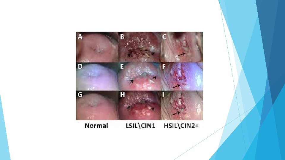
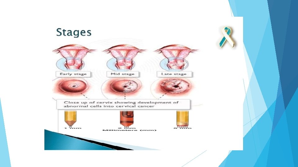
Spectrum of Changes in Cervical Squamous Epithelium Caused by HPV Infection *CIN = cervical intraepithelial neoplasia Normal. Cervix HPV Infection/ CIN* 1 /CIN 2 / CIN 3 / Cervical cancer
Natural histrory % Regress Persist Progress to CIS Progress to Invasion CIN 1 60 30 10 1 CIN 2 40 35 20 5 CIN 3 30 <56 - 18 (5 y), 36(
Additional Risk Factors for CX Cancer Early onset sexual activity Multiple sexual partners High-risk sexual partner History of a sexually transmitted infection Immunosupression Low socio-economic status Previous history of vulvar, vaginal or cervical squamous dysplasia
Diagnosis of cervical cancer Cervical cytology (Pap smear) ; results may suggest invasive cancer Diagnosis must be confirmed by a biopsy When a tumor is clinically evident, a biopsy is usually sufficient
Histologic Subtypes Squamous-Cell Carcinoma Keratinizing or Nonkeratinizing Verrucous Papillary transitional Lymphoepithelioma-like Adenocarcinoma Mucinous Endometrioid Clear Cell Serous Mesonephric Well differentiated villoglandular Minimal deviation (adenoma malignum) Other epithelial – Adenosquamous – Glassy Cell – Carcinoid Tumor – Neuroendocrine – Small-cell – Undifferentiated
Patterns of Spread Local Invasion Lymphatic Risk relates to depth of invasion , Pelvic nodes before paraaortic or supraclavicular • Hematogenous – More likely in adenocarcinoma, neuroendocrine or small cell tumors • Intraperitoneal – Unknown incidence – Poor prognosis
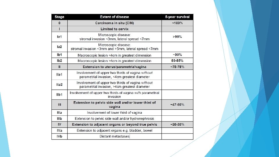
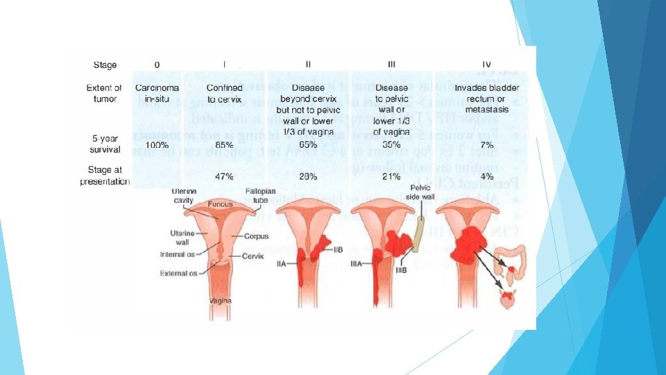
Cervical Cancer Staging The International Federation of Gynecology and Obstetrics (FIGO) staging system is exclusively based on clinical evaluation This allows staging to occur in low resource settings
What are the symptoms of cervical cancer? Early stage disease: • Often no symptoms! • Vaginal discharge • Abnormal bleeding – Post-coital bleeding – Abnormal menses – Post-menopausal bleeding Late stage disease: • Pelvic or lower back pain • Sciatica • Weight loss • Bowel or bladder fistula
How is cervical cancer staged? Physical exam Palpation and inspection of primary tumor Palpation of groin and supraclavicular lymph nodes Rectovaginal exam Exam under anesthesia • Diagnostic exam(s) – Chest X-ray – Intravenous pyelogram (IVP) • Procedures – Conization – Cystoscopy – Proctosigmoidoscopy
Imaging Modalities Prognosis- NOT Staging • Computed Tomography (CT scan) • Magnetic Resonance Imaging (MRI) – May help define extent of disease in cervix and parametria • Positron Emission Tomography (PET) – Superior for detecting metastatic disease compared to CT or MRI • Surgical “Staging” – The “gold standard” for lymph node evaluation
Stage Determines Treatment Early Stage (I-IB 1 [IB 2 -IIa]) Primary Surgery Chemoradiation • Locally Advanced(IB 2 -IVA) – Primary Chemoradiation • Disease with Distant Metastases (IVB) – Systemic chemotherapy
Treatment of Early Stage (I-IIA)/ SURGERY • Pros 1. Preserves ovarian function 2. Opportunity for pathologic information 3. Preferred for women at risk for irradiation complications • Cons 1. Surgical morbidity and mortality 2. Risk of general anesthesia 3. Risk of bleeding/transfusion 4. Possibility of long-term bowel and bladder problems 5. Possible need for post-op XRT
RADIATION THERAPY Pros 1. Avoids risks of surgical morbidity 2. blood loss/transfusion 3. 4. general anesthesia Allows for outpatient treatment • Cons 1. Permanent ovarian failure 2. Vaginal shortening and fibrosis 3. Possibility of long-term bowel and bladder problems 4. Risk of bladder or bowel fistula, or rectal stricture
Surgical options for early stage cervical cancer Stage IA 1 Stage IA 2 -IB 1 Cervical Cancer Candidates for Radical Trachelectomy Criteria for eligibility: Confirmed cervical cancer Stage IA 1 with LVSI, IA 2 -IB 1 Lesion 2 cm Post conization 4 -6 weeks BMI < 35 kg/m 2 Desire for future fertility No evidence of impaired fertility
Radical Trachelectomy Obstetrical outcomes 62% of pregnancies reach 3 rd trimester (65% term) 1 st trimester loss: 16 -20% (Equal to general population) 2 nd trimester loss: 9% (4% in general population) Fertility outcomes: 50 -60% of women attempting will succeed spontaneously
Prognostic Factors Status of lymph nodes Size of primary tumor Depth of stromal invasion +/- lymph-vascular space (LVSI) invasion +/- parametrial extension Histologic cell type Close vaginal margines
Indications for Adjuvant Therapy Women with one or more of the findings below are considered to be at HIGH risk for recurrent disease Positive or close resection margins Positive lymph nodes Parametrial involvement
IIB-IVA (Locally Advanced) Cervical Cancer Radiation therapy Addition of chemotherapy can reduce the risk of death by 30 -50% Recommended Tx: – External beam radiation – Concurrent cisplatin (40 mg/m 2 per week) – Intracavitary brachytherapy
Post-treatment Follow Up Clinical Evaluation – Review of symptoms, physical exam with attention to supraclavicular and inguinal lymph nodes, rectovaginal exam, abdominal exam, +/- cytology) – every 3 months for one year – every 4 months for one year – -every 6 months for 3 years – annually • Annual Chest X-Ray • Other studies only as clinically indicated
Recurrent or Metastatic (IVB) Cervical Cancer Rarely- recurrent disease can be cured Surgery for those previously irradiated Radiation for those with previous surgery Triad of symptoms: 1) 2) 3) ureteral obstruction(hydronephrosis) 2)sciatica 3)lower extremity edema suggest sidewall involvement and surgery is NOT advised When curative surgery or XRT not possible PALLIATION is the goal Chemotherapy
Pelvic Exenteration Anterior Pelvic Exenteration: Pelvic reproductive organs Bladder and distal ureters Entire pelvic floor Preservation of the rectum
Pelvic Exenteration Posterior Pelvic Exenteration: Pelvic reproductive organs Rectum and anus Entire pelvic floor Preservation of the bladder and the ureters
Pelvic Exenteration Reconstructive Phase: Urostomy Colostomy Vaginal reconstruction
Prognosis in Recurrent Disease 80% of recurrences occur within 2 years of primary Depends on site of recurrence and ability to pursue potentially curative therapy Overall, poor prognosis if curative therapy not possible Favorable prognostic factors 1. Localized, central pelvic recurrence 2. Disease-free interval >6 months 3. Size < 3 cm 4. No sidewall fixation