ORAL ULCERATION AND VESICULOBULLOUS LESIONS Ulcer Is a

ORAL ULCERATION AND VESICULOBULLOUS LESIONS

Ulcer : Is a localized defect of the surface in which the covering epithelium is destroyed leaving an inflamed area of exposed connective tissue. Erosion: Is a superficial ulcer.

Traumatic Ulceration 1 -Mechanical: Biting, sharp cusps, outstanding teeth or ill fitting intraoral appliances. 2 -Chemical: Irritant or caustic agents used in dental practice; also some antiseptic mouth washes (not diluted) and misused aspirin tablets 3 -Thermal: Very hot food or drink
 a manifestation of stress, anxiety or sever emotional disturbance.")
4 -Factitious ulcer: (self-inflicted ulcer) a manifestation of stress, anxiety or sever emotional disturbance. 5 -Radiation: immediate effects include erythema, radiation mucositis and ulceration. 6 -Eosinophilic ulcer: associated with trauma and crush injury to muscle

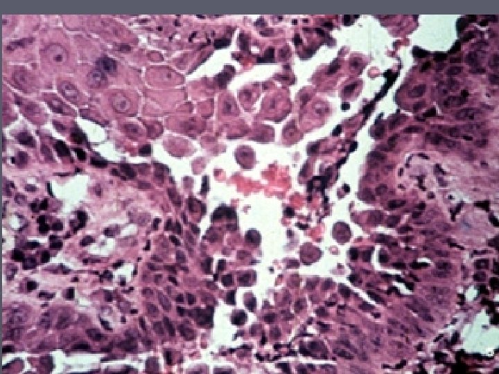
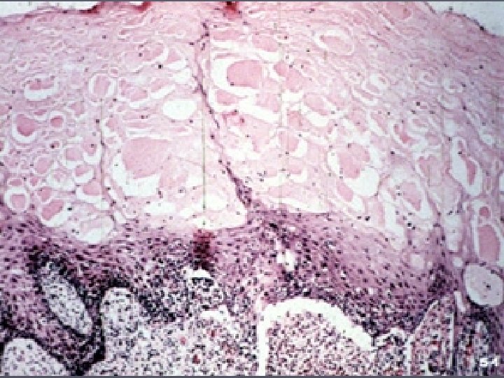
Vesiculobullous lesions Groups of diseases characterized by the appearance of vesicles and bullae in oral cavity and skin. Vesicles are fluid filled blisters, measures less than 5 mm in diameter, bullae are many vesicles cluster together and are more than 5 mm in diameter.

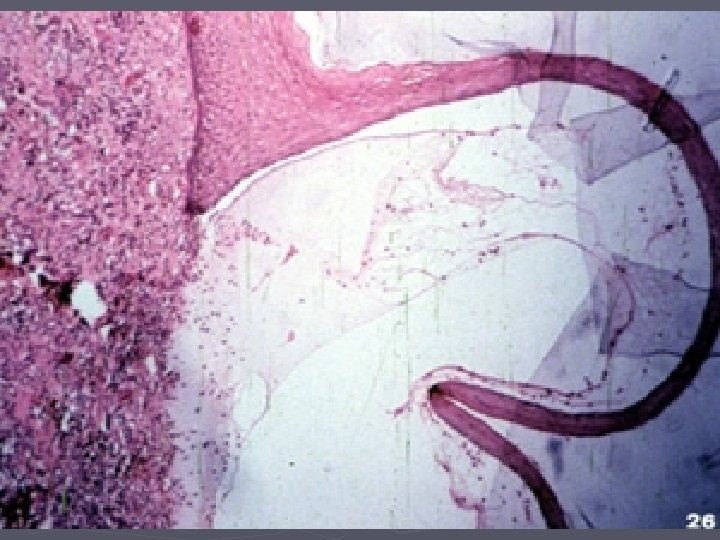
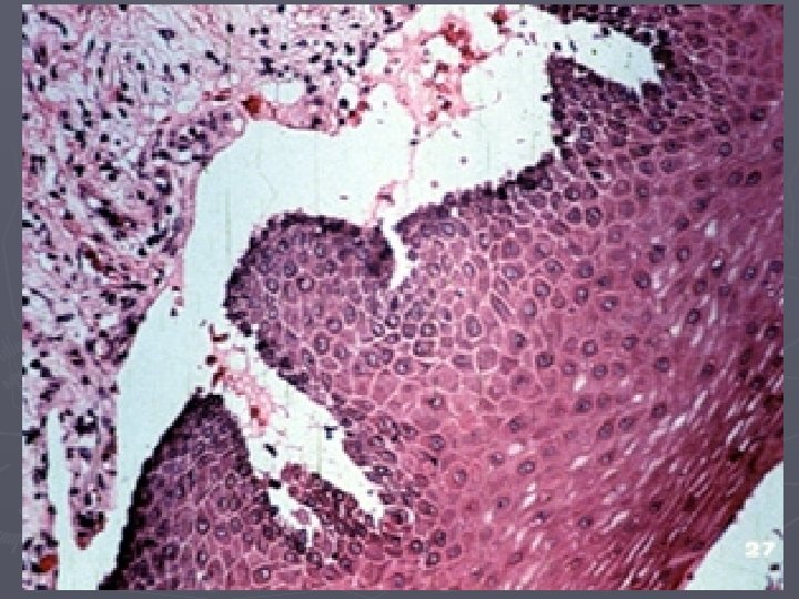
Vesicles are of 2 types: Subepithelial vesicles: Accumulation of fluid beneath stratum basale, so that all layers of epithelium raise. Intraepithelial vesicles: Fluid collection in epithelial layer usually stratum spinosum.

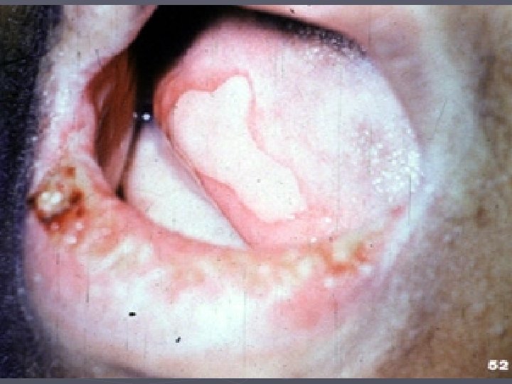
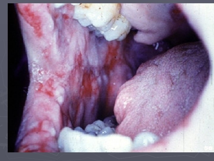
► Pemphigus: Types: pem. vulgaris, pem. foliaceus, pem. Vegetans and pem. erythematosus. ► Cicatricial pemphigoid (mucous membrane pemphigoid) ► Bullous pemphigoid ► Erythema multiforme ► Bullous-like lichen planus ► Epidermolysis bullosa ► Lupus erythematsus ►

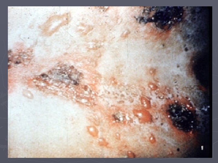
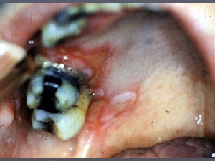
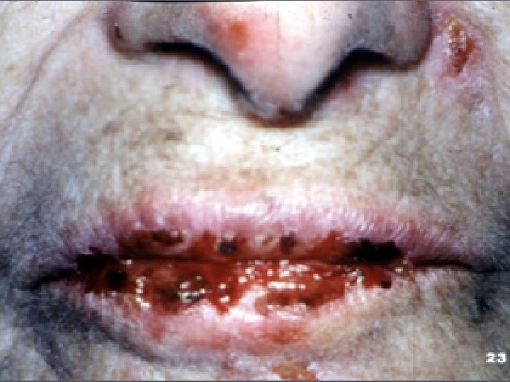
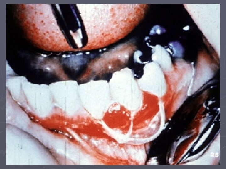
PEMPHIGUS Is a serious autoimmune chronic disease characterized by wide spread bullous eruption involve the skin and mucous membrane. PV is an immune pathologic dermatological disease characterized by bullous formation, oral lesion may be present at same time in the course of disease and may actually precede the development of skin lesion.

Clinical features: In adult and predominantly in women. The average at diagnosis is 50 years, The course of disease varies and may lead to recovery in few a days or run in chronic course if left untreated. The disease may be fatal.

When epithelium of skin or mucous membrane is rubbed by wooden spatula, the epithelium slough off leaving a raw eroded area), this is known as Nikolsky’s sign, this is characteristic of pemphigus, caused by perivesicular oedema which destructs the junction between epithelium and dermis.


 a chronic blistering mucocutaneous autoimmune disease (subepithelial vesicle). Clinical")
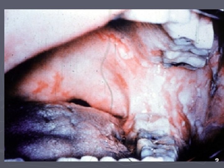
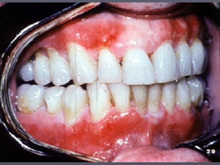
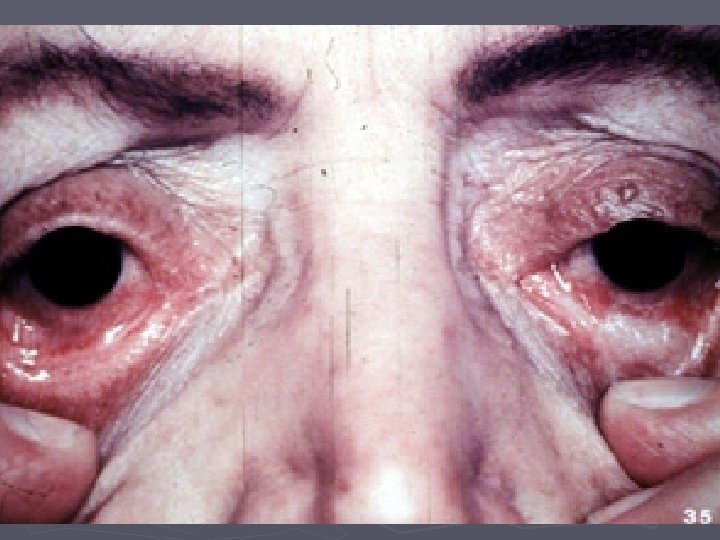
Cicatricial Pemphigoid (mucous membrane pemphigoid) a chronic blistering mucocutaneous autoimmune disease (subepithelial vesicle). Clinical feature: Usually affects adults with an average of 60 years. more frequently in female, in oral mucous membrane, eyes, genitalia and also on skin.

Bullous pemphigoid Clinically: Typically developed in older people between 60 -80 years, no sex predilection is seen, oral mucosal involvement is uncommon, healing takes place without scar formation

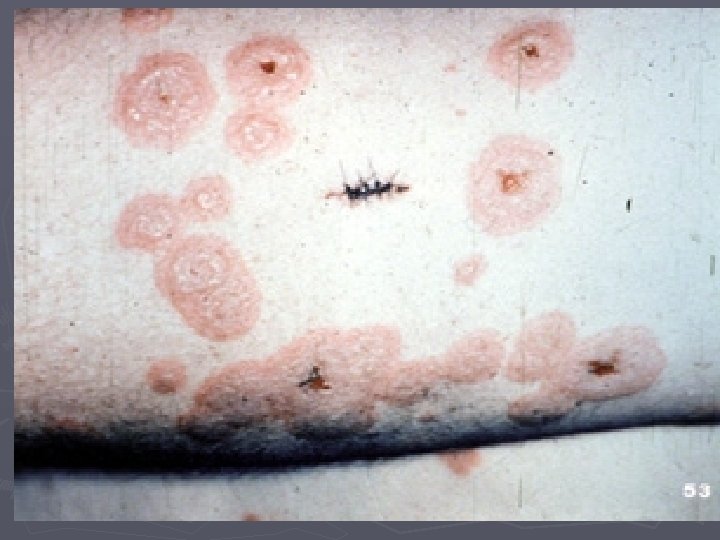
Erythema Multiforme blistering ulcerative mucocutaneous condition of unknown aetiology, it thought to be arisen from: 1 -Viral infection: e. g. herpes simplex 2 - Drug and medication 3 -Vaccine such as small pox vaccine. 4 -Radiation therapy.

5 -Crohn’s disease, ulcerative colitis and infectious mononucleosus. 6 -Stress. 7 -Malignant lesion. 8 -Auto immune disease. 9 -Bacterial, fungal and viral infection. 10 -Idiopathic cause.

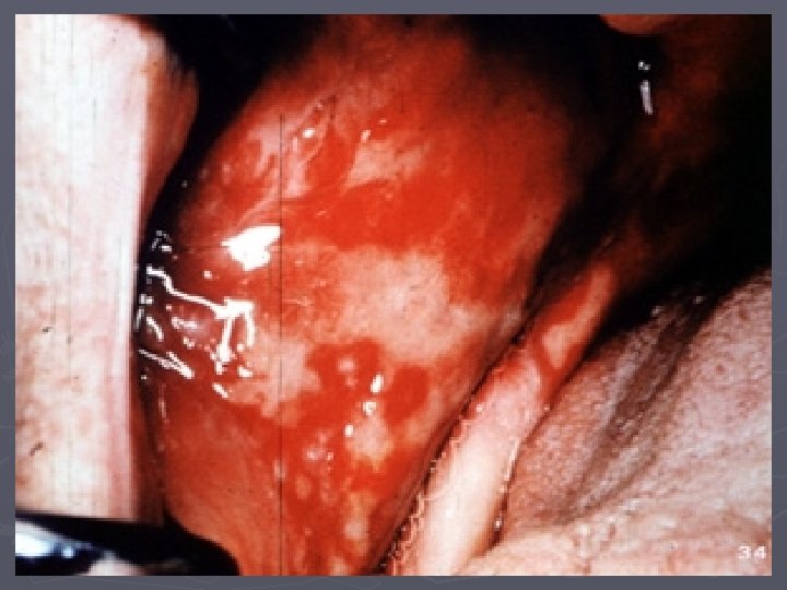
Clinical feature: in young adult and usually has acute onset, affect male more than female, slightly elevated temperature is a common effect. On skin maculopapular rashes in erythematous circular with circumferentic hollow, the so called target-iris lesion. The lips are almost constantly affected with painful erythematous erosion.

Although the disease is self limiting and generally last for 10 -14 days The more sever form known as Stevens. Johnson syndrome or called erythema multiforme major, usually triggered by drug rather than infection; there is involvement of skin, oral cavity, eye and genitalia, with fever, malaise and photophopia.


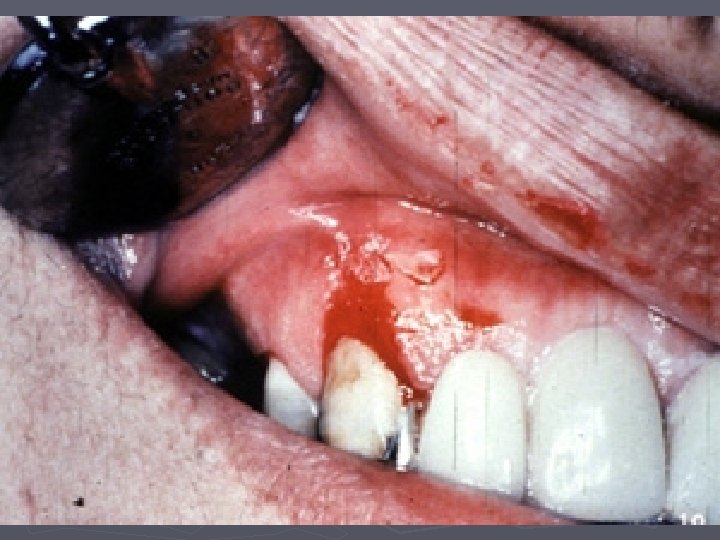
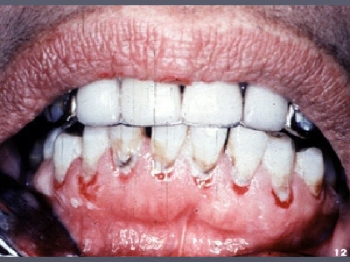
Recurrent aphthous stomatitis It is one of the most common oral mucosal pathologic conditions, characterized by the development of painful recurrent solitary or multiple erosion or ulceration of the oral mucosa.

aphthous ulcer The precipitating factors which precede the out break of aphthous ulcer: 1 -Hereditary predisposition: the familial history found in 45% of patients 2 -Trauma, such as during tooth extraction, dental procedure, self induced bite and tooth brushing. 3 -Hormonal influences

4 -Stress. 5 -Allergic factor, hypersensitivity to certain food or drug. 6 - Iron, B 12 and folic acid deficiency, 7 -Bacteria and viral infection. 8 -Gastrointestinal disturbance.

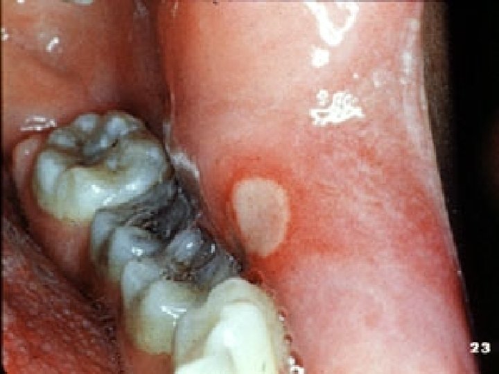
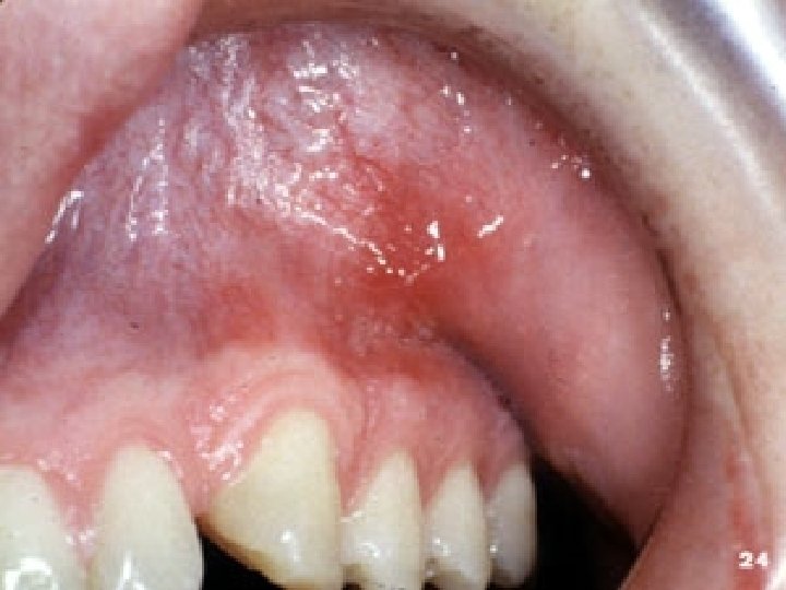
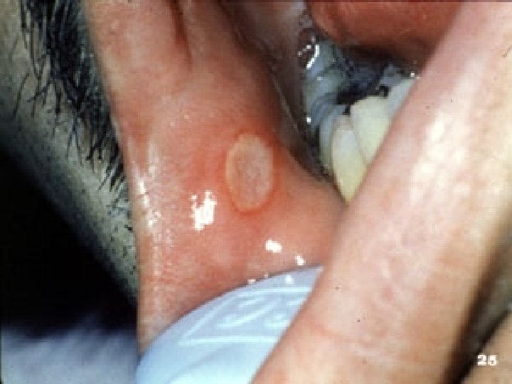
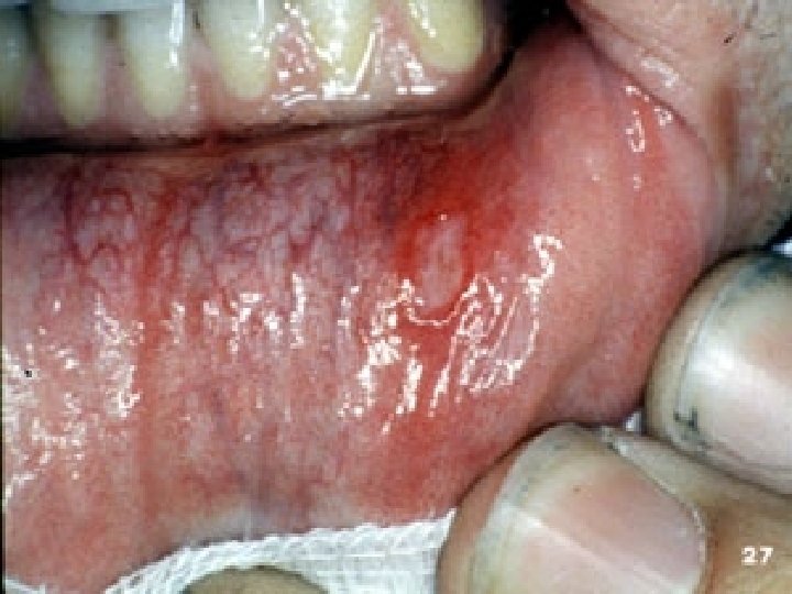
Minor aphthous ulcer Occurs in woman more than man, arises almost on a non-keratinized mucosa, the lesion may be preceded by prodromal symptoms of burning, itching with development of erythematous macule. The macule develops an ulcer that is covered by a yellowish white movable fibrino purulent membrane and it is encircled by an erythematous hollow.

Measures between 3 -10 mm in diameter and heals without scaring in 7 -14 days, the most common sites are the buccal and labial mucosa followed by ventral surface of the tongue, mucobuccal fold, floor of the mouth and soft palate. The recurrence rate is highly variable ranging from one ulceration every few years up to two episodes per month.

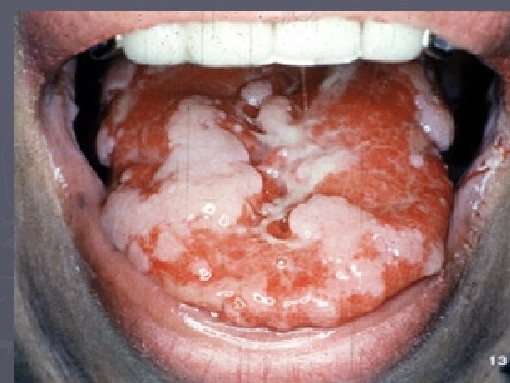
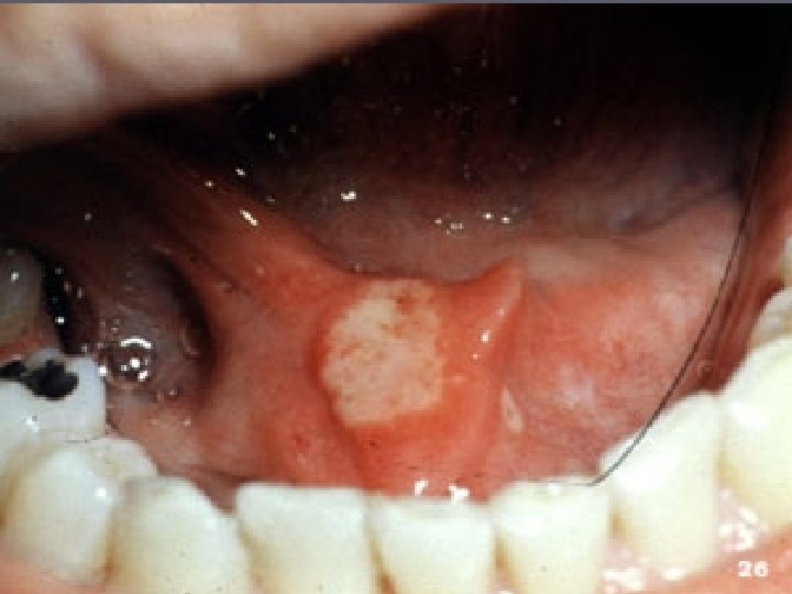
Major aphthous ulcer Usually greater than 10 mm in diameter. may occur at any of the sites of minor aphthae but may also involve the keratinized oral mucosa. It is commonly affect the soft palate, tonsillar areas, and oropharynx. The number of ulcers varies from one to ten and they may take 4 -6 wks to heal, and may heal with scarring.

tend to recur at less than monthly intervals, so that in severe cases ulceration of the oral cavity is virtually continuous and may be associated with severe discomfort and with difficulty in eating and speaking. Extend deeper and may present as crater-like ulcers with rolled margins which are indurate on palpation because of underlying fibrosis.

Herpetiform aphthous ulcer The individual lesions are small 1 -3 mm in diameter and as may as 100 may be present in a single ocurrence, the cause of these small size and large number of lesions bear a superficial resemblances to primary herpes simplex virus infection.

The ulceration heals within 7 -10 days, but the recurrence tends to be closely spaced. Any oral mucosal surface may be involved, but the non-keratinized movable mucosa is mostly affected, there is a female predominance and typically the onset is in adult group.
- Slides: 54