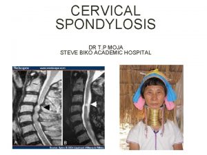
Cervical Spondylosis Cervical disc Cervical Spondylotic myelopathy Anatomy


- Slides: 38


Cervical Spondylosis Cervical disc Cervical Spondylotic myelopathy

Anatomy Of The Cervical Spine Adult cervical spine dimensions have considerable variation. Normal size: ü Antero-posterior (at C 5): 14 mm minimum ü Width: 25 mm (average) ü Spinal cord: AP 8 mm /width 13 mm at C 5 (Remainder of the canal is occupied by dura, CSF , epidural fat and veins. ) Normal cervical disc: ü Formed of nucleus bulposes and anulus fibrosus. ü 88% water in infancy, 65% in elderly. ü Height 45% of vertebral body.

Anatomy Of The Cervical Spine

Cervical Spondylosis (definition) A non specific chronic degenerative condition affecting the cervical spine components (vertebrae, discs, ligaments, joints, blood vessels, nerve roots and spinal cord) which may result in deformity , instability, myelopathy and radiculopathy.

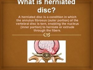
(Cervical disc disease (definition Rupture of fibrocartilagenous material (annulus fibrosis) that surrounds the intervertebral disk. This rupture involves the release of the disk's center portion containing a gelatinous substance called the nucleus pulposus. Pressure from the vertebrae above and below may cause the nucleus pulposus to be forced outward, placing pressure on a spinal nerve and causing considerable pain and damage to the nerve (compression of the cord or root)

Cervical Spondylosis (incidence) Radiographic evidence in 20 -25% of population by the age of 50 y and 70% by the age of 65 y Cervical spondylotic myelopathy is the commonest cause of acquired spstic paraparesis in middle and old age. Starts earlier in men than women C 5/6 &C 6/7 levels are commonest to be involved C 5&C 6 roots are comonly affected.

Cervical Spondylosis (pathology) Vertebrae: § Marginal osteophytes: ü Anterior (may lead to dysphagia if extensive) ü Posterior &lateral (leads to Neural foraminal encroachment and canal stenosis. § Spontaneous fusion: between osteophytes and adjacent vertebrae.

Cervical Spondylosis (pathology)

Cervical disc (pathology) • Discs: üDehydration. üFragmentation. üLoss of disc height. üDisc herniation. bulging, protrusion or extrusion

Cervical disc (pathology)

Cervical disc (pathology)

Cervical Spondylosis (pathology) • Ligaments: üThickening üOssification üBuckling ? üRupture.

Cervical Spondylosis (pathology)

Cervical Spondylosis (pathology)

Cervical Spondylosis (pathology) • Joints: üReduced mobility üHypertrophy üSublaxation. üInflamation.

Cervical Spondylosis (pathology)

Cervical Spondylosis (pathology) • Neural foramina üNarrowing. üRoot compression. üRoot ischemia. üRadicular artery and vein compression

Cervical Spondylosis (pathology) • Spinal cord: üThecal indentation. üCord compression üischemia / edema / infarction üGliosis. üSyrinx.

Clinical presentation • History : üThe course may be slowly progressive , the patient may be asymptomatic or has mild neck pain. üAcute on top of chronic deterioration may occurs , and may be predisposed by trauma.

Clinical (cont. ) • Presentation: ü Muscloskeletal : Ø Pain (neck pain, occipital pain, headache, shoulder pain). Ø Diminished cervical range of motion. Ø Dysphagia. ü Radiculopathy : (in upper limbs) Ø Sensory : ( with dermatoms) Radicular pain (brachialgia) Parathesia. Ø Motor: Weakness Atrophy ü Myelopathy : (in upper and lower limbs) Ø weakness Ø Spasticity and gait disturbance. Ø Impaired fine motor movements. Ø Sphincter dysfunction and impotence.

Clinical (cont. )

Clinical (cont. ) • Examination: ü Local: Ø Decreased range of motion especially with neck extension Ø L’hermitte sign. ü Neurological : Ø Motor: - Spastic quadriparisis or paraparisis (below the level of compression) - Weakness taking myotomal distribution (according to level of compression) Ø Reflexes: - Hyper-reflexia , clonus (below the level). - Pathological reflexes (hoffman sign, pectoralis reflex, adductor reflex, babiniski reflex) - Hypo-reflexia (at the level) Ø Sensory: Hypothesia with dermatomal distribution or with sensory level

• Differential diagnosis

Correlation between radiological and clinical findings is mandatory.

Imaging • PXR

Imaging • CT Scan

Imaging • CT Scan reconstruction

Imaging • MRI

Imaging

Management • Conservative management: ü For relief of symptoms ØBed rest, neck massage, heat application, analgesics, anti-inflamatory medications, muscle relaxants. ØNeck immobilization (neck collar). ØPhysiotherapy ? ü Minor neurological deficit can also be treated. ü Frequent monitoring is required. • Surgical management.

Management Surgical management • Indications: ü ü Intractable pain due to identified cause. Progressive neurologic deficite. Root compression clinicaly correlated to radiological finding. Instability • Aim of surgery ü Relieve pain and neuronal compression. ü Achieve stability. • Choice of procedure ü Depends upon findings on imaging ü Level of disease process. ü Extent of pathological changes.

Management • Surgical procedures ü Posterior approaches: Ø Laminectomy. Ø Posterior foraminotomy. Ø Laminoplasty. Ø With or without fixation with plate and screws. NB. Posterior approaches for posterior pathology. ü Anterior approaches: Ø Anterior cervical discectomy/osteophytectomy and fusion. Ø Corpectomy with fusion and fixation. Ø Anterior foraminotomy. Ø Cervical arthroplasty. (artificial disc and dynamic implants) ü Percutaneous facet injection or radiofrequency.

Management

Management

Management

Take home messages • Correlation • Diffrenttial • Different approaches acc to path

 Myelopathy vs radiculopathy
 Cervical myelopathy wayne
 L
L Uncovertebralis
 Spondylolisthesis
 Spondylosis definition medical
 Spinal cord spasticity
 Surfer's myelopathy
 Degenerative myelopathy wayne
 Thick disc
 Platelets yellow
 Miller disc method
 Tidewater disc golf course
 Examples of optical storage devices
 Aria totala con circular drept
 Elliquence disc fx
 Reticulocyte normal
 Plasma disc decompression
 Extended disc sverige
 Device handler seek strategies
 Hubless rotors are held onto the hub by
 Merkel disc receptor
 Ontario disc sports association
 Neurulation
 Parts of a compound light microscope and their functions
 Disc profil
 Dorsiflexion plantar
 Brown dwarf
 Champagne cork appearance of optic disc
 Cow eye
 Www.123 test.com/disc-personality-test/
 123 disc personality test
 Sound-on-disc
 Linear momentum in circular motion
 Bronze age sun disc
 Choroid in cow eye
 Taking flight with disc
 Chorionic villus
 Broad-based disc bulge
