Wacky Sports Injuries Spine Injuries in Surfers Jaspal


 cervicodorsal")


: 288– 293. PMCID: PMC 2031959 Nontraumatic Myelopathy Associated")


- Slides: 30
Wacky Sports Injuries: Spine Injuries in Surfers Jaspal R. Singh, MD Assistant Professor of Rehabilitation Medicine Director of Interventional Spine
Disclosures Jaspal R. Singh, M. D. • Consultant- Physician’s Pharmaceutical Solutions • Consultant- Kimberly Clark
Cases 1. 2. 3. 4. Surfer’s Myelopathy Nontraumatic Myelopathy Complete Paraplegia Annular Tears
History • First reported in 2004 as a series of nine cases • Pearce; Spine 2004; Vol 29; No: 16
Surfers Myelopathy • Atraumatic injury to the cord • Affects first-time surfers – Hyperextension moment in a “predisposed” individual • MRI shows signal change in the affected portions of the cord
Thompson et al in 2004 • 9 Cases – 9 presented with back pain – 8 with paraparesis – 8 with urinary retention – 3 with sensory disturbances – 1 with paraplegia
2012 Review • • • 19 cases 15 -46 yo Novice surfers All had lower back discomfort 10 -60 minute onset of weakness and paresthesias Within minutes of onset, unable to walk
MRI Findings • • • All had hyperintense T 2 signal from mid- to lower thoracic level to the conus No segmental image Restricted diffusion in 6/10 patients No evidence of aortic injury Proposed mechanism is Artery of Adamkiewicz vasospasm
Clinical Presentation • • Average of 25 New to surfing Initial back pain Relatively rapid progression of neurological symptoms (<24 hours)
Reported Outcomes • At time of discharge: 9 cases – 3 patients had complete recovery – 4 patients had “mild” weakness but no sensory deficits – 3 had urinary retention – 1 patient remained paraplegic
Proposed Mechanisms • • • Hyperextension leading to ischemia? – Watershed zones within the cord Concussive forces of the waves? – Less likely given the nature of the presentation Thrombotic events?
Risk Factors • • Thin body habitus Underdeveloped back musculature Recent long-distance travel Dehydration
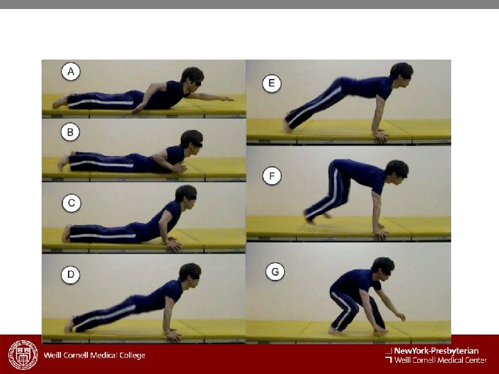
Why Thoracic Spine • • three regions of anterior vascular supply – (1) cervicodorsal region; – (2) intermediate region (midthoracic area), from T 4 to T 7 or T 8 – (3) inferior dorsolumbar region midthoracic area is poorly vascularized the lower thoracic to the lumbar area is mainly supplied by a single Adamkiewicz artery. Among surfers, the technique of standing up on a surfboard is called “popping up. ” – first, pushing up one’s torso by extending both arms from a prone “paddling” position – second, crouching on the surfboard and sliding the legs under the torso – third, standing to a half-rising position, which is called the “riding” position – Insert VIDEO and PICS – not only the continued hyperextended posture of paddling but also repetitive mechanical stress caused by several tries of popping up may contribute to its pathogenesis.
• • • thin and underdeveloped back musculature is a possible risk factor for surfer’s myelopathy poor stability of the spine may result in accidental overextension or overflexion recommend that novice surfers take mandatory rest periods during surfing lessons – (e. g. , 10 mins of rest every 45 mins) – the time of lessons should be limited (e. g. , maximum of 90 mins) – instructors be educated as to the early detection of students’ back pain
Conclusion • • Surfing is a popular sport worldwide The etiology of surfer’s myelopathy remains enigmatic resulting disability can be devastating early detection and early treatment are necessary for the prevention of neurologic deterioration. Awareness among clinicians and surfers is desirable. Immediate imaging (e. g. , MRI with diffusion-weighted images, magnetic resonance angiography, and computed tomographic angiography) is desirable for the further elucidation of its pathogenesis. Aggressive hydration, induced hypertension, and empiric corticosteroids are recommended as acute treatments for spinal cord ischemia. In addition, adequate rehabilitation for the neurologic deficits is indispensable.
Complete Paraplegia • • • Three patients with diagnoses of surfer’s myelopathy (24– 31 yrs old; two men, one woman) novice surfers rapid progression of paraplegia after back pain while taking surfing lessons Despite months of rehabilitation – in all three patients, complete paraplegia (T 9–T 12) and bladder-bowel dysfunction remained. neurologic outcome of surfer’s myelopathy is potentially catastrophic Complete Paraplegia Resulting from Surfer's Myelopathy. Takakura, Tomokazu; Yokoyama, Osamu; Sakuma, Fujiko; Itoh, Ryousuke; Romero, Ray American Journal of Physical Medicine & Rehabilitation. 92(9): 833 -837, September 2013.
FIGURE 2. Case 1. Midsagittal T 2 WI magnetic resonance image of thoracolumbar spinal cord 4 hrs after onset. Increased signal and mild enlargement of lower thoracic cord to the conus medullaris are observed (arrows). © 2013 by Lippincott Williams & Wilkins. Published by Lippincott Williams & Wilkins, Inc. 2
FIGURE 3. Case 1. Axial T 2 WI magnetic resonance image of thoracic spinal cord at T 9 spine level 4 hrs after onset. Massive increased signal of central cord, which involves both gray and white matter, is observed (arrow). © 2013 by Lippincott Williams & Wilkins. Published by Lippincott Williams & Wilkins, Inc. 2
FIGURE 4. Case 1. Midsagittal T 2 WI magnetic resonance image of thoracolumbar spinal cord at day 110. Marked atrophy of spinal cord below T 11 to the conus medullaris (arrows) are observed. © 2013 by Lippincott Williams & Wilkins. Published by Lippincott Williams & Wilkins, Inc. 2
FIGURE 1. Case 1. Midsagittal T 2 WI magnetic resonance image of thoracic spinal cord 4 hrs after onset. Increased signal and mild enlargement of lower thoracic cord below the level of T 8 vertebra are observed (arrows). In addition, dorsal-dephasing artifacts are seen in the midthoracic cord. © 2013 by Lippincott Williams & Wilkins. Published by Lippincott Williams & Wilkins, Inc. 2
J Spinal Cord Med. 2007; 30(3): 288– 293. PMCID: PMC 2031959 Nontraumatic Myelopathy Associated With Surfing Israel Avilés-Hernández, MD, 1, 2 Inigo García-Zozaya, MD, 2 and Jorge M De. Villasante, MD 2 • • Results: A 37 -year-old man developed T 11 American Spinal Injury Association (ASIA) A paraplegia shortly after surfing. The clinical history and magnetic resonance imaging findings were compatible with an ischemic insult to the distal thoracic spinal cord. Our patient did not have any of the proposed risk factors associated with this condition, and, contrary to most reports, he sustained a complete spinal cord lesion without neurological recovery by 8 weeks post injury. Conclusions: Surfer's myelopathy, because of its proposed mechanism of injury, is amenable to medical intervention. Increased awareness of this condition may lead to early recognition and treatment, which should contribute to improved neurological outcomes.
• Sagittal T 1 W and T 2 W images at 15 hours after the beginning of symptoms demonstrated mild fusiform expansion of the distal spinal cord and increased T 2 W signal (arrows).
• Sagittal T 2 W MRI on day 2 demonstrated cephalad progression of increased T 2 W signal extending from the tip of the conus to level T 10 (arrows).
• T 1 W sagittal image without IV contrast medium at 4 weeks demonstrated increased T 1 W signal at the distal spinal cord consistent with hemorrhagic products (arrow).
Annular Tears while Surfing
L 3 -4
L 4 -5
L 5 -1