Cell Theory Wacky History of Cell Theory https


 observed cells and cell division, and")

")


 send electrons through a specimen Shows")

- Slides: 14
Cell Theory
Wacky History of Cell Theory • https: //www. youtube. com/watch? v=4 Op. Bylw H 9 DU (6 min) • Let’s review…
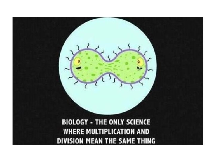
Cell Theory: 3 Main Ideas • All living things are composed of cells • Cells are the basic units of structure and function in living things • All cells are produced from other cells
The Role of Microscopes • Many centuries ago Greek philosophers thought that organisms appeared from non-living or rotting material, and idea called spontaneous generation Example: A Recipe for Bees (Roman, 2000 years old) 1. Kill a bull during the first thaw of winter 2. Build a shed 3. Place the dead bull on branches and herbs inside the shed 4. Wait for summer. The decaying body of the bull will produce bees
• In 1665 Robert Hooke created and used a simple microscope to observe a piece of cork – noticed it was made up of air-filled sacs that he called cells.
• In 1838, with more advanced microscopes, German biologists Schleiden and Schwann recognized that all plant and animal cells have a nucleus and other key organelles. They proposed cells as the basic unit of life.
• Finally Rudolf Virchow (and Robert Remak) observed cells and cell division, and determined that all cells come from other cells. Our understanding of cell theory is dependent on the microscope!
Microscopes • Microscopes are devices that produce magnified images of structures that are too small to see with the “naked eye” • Two main types: – Light microscopes which produce magnified images by focusing visible light rays – Electron microscopes produce magnified images by focusing beams of electrons
Light Microscopes • Most commonly used microscopes (the ones we have in the lab) • Can produce clear images of objects at 1000 times the magnification • Compound Light Microscopes allow light to pass through the specimen and use two lenses to form the image • Can observe cells and tiny organisms while they are still alive
Light Microscopes • Chemical stains and fluorescent dyes can be used to highlight structures and processes inside cells • Video cameras and computer processing can produce moving 3 D images
Electron Microscopes • To study objects smaller than 0. 2 micrometers (one fiftieth the diameter of a cell), an electron microscope must be used. • Can produce images 1000 times more detailed than a light microscope
Two Main Types • Transmission Electron Microscopes (TEM) send electrons through a specimen Shows detail inside the cell • Scanning Electron Microscopes (SEM) send electrons across the surface of a specimen Produces realistic three-dimensional images of objects • Both require a vacuum environment, so samples must be preserved and dehydrated – Therefore cannot be used to observe living cells.
Microscope Parts • Use the Information on pg 1070 to fill out the diagram of the compound microscope