CELL DISCOVERY AND THEORY History of Cell Theory


- Slides: 13

CELL DISCOVERY AND THEORY

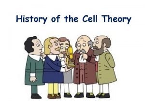
History of Cell Theory • The invention of the microscope lead to the discovery of cells. • In 1665, Robert Hooke made a simple microscope and observed cork, the dead cells of oak bark. ØHe named the tiny chambers that he saw “cells” because he thought they looked like the small rooms in a monastery. cork cells, 100 X

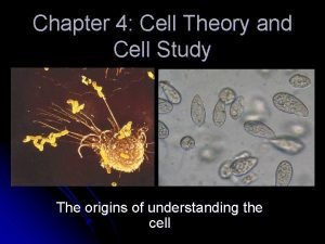
History of Cell Theory • In 1683, Anton van Leeuwenhoek observed living, single-celled, animal-like organisms (now called protozoans) in pond water.

History of Cell Theory • In 1838, Matthias Schleiden demonstrates that all plants are composed of cells. • In 1839, Theodor Schwann concluded that all animal tissues are made of cells. • In 1855, Rudolph Virchow proposed that all cells are produced from the division of existing cells. Matthias Schleiden Theodor Schwann Rudolph Virchow

History of Cell Theory includes the following three principles: 1. All living organisms are composed of one or more cells. 2. Cells are the basic unit of structure, function, and organization of all living organisms. 3. Cells arise only from previously existing cells, with cells passing copies of their genetic material on to their daughter cells.

History of Cell Theory • From 1880 -1890, Louis Pasteur and Robert Koch, using compound microscopes, pioneered the study of bacteria (prokaryotic cells). Louis Pasteur Robert Koch

History of Cell Theory • In 1939, Ernest Everett Just writes the textbook Biology of the Cell Surface after years of studying the structure and function of cells. • 1970: Lynn Margulis proposes the endosymbiont theory, the idea that some organelles in eukaryotes were once free-living prokaryotes. Ernest Everett Just Lynn Margulis

Microscopes Compound Light Microscopes • Uses visible light to produce a magnified image • Total magnification is the power of the ocular (or eyepiece) times the power of the objective • Maximum magnification without blurring is around 1000 X.

Electron Microscopes use electrons rather than light to generate an image Transmission Electron Microscope (TEM) • TEM uses magnets to aim a beam of electrons at thin slices of cells • Can magnify up to 500, 000 X but specimen must be dead, sliced very thin, and stained with heavy metals TEM images (kidney, above; Golgi apparatus in plant, below

Electron Microscopes Scanning Electron Microscope (SEM) • SEM directs electrons over the surface of a specimen, producing a 3 SEM images (clockwise order from top, wasp head; walnut leaf -D image underside; anthers and pollen) • As with the TEM, only nonliving cells and tissues can be observed.

Electron Microscopes Scanning Tunneling Electron Microscope (STM) • Involves bringing the charged tip of a probe extremely close to the specimen so that the electrons can “tunnel” through the small gap between the specimen and the tip • Creates 3 -D images of objects as small as atoms • Can be used with live specimens STM image (cobalt phthalocyanine on a surface) STM equipment

Electron Microscopes Scanning Tunneling Electron Microscope (STM) image of a DNA molecule

Scanning Probe Microscope Atomic Force Microscope (AFM) • Measures various forces between the tip of a probe and the cell surface AFM of Zinc Oxide (Zn. O)
 Chapter 7 section 1 cell discovery and theory
 Chapter 7 section 1 cell discovery and theory
 Section 1 cell discovery and theory
 Section 1 cell discovery and theory
 History of atomic theory
 Chapter 4 cell theory and cell study
 Robert hooke 1665 discovery
 Office cell
 Wacky history of cell theory
 Matthias schleiden
 Robert hooke timeline
 Discovery learning theory
 Conditional matrix grounded theory
 Discovery learning theory
