Imaging at Lafayette Surgical Specialty Hospital SPINE IMAGING


, herniate (v): 1. Localized displacement of disc material")
: The cross sectional area of disc material at")
, bulge (v): 1. A disc in which the")
; < 3 MM")
, protrude (v): 1. A herniated disc in which")


, extrude (v): A herniated disc in which, in")


, sequestrate (v); (var: sequestered disc): An extruded disc")


: Reactive vertebral body modifications associated with")

, degenerate (v): 1. Changes in a disc characterized")
")


,")


- Slides: 52
Imaging at Lafayette Surgical Specialty Hospital
SPINE IMAGING Fundamentals and Standardized Nomenclature Presented by: Dr. Rodney Burns S res
SPINE NOMENCLATURE S SPINE Volume 26, Number 5, pp E 93–E 113 S © 2001, Lippincott Williams & Wilkins, Inc. S Nomenclature and Classification of Lumbar S Recommendations of the Combined Task Forces of the North S American Spine Society, American Society of Spine Radiology, and S American Society of Neuroradiology
Overview • Diagnostic Approach • Terminology and Concepts • Common Spine Pathology
Standard Spine Sequences • T 2 -weighted: Axial and Sagittal Pathology • T 1 -weighted: Sagittal +/- axial Anatomy • STIR or T 2 W fat sat: Sagittal Bone edema
Radiologist Approach to Spine MRI • Alignment • Bones • Spinal Cord • Spinal Canal • Disc spaces • Neural foramen, facet joints • Extraspinal tissue
Terminology • herniated disc, herniation (n), herniate (v): 1. Localized displacement of disc material beyond the normal margins of the intervertebral disc space. 2. (Non-Standard) [Any displacement of disc tissue beyond the disc space]. Note: Localized means, by way of convention, less than 50% (180 degrees) of the circumference of the disc. Disc material may include nucleus, cartilage, fragmented apophyseal bone, or fragmented anular tissue. The normal margins of the intervertebral disc space are defined, craniad and caudad, by the vertebral body end-plates and peripherally by the edges of the vertebral body ring apophyses, exclusive of osteophytic formations. Herniated disc generally refers to displacement of disc tissues through a disruption in the anulus, the exception being intravertebral herniations (Schmorl's nodes) in which the displacement is through vertebral end-plate. Herniated discs in the horizontal (axial) plane may be further subcategorized as protruded or extruded. Herniated disc is sometimes referred to as "herniated nucleus pulposus, " but the term herniated disc is preferred because displaced disc tissues often include cartilage, bone fragments, or anular tissues. The term "ruptured disc" is used synonymously with herniated disc, but is more colloquial and can be easily confused with violent, traumatic rupture of the anulus or end-plate. The term "prolapse" has also been used as a general term for disc displacement, but its use has been inconsistent. The term herniated disc does not infer knowledge of cause, relation to injury or activity, concordance with symptoms, or need for treatment.
Terminology • base (of displaced disc): The cross sectional area of disc material at the outer margin of the disc space of origin, where disc material displaced beyond the disc space is continuous with disc material within the disc space. In the cranio-caudal direction, the length of the base cannot exceed, by definition, the height of the intervertebral space.
Terminology • bulging disc, bulge (n), bulge (v): 1. A disc in which the contour of the outer anulus extends, or appears to extend, in the horizontal (axial) plane beyond the edges of the disc space, over greater than 50% (180 degrees) of the circumference of the disc and usually less than 3 mm beyond the edges of the vertebral body apophyses. 2. (Non-Standard) [A disc in which the outer margin extends over a broad base beyond the edges of the disc space. ] 3. (Non-Standard) [Mild, smooth displacement of disc, whether focal or diffuse. ] 4. (Non-Standard) [Any disc displacement at the discal level. ] >Note: Bulging is an observation of the contour of the outer disc and is not a specific diagnosis. Bulging has been variously ascribed to redundancy of anulus secondary to loss of disc space height, ligamentous laxity, response to loading or angular motion, remodeling in response to adjacent pathology, unrecognized and atypical herniation, and illusion from volume averaging on CT axial images. Bulging may or may not represent pathologic change, physiologic variant, or normalcy. Bulging is not a form of herniation; discs known to be herniated should be diagnosed as herniation or, when appropriate, as specific types of herniation. See: herniated disc, protruded disc, extruded disc.
Generalized Disc Bulging Disc base >50% (>180 degrees); < 3 MM
Terminology • protruded disc, protrusion (n), protrude (v): 1. A herniated disc in which the greatest distance, in any plane, between the edges of the disc material beyond the disc space is less than the distance between the edges of the base in the same plane. 2. (Non-Standard) [A disc in which disc tissue beyond the disc space is contained within intact anulus]. 3. (Non. Standard) [Any, or unspecified type of, disc herniation. ] Note: The test of protrusion is that there must be a localized (less than 50% or 180 degrees of the circumference of the disc) displacement of disc tissue so that the distance between the edges of the displaced portion must not be greater than the distance between the corresponding edges of the base. A disc that has broken through the outer anulus at the apex, but maintains a broad continuity at the base, is protruded and uncontained. While sometimes used as a general term in the way herniation is defined here, the use of the term protrusion is best reserved for sub-categorization of herniations meeting the above criteria. See: extruded disc.
Terminology • broad-based protrusion: Herniation of disc material extending beyond the outer edges of the vertebral body apophyses over an area greater than 25% (90 degrees) and less than 50% (180 degrees) of the circumference of the disc. See protrusion. Note: Broad based protrusion refers only to discs in which disc material has displaced in association with localized disruption of the anulus and not to generalized (over 50% or 180 degrees) apparent extension of disc tissues beyond the edges of the apophyses. If the base is less than 25%, it is called "focal protrusion. " Apparent extension of disc material, formation of additional connective tissue between osteophytes, or overlapping of non-disrupted tissue beyond the edges of the apophyses of over 50% of the circumference of the disc may be described as bulging. See: bulging disc, focal protrusion.
Broad Based Protrusion/ Herniation Disc base 25 -50% in width
Terminology • focal protrusion: Protrusion of disc material so that the base of the displaced material is less than 25% (90 degrees) of the circumference of the disc. Note: Focal protrusion refers only to herniated discs that are not extruded and do not have a base greater than 25% of the disc circumference. Herniated discs with a base greater than 25% are "broad-based protrusions. "
Terminology • extruded disc, extrusion (n), extrude (v): A herniated disc in which, in at least one plane, any one distance between the edges of the disc material beyond the disc space is greater than the distance between the edges of the base in the same plane, or when no continuity exists between the disc material beyond the disc space and that within the disc space. Note: The preferred definition is consistent with the common language image of extrusion as an expulsion of material from a container through and beyond an aperture. Displacement beyond the outer anulus of disc material with any distance between its edges greater than the distance between the edges of the base distinguishes extrusion from protrusion. Distinguishing extrusion from protrusion by imaging is best done by measuring the edges of the displaced material and remaining continuity with the disc of origin, whereas relationship of the displaced disc material to the aperture through which it has passed is more readily observed surgically. Characteristics of protrusion and extrusion may co-exist, in which case the disc should be subcategorized as extruded. Extruded discs in which all continuity with the disc of origin is lost may be further characterized as sequestrated. Disc material displaced away from the site of extrusion may be characterized as migrated. See: herniated disc, migrated disc, protruded disc.
Focal Extrusion Depth of disc is greater than width
Protrusion vs. Extrusion
Terminology • sequestrated disc, sequestration (n), sequestrate (v); (var: sequestered disc): An extruded disc in which a portion of the disc tissue is displaced beyond the outer anulus and maintains no connection by disc tissue with the disc of origin. Note: An extruded disc may be subcategorized as "sequestrated" if no disc tissue bridges the displaced portion and the tissues of the disc of origin. If there is a fragment of disc tissue that is not continuous with parent nucleus, but still contained, even in part, by anular tissues, the disc may be characterized as protruded or extruded, but not as sequestrated. If even a tenuous connection by disc tissue remains between a displaced fragment and disc of origin, the disc is not sequestrated. If a displaced fragment has no connection with the disc of origin, but is contained within peridural membrane or under a portion of posterior longitudinal ligament that is not intimately bound with the anulus of origin, the disc is considered sequestrated. If the fragment is attached to the disc of origin by scar, or other non-discal tissue, or is merely in apposition to the disc of origin and not connected by disc tissue, it is considered sequestrated. Sequestrated and sequestered are used interchangeably.
Disc Sequestration
Terminology • radial fissure or tear: Disruption of anular fibers extending from the nucleus outward toward the periphery of the anulus, usually in the vertical (cranio-caudal) plane, with occasional horizontal (transverse) components. Note: Occasionally a radial fissure extends in the transverse plane to include avulsion of the outer layers of anulus from the apophyseal ring. See: concentric tears, radial tears.
Radial Tear – Anular Fissure
Terminology • vertebral body marrow changes (Modic's classification): Reactive vertebral body modifications associated with disc inflammation and degenerative disc disease, as seen on MR images. Type 1 refers to decreased signal intensity on T 1 weighted spin-echo images and increased signal intensity on T 2 -weighted images, indicating bone marrow edema associated with acute or sub-acute inflammatory changes. Types 2 and 3 indicate chronic changes. Type 2 refers to increased signal intensity on T 1 -weighted images and isointense or increased signal intensity on T 2 -weighted images, indicating replacement of normal bone marrow by fat. Type 3 refers to decreased signal intensity on both T 1 and T 2 weighted images, indicating reactive osteosclerosis.
Modic Types I-III
Terminology • degenerated disc, degeneration (n), degenerate (v): 1. Changes in a disc characterized by desiccation, fibrosis and cleft formation in the nucleus, fissuring and mucinous degeneration of the anulus, defects and sclerosis of end-plates, and/or osteophytes at the vertebral apophyses. 2. Imaging manifestations commonly associated with such changes. 3. (Non. Standard) [Changes in a disc related to aging. ] Note: Either of the first two definitions may be correct, depending upon context. Clinical features must be considered to determine whether degenerative changes are pathologic and what may or may not have contributed to their development. The term degenerated disc, in itself, does not infer knowledge of cause, relationship to aging, presence of symptoms, or need for treatment. See intervertebral osteochondrosis, spondylosis deformans.
DDD (Degenerative Disc Disease)
Focal Protrusion
Extrusion
Sequestered / Extruded Fragment
Discography CLASSIFICATION OF DISC MORPHOLOGY: • Grade 0 = Normal nucleus Grade 1 = Radial fissure involving inner third of annulus Grade 2 = Fissure extends to middle third of annulus Grade 3 = Extension to outer third of annulus (not more than 30°of disc circumference) Grade 4 = A grade III tear involving more than 30° of disc circumference Grade 5 = Full thickness radial tear of annulus with leak of contrast outside of disc
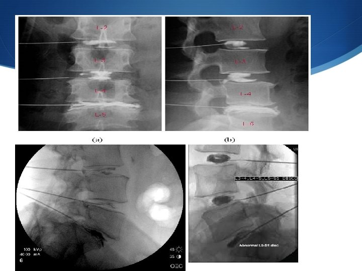
Discography INDICATIONS: • Provocative test to reproduce discogenic back pain in order to identify the source of pain and the symptomatic disc level, as well as define the morphologic abnormalities of the discs. A common clinical scenario is severe DDD at L 5 -S 1, with upcoming discectomy planned. However the patient also has mild to moderate DDD at L 4 -L 5 and a normal disk at L 3 -L 4. The three level discogram helps the surgeon decide if he can simply do a microdiscectomy at L 5 -S 1 or if he must do a bigger procedure including L 4 -L 5.
Discography
DISCOGRAM S
Discogram grading system
CT scan discogram
SPONDY – WHAT? • Spondylosis • Spondylolisthesis • Spondylolysis
Spondylosis • Lumbar spondylosis, as shown in the image below, describes bony overgrowths (osteophytes), predominantly those at the anterior, lateral, and, less commonly, posterior aspects of the superior and inferior margins of vertebral centra (bodies). This dynamic process increases with, and is perhaps an inevitable concomitant, of age.
Spondylolisthesis • Spondylolisthesis is a condition in which one of the bones of the spine (vertebrae) slips out of place onto the vertebra below it. If it slips too much, the bone might press on a nerve, causing pain. Usually, the bones of the lower back are affected. • The word spondylolisthesis comes from the Greek words spondylos, which means "spine" or "vertebra, " and listhesis, which means "to slip or slide.
Spondylolisthesis Types There are different types of spondylolisthesis. The more common types include. • Congenital spondylolisthesis — Congenital means "present at birth. " Congenital spondylolisthesis is the result of abnormal bone formation. In this case, the abnormal arrangement of the vertebrae puts them at greater risk for slipping. • Isthmic spondylolisthesis — This type occurs as the result of spondylolysis, a condition that leads to small stress fractures (breaks) in the vertebrae. In some cases, the fractures weaken the bone so much that it slips out of place. • Degenerative spondylolisthesis — This is the most common form of the disorder. With aging, the discs — the cushions between the vertebral bones — lose water, becoming less spongy and less able to resist movement by the vertebrae. Less common forms of spondylolisthesis include: • Traumatic spondylolisthesis, in which an injury leads to a spinal fracture or slippage • Pathological spondylolisthesis, which results when the spine is weakened by disease — such as osteoporosis — an infection, or tumor • Post-surgical spondylolisthesis, which refers to slippage that occurs or becomes worse after spinal surgery
Spodylolisthesis Grading Slippage is graded I through IV: • Grade I — 1 percent to 25 percent slip • Grade II — 26 percent to 50 percent slip • Grade III — 51 percent to 75 percent slip • Grade IV — 76 percent to 100 percent slip Generally, Grade I and Grade II slips do not require surgical treatment and are treated medically. Grade III and Grade IV slips might require surgery if persistent, painful, slips are present.
Degenerative Spondylolisthesis
Spondylolysis • Spondylolysis is a specific defect in the connection between vertebrae, the bones that make up the spinal column. This defect can lead to small stress fractures (breaks) in the vertebrae that can weaken the bones so much that one slips out of place, a condition called spondylolisthesis. Spondylolysis is a very common cause of low back pain. • The word spondylolysis comes from the Greek words spondylos, which means spine or vertebra, and lysis, which means a break or loosening.
Spondylolysis continued. . . Many people with spondylolysis have no symptoms and don’t even know they have the condition. When symptoms do occur, low back pain is the most common. The pain usually spreads across the lower back, and might feel like a muscle strain. The pain is generally worse with vigorous exercise or activity. Symptoms often appear during the teen-age growth spurt. The typical age of a person diagnosed with spondylolysis is 15 to 16 years. What causes spondylolysis? • Spondylolysis results from a weakness in a section of the vertebra called the pars interarticularis, the thin piece of bone that connects the upper and lower segments of the facet joints. Facet joints link the vertebrae directly above and below to form a working unit that permits movement of the spine. • The exact cause of the weakness of the pars interarticularis is unknown. One theory points to genetics (heredity) as a factor, suggesting that some people are born with thin vertebrae, which places them at higher risk for fractures. Another theory suggests that repetitive trauma to the lower back can weaken the pars interarticularis. How common is spondylolysis? • Spondylolysis affects about 3 percent to 7 percent of Americans. The condition is a common cause of low back pain in children and the most likely cause of low back pain in people younger than 26 years of age. Spondylolysis is more common in children and teens participating in sports that place a lot of stress on the lower back or cause a constant over-stretching (hyperextending) of the spine, such as gymnastics, weightlifting, and football. It is seen more often in males than in females.
Spondylolysis • Left, The pars interarticularis is found in the posterior portion of the vertebra. Center, Spondylolysis occurs when there is a fracture of the pars portion of the vertebra. Right, Spondylolisthesis occurs when the vertebra shifts forward due to instability from the pars defect.
Spondylolysis
Spondylolysis
Summary • Anular tears are also called anular fissures and are separations between anular fibers, avulsion of fibers from their vertebral body insertions, or breaks through fibers involving one or many layers of the anular lamellae. The terms 'tear' or 'fissure' does not imply that the lesion is consequent to trauma. In case of a traumatic event, the term 'rupture' is appropriate. • Disc Herniation is displacement of disc material beyond the limits of the intervertebral disc space. A herniated disc can be contained (covered by outer anulus fibrosus) or uncontained.
Summary • Protrusion indicates that the distance between the edges of the disc herniation is less than the distance between the edges of the base. • Extrusion is present when the distance between the edges of the disc material is greater the distance at the base.
Summary • Focal herniation is a herniated disc less than 90˚ of the disc circumference. • Broad-based herniation is a herniated disc in between 90˚ 180˚ of the disc circumference. • Bulging Disc is the presence of disc tissue 'circumferentially' (180˚-360˚) beyond the edges of the ring apophyses and is not considered a form of herniation.
My Back Hurts!
Thank You/Questions