Tumefactive demyelinating lesions TDLs Tumefactive demyelinating lesions 1979Van
 影像特征以及鉴别诊断")


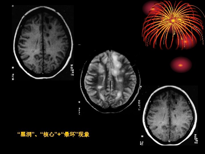
只有7%出 现非闭合性环形增强 • TDLs水肿程度及占位效应相对较轻 Distinguishing Tumefactive Demyelinating Lesions from Glioma or")


 of the")
- Slides: 52
肿瘤样脱髓鞘病变 (Tumefactive demyelinating lesions ,TDLs ) 影像特征以及鉴别诊断
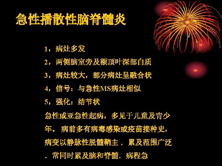
Tumefactive demyelinating lesions • 1979年,Van Dor Velden首次对该病进行了报告。 • 关于这种临床综合症是否属于一种独立的疾病仍然存在争 论,目前归类为多发性硬化和急性播散性脑脊髓炎之间的 独立中间型。 • 肿瘤样炎性脱髓鞘性病变(tumor-like inflammatory demyelinating diseases, TIDD) • 肿瘤样脱髓鞘病变(tumor-like masses of demyelination) • 脱髓鞘假瘤(demyelinating pseudotumor lesion)、 • 假瘤样脱髓鞘病(pseudotumor formus of demyelinating disease
A previously healthy 31 -year-old woman presented with a 2 -week history of progressive left hemiparesis. NEUROLOGY, 2007
21 -year-old woman presenting with new-onset seizure and biopsy-proven tumefactive demyelinating lesion.
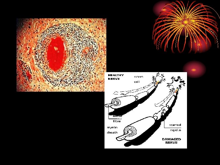
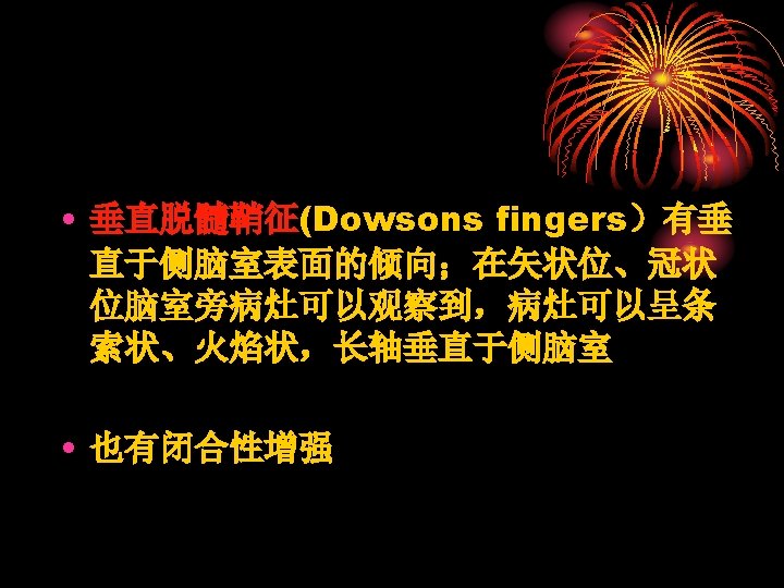
Multiple Sclerosis – Dawson’s Fingers
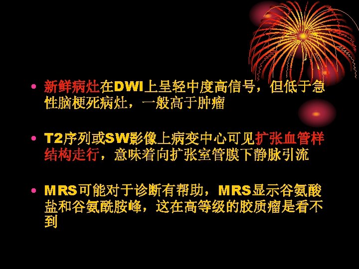
激素治 疗后好 转 2 months after corticosteroid therapy 50 -year-old man presenting with slurred speech and
• TDLs非闭合性增强是鉴别诊断的依据之 一;非脱鞘病(炎症、肿瘤等)只有7%出 现非闭合性环形增强 • TDLs水肿程度及占位效应相对较轻 Distinguishing Tumefactive Demyelinating Lesions from Glioma or Central Nervous System Lymphoma: Added Value of Unenhanced CT Compared with Conventional Contrast-enhanced MR Imaging Radiology 2009, 251(2): 467 -484
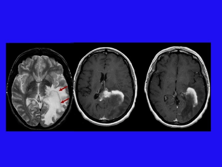
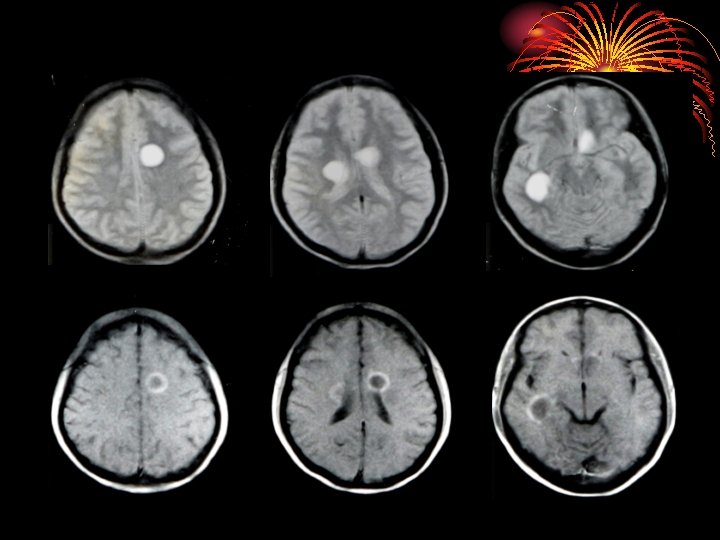
TDLs MR imaging and CT findings in 30 -year-old woman with TDL. A, Axial T 2 -weighted and, B, con- trast-enhanced axial T 1 -weighted MR images show a round mass with complete rim enhancement and perilesional edema in left frontal white matter. The signal intensity of the rim is isointense to gray matter on the T 2 -weighted image (arrow). C, Unenhanced axial CT image shows hypoattenuation (grade 1) of the rim; the margin of the enhanced rim on the MR image is not discernible on unenhanced CT image.
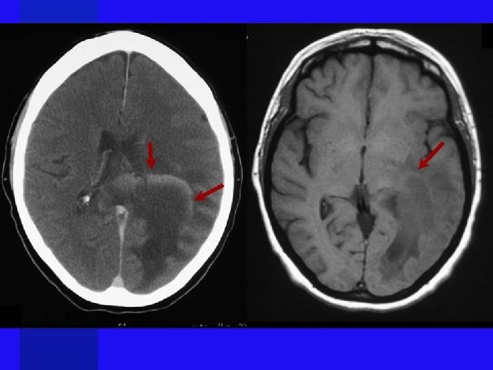
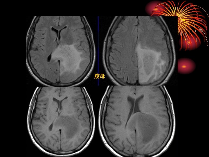
胶母 MR imaging and CT findings in 54 -year-old woman with glioblastoma. A, Axial T 2 -weighted and, B, contrast-enhanced axial T 1 -weighted MR images show a round cystic mass with complete rim enhancement and peritumoral edema in the subcortical white matter of the right frontal lobe. The signal intensity of the rim is isointense to gray matter on the T 2 -weighted image (arrow). C, Unenhanced axial CT image demonstrates isoattenuation (grade 2) of the rim (arrowhead).
TDLs
胶母 Glioblastoma Multiforme: no dark line of advancing demyelination
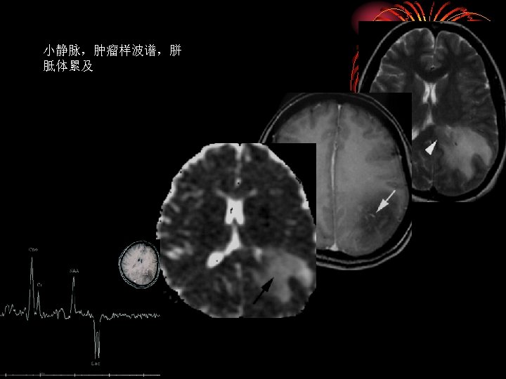
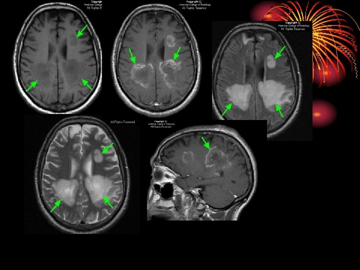
TDL MR imaging and CT findings in 32 -year-old man with TDL. A, Axial T 2 -weighted and, B, contrast- enhanced axial T 1 -weighted MR images demonstrate white matter lesions with heterogeneous enhancement in the parietal lobe and corpus callosum. The signal intensity of the enhancing components of the right parietal lobe is mixed (isointense plus hyperintense) on the T 2 -weighted image. C, Unenhanced axial CT image shows hypoattenuation (grade 1) of both the enhanced and unenhanced components of the lesions (arrows).
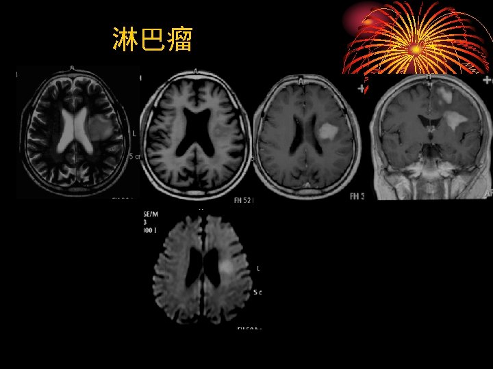
与淋巴瘤鉴别 MR imaging and CT findings in 65 -year-old woman with lymphoma. A, Axial T 2 -weighted and, B, contrast-enhanced axial T 1 -weighted MR images demonstrate bilateral lesions with diffuse enhancement in the white matter of both parieto-occipital lobes. Signal intensities of the enhancing lesions are hyperintense on the T 2 -weighted image. C, Unenhanced axial CT image demonstrates isoattenuation (grade 2) of the lesion in the left parietal lobe (arrowhead).
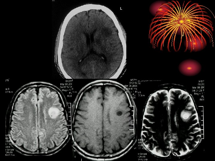
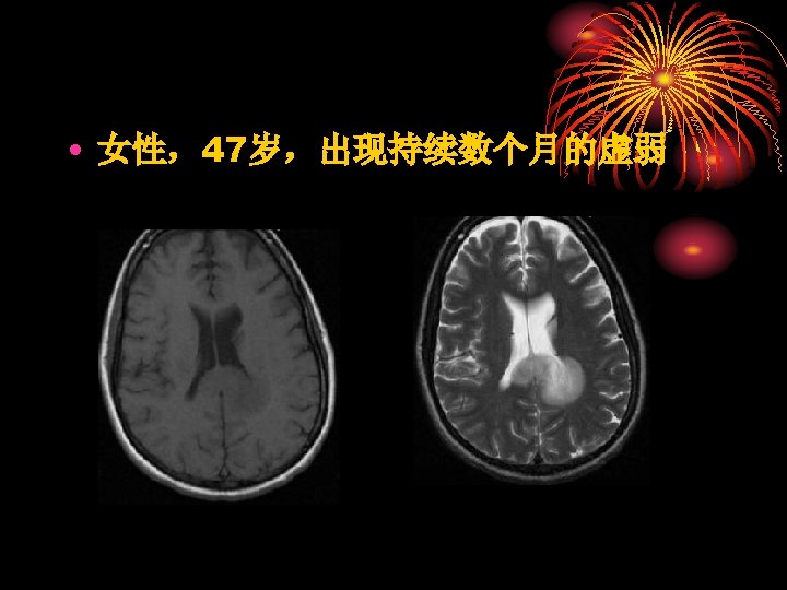
The mass has well defined borders and partially effaces the atrium (trigone) of the left lateral ventricle. There is mild patchy enhancement. DWI images demonstrate increased signal throughout, but only the even more hyperintense rim demonstrates true restricted diffusion on ADC images. The remainder of the mass is increased signal on ADC images, indicating increased diffusivitiy.