
EARLY PREGNANCY BLEEDING Dr Lama AlMehaisen MD MRCOG
• EARLY PREGNANCY BLEEDING Dr Lama Al-Mehaisen, MD, MRCOG Associated professor /obstetrics and gynaecology • • urogynaecology
INCIDENCE Early pregnancy Bleeding (EPB) is the most common complication in early pregnancy. The incidence of EPB is as high 10 percent The incidence of second-trimester loss up to 20 weeks gestation is less than 1 percent The rate of pregnancy Bleeding appears to be further influenced by maternal age and history of prior pregnancy loss.
Early pregnancy loss nonviable, intrauterine pregnancy within the first trimester (up to 12+6 weeks from the last menstrual period) terminology has included "miscarriage, " "blighted ovum, " "spontaneous abortion, " and "missed abortion Early second-trimester pregnancy loss is one that occurs after 13+0 and prior to 24+0 weeks of gestation By convention, pregnancies lost after 24 weeks are typically referred to as stillbirth or fetal death. Stillbirth or fetal death Pregnancy loss that occurs at 24 weeks gestation or later, or at a weight of 350 grams or greater, is generally referred to as a stillbirth or fetal death,
FIRST TRIMESTER BLEEDING Vaginal bleeding is common in the first trimester (up to 12+6 weeks). It may be any combination of light or heavy, intermittent or constant, painless or painful. Causes The four major sources of bleeding in early pregnancy are: Ectopic pregnancy Early pregnancy loss (threatened, inevitable/incomplete, complete. , , ) Implantation of the pregnancy Molar pregnancy Cervical, vaginal, or uterine pathology (eg, polyps, inflammation/infection, trophoblastic disease)
Bleeding related to early pregnancy loss (also called miscarriage or spontaneous abortion) is the most common nontraumatic cause of first trimester bleeding (prevalence: 15 to 20 %of pregnancies). Although bleeding may be heavy, almost all women remain hemodynamically stable; only about 1 percent of expectantly managed women require blood transfusion. Ectopic pregnancy is much less common (prevalence: 2 % of pregnancies), but the most serious etiology of first trimester bleeding as rupture of the extrauterine pregnancy is a potentially life-threatening complication; therefore, this diagnosis must be excluded in every pregnant woman with bleeding
Women may also note a loss or reduction of pregnancy symptoms, such as decreased breast tenderness and/or nausea and vomiting. Some women are asymptomatic, and E. P. loss is discovered incidentally or on routine ultrasound in early pregnancy. The volume of vaginal bleeding during EPB varies considerably, and women often report passing clots or tissue. The pain that occurs with EPB is often crampy in nature and can be mild to severe, especially during passage of gestational tissue. The pain can be constant or intermittent and is often associated with vaginal bleeding. As bleeding and cramping are also symptoms of other early pregnancy complications, including ectopic and molar pregnancy, pregnant women with vaginal bleeding or pelvic pain should be evaluated.
Complications of EPB/EBL Women with hemorrhage typically present with heavy vaginal bleeding combined with orthostatic vital signs, anemia, and/or tachycardia. The overall risk of this is low, approximately 1 percent Infectious complications can occur spontaneously or can follow medical or surgical interventions Signs and symptoms include abdominal or pelvic pain, uterine tenderness, purulent discharge, and/or systemic signs of infection, such as fever, tachycardia, or hypotension. Some women who have incomplete miscarriage develop an infection in the uterus with retained products of conception. This is known as a septic miscarriage. Symptoms include fever, chills, flulike aches, abdominal pain, vaginal bleeding, and vaginal discharge, which may be thick and may have a foul odor septic
Women who may be less likely to expect or recognize their pregnancy early in gestation, such as adolescents perimenopausal women with irregular menses might also be at higher risk of later presentation and/or complications.
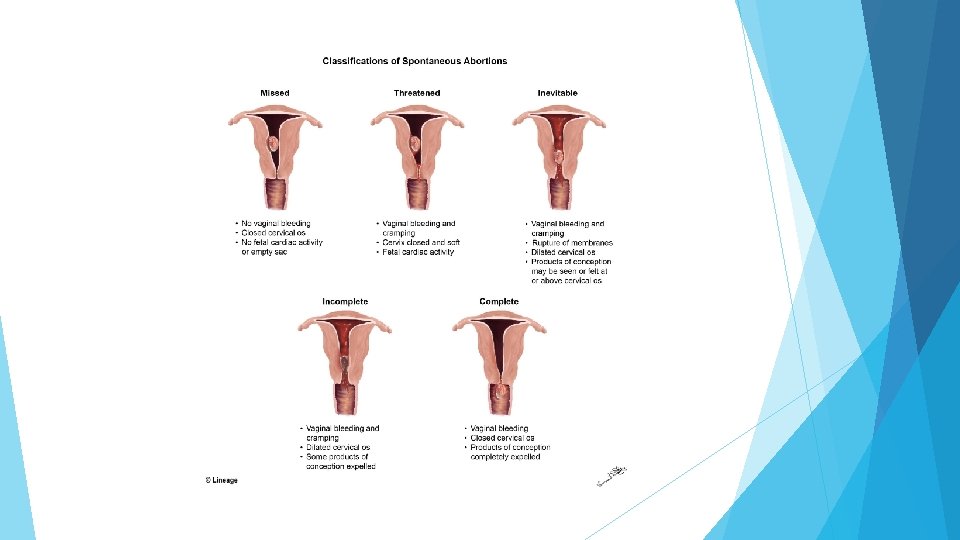
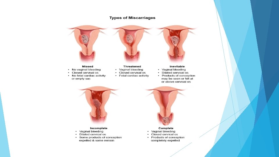
Types of miscarriage Complete early pregnancy loss / miscarriage When an early pregnancy loss occurs before 13 weeks of gestation, it is common for the entire contents of the uterus to be expelled, thereby resulting in complete pregnancy loss. If this has occurred . the uterus is small on physical examination well contracted with a closing or closed internal cervical os scant vaginal bleeding only mild cramping. Ultrasound will reveal an empty uterus and no extrauterine gestation.
A complete early pregnancy loss can be distinguished from an ectopic pregnancy by examining the tissue that was passed to confirm products of conception by demonstrating falling rather than rising or plateaued h. CG levels and by patient description of diminishing bleeding and pain. No further intervention is needed for complete early pregnancy loss if chorionic villi are identified by pathologic examination of the products of conception. However, if no villi are identified or no specimens are available for pathologic examination, then serum h. CG levels should be followed serially until the level is undetectable
Inevitable early pregnancy loss When early pregnancy loss is inevitable, the internal os of the cervix is dilated, vaginal bleeding is increasing, and painful uterine cramps/contractions are present. The gestational tissue often can be felt or seen at the internal cervical os; passage of this tissue typically occurs within a short time. Management may be expectant, or a medical or surgical intervention to complete the process can be undertaken. At a more advanced stage, the membranes may rupture and the fetus may be passed, but significant amounts of placental tissue can be retained, resulting in an incomplete early pregnancy loss.
Incomplete early pregnancy loss This is most common in the late first trimester and early second trimester. On examination, the internal cervical os is open gestational tissue may be observed in the cervical canal the uterine size is smaller than expected for gestational age, but not well contracted. The amount of bleeding varies, but can be sufficiently severe to cause hypovolemic shock. Painful cramps/contractions are often present. Ultrasound reveals tissue in the uterus. Medical or surgical evacuation is generally performed.
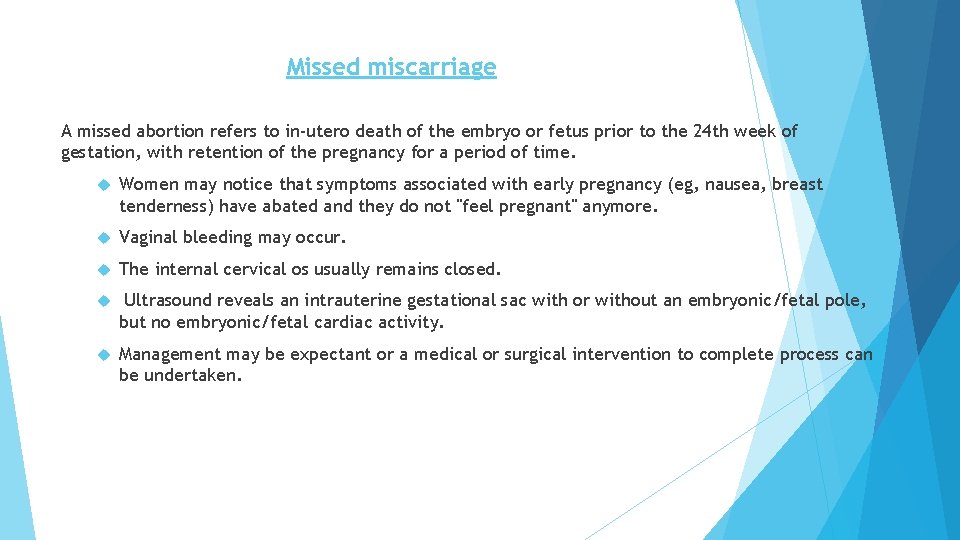
Missed miscarriage A missed abortion refers to in-utero death of the embryo or fetus prior to the 24 th week of gestation, with retention of the pregnancy for a period of time. Women may notice that symptoms associated with early pregnancy (eg, nausea, breast tenderness) have abated and they do not "feel pregnant" anymore. Vaginal bleeding may occur. The internal cervical os usually remains closed. Ultrasound reveals an intrauterine gestational sac with or without an embryonic/fetal pole, but no embryonic/fetal cardiac activity. Management may be expectant or a medical or surgical intervention to complete process can be undertaken.
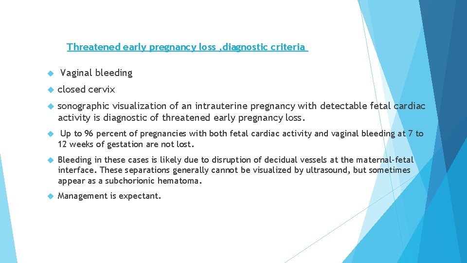
Threatened early pregnancy loss , diagnostic criteria Vaginal bleeding closed cervix sonographic visualization of an intrauterine pregnancy with detectable fetal cardiac activity is diagnostic of threatened early pregnancy loss. Up to 96 percent of pregnancies with both fetal cardiac activity and vaginal bleeding at 7 to 12 weeks of gestation are not lost. Bleeding in these cases is likely due to disruption of decidual vessels at the maternal-fetal interface. These separations generally cannot be visualized by ultrasound, but sometimes appear as a subchorionic hematoma. Management is expectant.
COMMON ETIOLOGY OF EARLY PREGNANCY LOSS/miscarriage 1 -Chromosomal abnormalities are present in up to 70 percent of pregnancy losses before 20 weeks The use of chromosomal microarray likely increased the ability to identify earlier preclinical losses that previously were undetected by karyotype.
2 -Maternal anatomic anomalies Anatomic anomalies, such as uterine leiomyomas (fibroids), polyps, adhesions, or septa, based on their size and position in relation to the developing pregnancy. the impact of fibroids on pregnancy loss likely varies by other factors, such as distortion of the uterine cavity and/or blood supply. 3 -Trauma Significant trauma. The developing embryo is relatively protected within the uterus in early pregnancy, but trauma that results in direct impact to the uterus can result in E. P. Loss. This can be due to violent trauma (gunshot wounds, penetrating injuries) or iatrogenic trauma, as with chorionic villus sampling and amniocentesis.
E. P. LOSS predisposing /risk factors 1. Increasing age Extremes of age increase the risk of pregnancy loss with age >35 years being the most significant risk factor because of the strong association with fetal chromosomal abnormalities EPL 2. risk does appear to rise with increasing paternal age as well Prior pregnancy loss appears to increase the risk of subsequent pregnancy loss, independent of maternal age. the risk of miscarriage increased among women whose prior pregnancy ended in a miscarriage
3 -Maternal medical conditions Infection 15 percent of EPL is associated with an infectious etiology ie Parvovirus B 19 Maternal cytomegalovirus (CMV) infection Obesity – Obesity is more strongly and consistently associated with pregnancy loss than either type 1 or type 2 diabetes. Diabetes – The effects of type 1 and type 2 diabetes on early pregnancy can be extreme, even resulting in lethal fetal anomalies or pregnancy loss. Euglycemia in the preconception and periconception time periods brings this risk back to baseline. Thyroid disease – Both hyper- and hypothyroidism have been associated with increased risk of pregnancy loss, Stress – Both acute and chronic stress can increase the risk of pregnancy loss Inherited thrombophilias – The effect of inheritable thrombophilias on EPL risk is unclear as the body of evidence conflicts. Pregnancy with intrauterine device (IUD) in place the risk of EPL appears to be higher for women who elect to leave the IUD in place rather than have it removed
4 -Medication and substance use (NSAIDs) aspirin and indomethacin are used for specific obstetric indications (preeclampsia prevention and treatment of acute preterm labor) while other NSAIDs, including ibuprofen and diclofenac, may increase EPL risk 5 -Environmental factors and exposures Exposure to ionizing radiation is associated with EPL , while excessive lead, arsenic, and air pollution exposure appear to increase the risk. 6 -Race and ethnicity increased risk of EPL in women of color compared with white women 7 -Subchorionic hematoma Subchorionic hemorrhage or hematoma is associated with increased risk of EPL, particularly when it amounts to 25 percent or more of the volume of the gestational sac women without with subchorionic hematoma had double the odds of EPL compared with women
DIAGNOSTIC EVALUATION History A history of vaginal bleeding and/or crampy pelvic pain in a woman with known pregnancy is suggestive Women present. with EPL may also note a reduction in pregnancy-related symptoms that were previously further evaluation with ultrasound or serial h. CG testing is required for definitive diagnosis. Physical examination Patients who present with bleeding in pregnancy first undergo a speculum examination to assess the source and quantity of bleeding. Signs suggestive of EPL include bleeding coming from the cervix and an open cervical os. A bimanual examination can also determine whether the cervix is open and whethere is tissue within the cervical canal. . Lastly, absence of fetal heart tones on handheld Doppler in a pregnancy of 12 weeks or greater should prompt further evaluation for pregnancy loss,
Imaging Women whose ultrasound demonstrates an intrauterine pregnancy but no fetal cardiac activity undergo repeat ultrasound at a future date to assess for interval change If transabdominal ultrasound is done instead, but is unable to demonstrate cardiac activity in an intrauterine pregnancy, then transvaginal ultrasound should be performed Women with a positive pregnancy test but no intrauterine pregnancy seen on ultrasound are diagnosed as having a pregnancy of unknown location and followed carefully with repeat imaging and possibly serial serum h. CG levels. These women may have an early intrauterine pregnancy, an ectopic pregnancy, or a molar pregnancy.
laboratory evaluation HCG ULTRASOUND ABDOMINAL /VAGINAL a dramatic drop in h. CG >25 percent over 48 hours in the setting of uterine bleeding is highly suggestive of EPL and may be especially helpful if ultrasound is not easily available. By contrast, h. CG testing is commonly performed as part of the assessment for pregnancy of unknown location and is often helpful in excluding ectopic pregnancy. .
In normal pregnancies, average h. CG levels plateau at approximately 8 to 12 weeks and then decline SERIAL BHCG is better indicator Serum progesterone has been used to assess pregnancy viability, as low serum progesterone levels are associated with E. P. Loss a cutoff of <35 nmol/L led to a positive predictive value for E. P. Loss, but it is not used for the diagnosis of EPL.
Diagnoses of early pregnancy loss A gestational sac ≥ 25 mm in mean diameter that does not contain a yolk sac or embryo. An embryo with a crown rump length (CRL) ≥ 7 mm that does not have cardiac activity. After a pelvic ultrasound showed a gestational sac without a yolk sac, absence of an embryo with a heartbeat in ≥ 2 weeks. After a pelvic ultrasound showed a gestational sac with a yolk sac, absence of an embryo with a heartbeat in 2 weeks.
Findings that are suspicious for, but not diagnostic of, pregnancy loss include: CRL <7 mm and no heartbeat. Absence of embryo with a heartbeat 7 to 13 days after a scan that showed a gestational sac without a yolk sac. Absence of embryo with a heartbeat 2 weeks after a scan that showed a gestational sac with a yolk sac. Absence of embryo ≥ 6 weeks after last menstrual period. Enlarged yolk sac (>7 mm).
Physiologic or implantation bleeding This is a diagnosis of exclusion. It is characterized by a small amount of spotting or bleeding approximately 10 to 14 days after fertilization (at the time of the missed menstrual period), is presumed to be related to implantation of the fertilized egg in the decidua (ie, lining of the uterus) No intervention is indicated.
Ectopic pregnancy
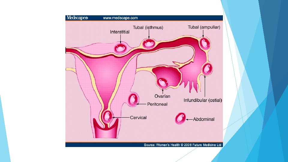
Ectopic pregnancy: Clinical manifestations and diagnosis An ectopic pregnancy is an extrauterine pregnancy. The majority of ectopic pregnancies occur in the fallopian tube (96 percent) other possible sites include: cervical, interstitial (also referred to as cornual; a pregnancy located in the proximal segment of the fallopian tube that is embedded within the muscular wall of the uterus), hysterotomy (cesarean) scar, intramural, ovarian, or abdominal. In addition, in rare cases, a multiple gestation may be heterotopic (include both a uterine and extrauterine pregnancy). The diagnosis of ectopic pregnancy is based upon a combination of measurement of the serum quantitative human chorionic gonadotropin and findings on transvaginal ultrasonography.
Ectopic pregnancy All women with early pregnancy bleeding and pain are assumed to have ectopic pregnancy until this diagnosis has been excluded by laboratory and imaging studies. Women with a history of ectopic pregnancy or other risk factors for the disorder are at highest risk An adnexal mass may or may not be seen. The presence of hemodynamic instability and a tender abdomen suggest the ectopic pregnancy has ruptured. Management of ectopic pregnancy is generally medical or surgical Expectant management can be dangerous for the mother, but may be possible in some cases. Even if an intrauterine pregnancy is diagnosed, the possibility of heterotopic pregnancy should be kept in mind, even though rare (1 in 30, 000 pregnancies). This is particularly important in women who conceived via an assisted reproductive technique (ART
CLINICAL PRESENTATION Ectopic pregnancy may be asymptomatic. The most common clinical presentation of ectopic pregnancy is first-trimester vaginal bleeding and/or abdominal pain. Clinical manifestations of ectopic pregnancy typically appear six to eight weeks after the last normal menstrual period, but may occur later, especially if the pregnancy is at an extrauterine site other than the fallopian tube. Normal pregnancy discomforts (eg, breast tenderness, frequent urination, nausea) are sometimes present in addition to the symptoms specifically associated with ectopic pregnancy. There may be a lower likelihood of early pregnancy symptoms in women with ectopic pregnancy because progesterone, estradiol, and human chorionic gonadotropin may be lower in ectopic pregnancy than in normal pregnancy. An ectopic pregnancy may be unruptured or ruptured at the time of presentation to medical care.
CLINICAL PRESENTATION Tubal rupture can result in life-threatening hemorrhage. Any symptoms suggestive of rupture should be noted. These include severe or persistent abdominal pain or symptoms suggestive of ongoing blood loss (eg, feeling faint or loss of consciousness). Vaginal bleeding The volume and pattern of vaginal bleeding vary, and there is no bleeding pattern that is pathognomonic for ectopic pregnancy. Bleeding may range from scant brown staining to hemorrhage. Bleeding is typically intermittent, but may occur as a single episode or continuously. The vaginal bleeding associated with ectopic pregnancy is typically preceded by amenorrhea.
DIAGNOSTIC EVALUATION OF ECTOPIC PREGNANCY Confirm that the patient is pregnant. Evaluate the patient for hemodynamic instability. Determine whether the pregnancy is intrauterine or ectopic Perform additional testing to guide further management Confirm pregnancy and ectopic pregnancy symptoms History — A menstrual history should be taken and the estimated gestational age should be calculated.
Risk factors for ectopic pregnancy should be elicited, including prior ectopic pregnancy, current use of an intrauterine device, prior tubal ligation, and in vitro fertilization (IVF)
SPECIAL ISSUES Multiple gestation In women with an intrauterine multiple pregnancy, the serum human chorionic gonadotropin (h. CG) level could be higher than 1500 milli-international units/m. L and yet ultrasound examination will not reveal an intrauterine pregnancy (IUP) Levels of over 9000 international units/L have been described for intrauterine triplet pregnancies unobserved by transvaginal ultrasound (TVUS) Heterotopic pregnancy The investigation for ectopic pregnancy can be terminated, under most circumstances, if a transvaginal sonogram reveals an IUP. Heterotopic pregnancy (combined intrauterine and extrauterine pregnancy) is rare, except among women conceiving through in vitro fertilization (IVF). The extrauterine pregnancy is usually in the fallopian tube, but can be at another location, such as the cervix.
Natural history of ectopic pregnancy If left untreated, an ectopic pregnancy in the fallopian tube can progress to a tubal abortion or tubal rupture, or it may regress spontaneously. Rupture Abortion Tubal abortion may be accompanied by severe intra-abdominal bleeding, necessitating surgical intervention, or by minimal bleeding, not requiring further treatment. Spontaneous resolution. Potential candidates are hemodynamically stable. Gestational products left in the fallopian tube may resorb completely or, less commonly, may cause tubal obstruction. Alternatively, a tubal abortion may occur.
Recurrent pregnancy loss
Recurrent pregnancy loss Defined as : Three consecutive pregnancy losses, before 12 weeks gestation RPL can be further divided into primary or secondary processes Primary RPL refers to pregnancy loss in women who have never carried to viability. In contrast, secondary RPL refers to pregnancy loss in a woman who has had a previous live birth. The prognosis for successful pregnancy is better with secondary RPL There is no specific term for describing women who have had multiple spontaneous miscarriages interspersed with normal pregnancies (ie, nonconsecutive pregnancy losses).
INCIDENCE Approximately 15 percent of pregnant women experience sporadic loss of a clinically recognized pregnancy. Just 2 percent of pregnant women experience two consecutive pregnancy losses only 0. 4 to 1 percent have three consecutive pregnancy losses.
RISK FACTORS AND ETIOLOGY Unfortunately, the cause of RPL can be determined in only 50 percent of patients General etiological categories of RPL include Anatomic/uterine Immunological genetic endocrine infectious thrombophilic environmental factors.
1 -Uterine factors/cervical Acquired and congenital uterine abnormalities are responsible for 10 to 50 percent of RPL Anomalies Congenital uterine anomalies ; Pregnancy loss may be related to impaired uterine distention or abnormal implantation due to decreased vascularity in a septum, increased inflammation, or reduction in sensitivity to steroid hormones The septate uterus is the uterine anomaly associated with the poorest reproductive outcome and the most common uterine abnormality associated with RPL Leiomyoma Submucous leiomyomas that protrude into the endometrial cavity can impede normal implantation as a result of their position, poor endometrial receptivity of the decidua overlying the myoma, or degeneration with increasing cytokine production Endometrial polyps
Intrauterine adhesions or synechiae lead to pregnancy loss because there is insufficient endometrium to support fetoplacental growth. The main cause of intrauterine adhesions is curettage for pregnancy complications. Cervical insufficiency Cervical insufficiency is a cause of recurrent mid-trimester, but not early, pregnancy loss. Defective endometrial receptivity Estrogen and progesterone prepare the endometrium for pregnancy Normal endometrial receptivity allows embryo attachment, implantation, invasion, and development of the placenta. These processes are likely to be disturbed when endometrial receptivity is defective
2 -Immunologic factors Antiphospholipid syndrome Several autoimmune diseases have been linked to poor obstetric outcome, but antiphospholipid syndrome (APS) is the only immune condition in which pregnancy loss is a diagnostic criteria for the disease. Five to 15 percent of patients with RPL may have APS 3 -Endocrine factors may account for 15 to 60 percent of RPL.
Endocrine factors Diabetes mellitus to increased frequencies of miscarriage and congenital malformations Polycystic ovary syndrome (PCOS) may be as high as 20 to 40 percent, which is higher than the baseline rate in the general obstetric population (10 to 20 percent) Thyroid antibodies and disease Antibodies (thyroid peroxidase or thyroglobulin), including those who are euthyroid increase risk of miscarriage Poorly controlled thyroid disease (hypo- or hyper-thyroidism) is associated with infertility and pregnancy loss. Excess thyroid hormone increases the risk of miscarriage independent of maternal metabolic dysfunction Hyperprolactinemia Normal circulating levels of prolactin may play an important role in maintaining early pregnancy. Treatment to lower prolactin concentration was associated with a higher rate of successful pregnancy (86 versus 52 percent). Prolactin levels during early pregnancy were significantly greater in women who miscarried Luteal phase defect Progesterone is required for successful implantation and maintenance of pregnancy; therefore, disorders related to impaired progesterone production or action are likely to affect pregnancy success Abnormal luteal-phase progesterone production may occur as the result of medical conditions such as elevated prolactin or abnormal thyroid function; women suspected to have one of these disorders are evaluated and treated for the underlying condition
4. Genetic factors Abnormalities of chromosome number or structure are the most common cause of sporadic early pregnancy loss, accounting for at least 50 percent A significant proportion of RPL may also be associated with structural or numerical chromosomal abnormalities (eg, aneuploidy, mosaicism, translocation, inversion, deletion, fragile sites) , Single-gene, X-linked, or polygenic/multifactorial disorders can also result in sporadic or recurrent miscarriage. Aneuploidy The risk of aneuploidy increases as the number of previous miscarriages increases
5. Thrombophilia and fibrinolytic factors Thrombosis of spiral arteries and the intervillous space on the maternal side of the placenta can impair adequate placental perfusion. resulting in late fetal loss, intrauterine growth restriction, placental abruption, or preeclampsia. There is a large and contradictory literature on the association between maternal inherited thrombophilia and RPL occurring in the first trimester 6. Environmental chemicals and stress Chemicals that have been associated with sporadic spontaneous pregnancy loss include anesthetic gases (nitrous oxide), arsenic, aniline dyes, benzene, ethylene oxide, formaldehyde, pesticides, lead, mercury, and cadmium
EARLY SECOND-TRIMESTER PREGNANCY LOSS
EARLY SECOND-TRIMESTER PREGNANCY LOSS Early second-trimester pregnancy loss, or fetal death, occurs in approximately 2 to 3 percent of pregnancies Conventionally, this is defined as fetal death between 13 and 24 weeks, and losses after 24 weeks gestation are defined as stillbirth, but this cutoff does not have a biological basis Known and suspected etiologies of second-trimester pregnancy loss include: Infection, including chorioamnionitis and maternal viral infection Chronic stressors, Uterine malformation Cervical insufficiency Fetal malformation or syndromes such as anencephaly, trisomies, renal agenesis, or hydrops Thrombophilias Abruption Premature Preterm preterm rupture of membranes labor
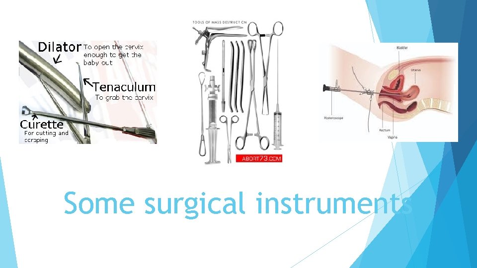
Some surgical instruments
- Slides: 51