CARDIAC MURMURS DR NIYAS K NASEER DM RESIDENT


 PR Mild or moderate PS Severe")


 AS PS HOCM(there is increase")


 MURMURS �Begin at a short interval following S 1, end before S")


 MURMURS Are pathologic Murmur begins immediately with S 1 and continues up")


![MITRAL STENOSIS Low pitch rough rumbling [sound of distant thunder] MDM Localized to apex,](https://slidetodoc.com/presentation_image_h2/a87b07dd6724197fffcff06a779f5842/image-92.jpg "MITRAL STENOSIS Low pitch rough rumbling [sound of distant thunder] MDM Localized to apex,")


![OTHER DIASTOLIC MURMURS Cabot– Locke Murmur- [Diastolic Flow murmur] The Cabot–Locke murmur is a](https://slidetodoc.com/presentation_image_h2/a87b07dd6724197fffcff06a779f5842/image-99.jpg "OTHER DIASTOLIC MURMURS Cabot– Locke Murmur- [Diastolic Flow murmur] The Cabot–Locke murmur is a")


![ALCAPA Murmur louder in diastole [LV contraction D/C flow] Do not peak at S](https://slidetodoc.com/presentation_image_h2/a87b07dd6724197fffcff06a779f5842/image-108.jpg "ALCAPA Murmur louder in diastole [LV contraction D/C flow] Do not peak at S")


- Slides: 118
CARDIAC MURMURS DR NIYAS K NASEER DM RESIDENT
SUBTITLES DEFINITION MECHANISM OF PRODUCTION CHARACTERISTICS OF MURMUR DYNAMIC AUSCULTATION SYSTOLIC MURMURS DIASTOLIC MURMURS CONTINUOUS MUMRURS
DEFINITION A cardiac murmur is defined as a relatively prolonged series of auditory vibrations of Varying intensity(loudness), frequency (pitch), quality, configuration, and duration
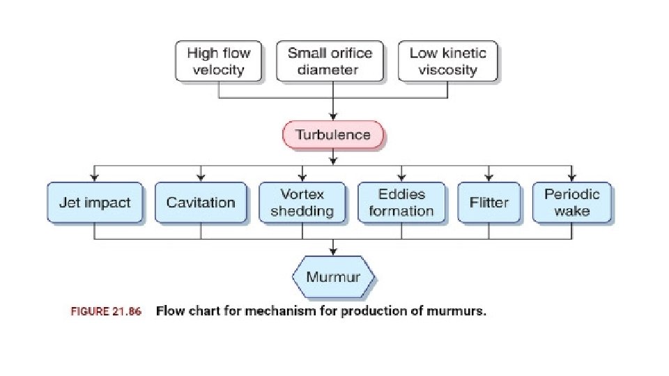
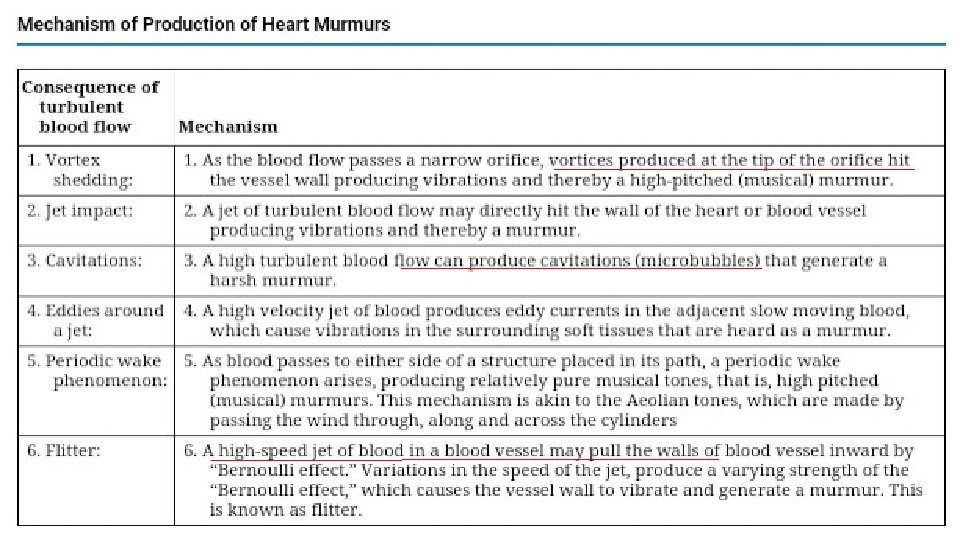
MECHANISM OF MURMUR PRODUCTION TURBULENT BLOOD FLOW
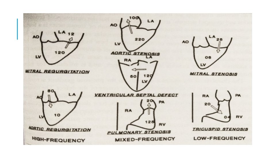
CHARACTERISTICS OF MURMUR 1. TIMING 2. LOCATION 3. DURATION 4. INTENSITY 5. FREQUENCY 6. CONFIGURATION 7. TRANSMISSION 8. DYNAMIC AUSCULTATION
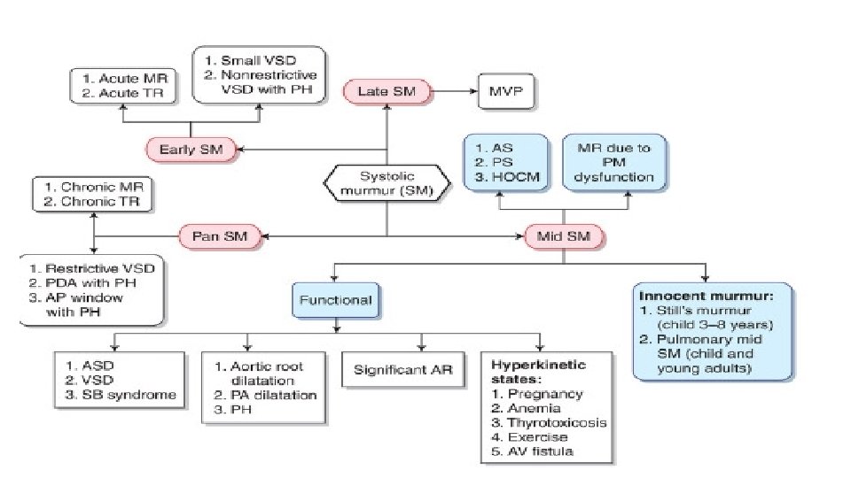
1. TIMING OF MURMUR SYSTOLIC MURMUR DIASTOLIC MURMUR SUBCLASSIFICATION-EARLY , MID , LATE OR PAN(HOLO) CONTINUOUS MURMUR
2. LOCATION OF MURMURS
3. DURATION OR LENGTH SHORT , LONG OR HOLO Depends on duration of events such as pressure gradient between the two sites or cardiac chambers reflects the severity of lesion in stenotic lesions
4. INTENSITY OR LOUDNESS
5. PITCH OF THE MURMUR pressure gradient between 2 chambers – flow velocity
6. CONFIGURATION OR SHAPE OF MURMUR
7. TRANSMISSION OF MURMUR Conduction with direct anatomical continuity SM of AS to carotids PSM of MR to left axilla Loud vs soft Loud-transmit widely Soft-confined High freq transmit proximally while low freq transmit distally MR to apex AS to base and carotid Low vs high freq murmurs Low freq-better transmitted to surface –thrill High freq –poorly transmitted – no
DYNAMIC AUSCULTATION
DEFINITION technique of altering circulatory dynamics by a variety of physiological manoeuvres and determining the effects of these manoeuvres on heart sounds and murmurs
INTERVENTION RESPIRATION POSITION PHYSICAL MANEUVERS PHARMACOLOGICAL
RESPIRATION - INSPIRATION ↓ INTRATHORACIC PRESSURE ↑ VENOUS RETURN to right side of the heart AND Dilatation of pulmonary vascular system causing decrease in pulmonary impedance there by increasing pulmonary hang out interval(>80 ms) ↑ stroke volume of right side accentuation of R side murmur
EXPIRATION ↓ lung volume → ↑ pulmonary venous flow left sided murmurs are loudest during expiration except MR which remain unchanged
MURMURS ACCENTUATED DURING Inspiration TS TR (Carvallo’s sign) PR Mild or moderate PS Severe PS no further increase in gradient Expiration • MS • AR • VSD • Pericardial rub (AP diameter)
In RV failure and PHT high Rt heart filling pressures , no increase in venous return with inspiration, hence no inspiratory augmentation of right sided murmurs and gallops Absence of respiratory influence is of no particular diagnostic value. Effects of inspiration may be accentuated by Muller maneuver.
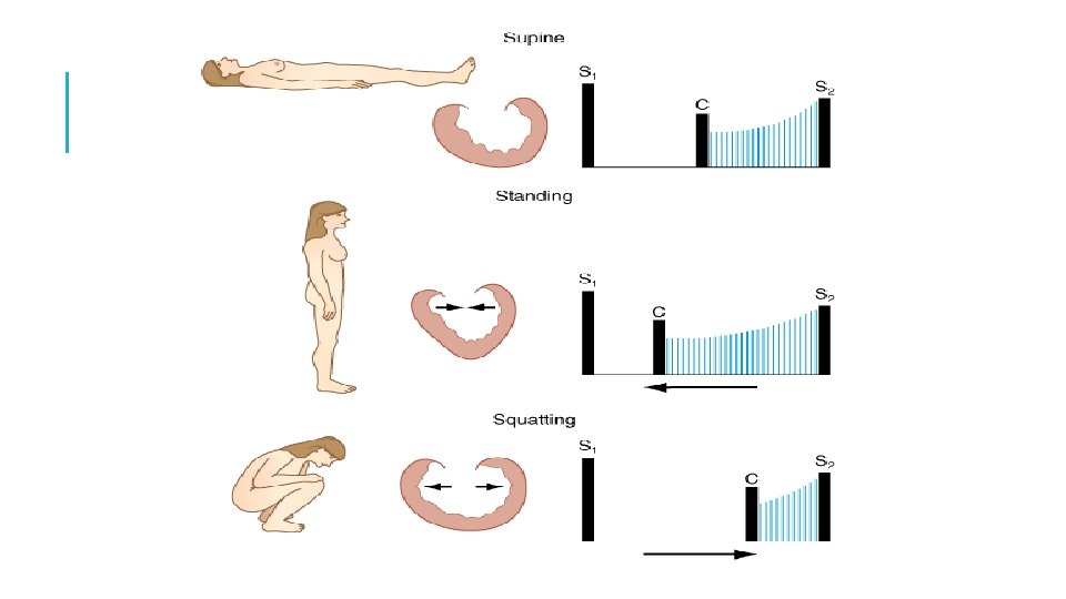
POSTURAL Auscultation is carried out immediately before and after the change in posture since effects may be quite transient persisting for only 10 – 15 heart beats
STANDING Rapid standing or sitting up from lying position or rapid standing from squatting posture results in § decreased venous return due to venous pooling in legs and splanchnic vessels leads toü decreased stroke volume ü decreased mean arterial pressure ü decrease in heart size ü followed by reflex increase in heart rate & systemic resistance
STANDING �All murmurs decrease except�ESM of HOCM becomes louder and longer �Click occurs earlier, murmur becomes longer in MVP while loudness shows variable response
SQUATTING Sudden change from standing to squatting leads to. Increases venous return & Stroke Volume Increase of systemic vascular resistance due to kinking of iliac artery and reduction of pressure of gravity Increase of systemic Arterial pressure with transient bradycardia
SQUATTING Increased venous return and CO - augments most murmurs (AS, PS, MR, AR, VSD) Right heart murmurs do so earlier Increased left ventricular volume - decreases murmur of HOCM and delayed murmur and click of MVP Ejection murmur of TOF↑ due to increase pulmonary blood flow and decrease in right to left shunt
OTHER POSTURAL CHANGES Left Lateral Position Causes closeness of heart to chest wall and transient rise of HR. So leads to – Increased murmur of MS, MR and austin flint murmur of AR Early appearance of click and systolic murmur of MVP due to increase HR. (increase HR & inotropism)
Sitting up and leaning forward causes more closeness of base of heart to chest wall so AR and PR murmurs more readly audible PRONE POSITION & KNEE CHEST POSITION Bring heart close to chest wall making pericardial rub more prominent
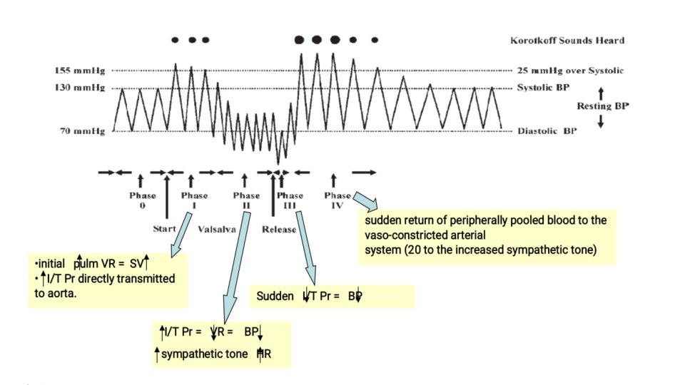
VALSALVA MANEUVER Described in 1704 –Antonio Maria Valsalva Relatively deep inspiration followed by forced exhalation against a closed glottis for 10 to 20 seconds Manometer method: Patient blows into the mercury manometer and maintains 40 mm. Hg for 15 seconds
VALSALVA EQUALENTS Patient pushes back against examiner’s hand which is pressed downward on mid abdomen. Caution : Not to be performed in IHD as it will reduce Coronary Blood Flow. It consists of 4 phases Clinically phase 1 and 3 are not perceptible at bedside
PHASES OF VALSALVA PHASE 1 -due to increase in intra thoracic pressure there is transient rise in LV output and systemic arterial pressure but there occurs fall in HR PHASE 2(stain phase)- decrease in venous return first to right then to left leads to decrease in systolic, diastolic and pulse pressure and reflex tachycardia
PHASES OF VALSALVA PHASE 3 - cessation of staining result in- ü sudden increase in systemic venous return ü but transient decrease in arterial pressure due to fall in intra-thoracic pressure §PHASE 4 - return to pre valsalva ü a transient overshoot of systemic arterial pressure ü Reflex bradycardia
PHASE BP HR 1 INCREASE DECREASE 2 DECREASE INCRESAE 3 DECREASE INCREASE 4 INCREASE DECREASE
EFFECTS ON MURMUR PHASE 2–as stroke volume fall - there is decrease in – systolic murmur of AS, PS, MR, TR diastolic murmur of AR PR MS TS §PHASE 2 - reduction in LV volume and size leads toincrease in systolic murmur of HOCM(increase LVOTO) increase in degree of MVP prolapse(louder and early midsystolic click and SM in MVP(increase in degree of prolapse)
PHASE 3 - sudden increase in Systemic venous Return leads to increase in right side murmurs PHASE 4 -left side murmur comes to control levels and may transiently increase
HEART FAILURE Phase 1 and 3 are normal but there is absence of decrease in arterial pressure tracing during phase 2 and overshoot of BP does not occur in phase 4 -since baroreceptors are not activated SQUARE WAVE RESPONSE
MULLERS MANEUVER Converse of Valsalva Maneuver Less frequently employed Forcibly inspires while the nose is held closed and mouth is firmly sealed for about 10 sec Augments murmur and filling sound originating in right side of the heart
ISOMETRIC EXERCISE Handgrip with both hands simultaneously for 20 -30 sec Use calibrated handgrip device or tennis ball or rolled up BP cuff. Measure the maximum effort Patient exerts 70 – 100% of this maximum for about 30 seconds CAUTION: Valsalva maneuver during handgrip should be avoided Avoid in those with ventricular arrhythmias and myocardial ischemia Contraindicated in recent myocardial infarction, uncontrolled hypertension, cerebrovascular disease, suspected aortic dissection
ISOMETRIC EXERCISE Hemodynamic changes: Significant increase in SVR Arterial pressure Heart rate LV filling pressure LV size
ISOMETRIC EXERCISE 1. Systolic Murmur of AS reduced due to reduced gradient across aortic valve 2. AR , MR , VSD – increased due to increase systemic vascular resistance 3. MDM of MS – increased due to Increased CO 4. Syst Murmur of HOCM reduced 5. MVP murmur + click delayed
CARDIAC CYCLE LENGTH CHANGES POST PVC AND AF CP-↑ preload will increases ventricular filling and size Also in addition there is secondary increase in ventricular contractility of new beat and transient increase in arterial pressure.
CARDIAC CYCLE LENGTH CHANGES
Increased ( L or R vent ejection murmurs ) AS PS HOCM(there is increase in SM due to increase LVOTO- known as BROKENBROUGH BRAUNWALD SIGN) MS click and late SM of MVP are delayed No change for MR , TR or VSD DM of AR increases due to transient rise in arterial pressure
AMYLNITRITE INHALATION Inhalation of Amyl Nitrate Crush ampoule in towel and take 3 -4 deep breaths over 10 – 15 secs Changes observedq < 30 secs : Systemic vasodilatation->decrease SVR q 30 – 60 secs : increase HR & CO § However majority of auscultatory changes are observed in first 30 sec
Due to increase in CO-increase SM of AS and PS SM of TR All functional SM DM of MS and TS DM of PR
Due to decrease in SVR following murmur are decreased- ü SM of MR ü DM and austin flint murmur of AR ü SM of TOF §Due to decrease LV volume and size – ü SM of HCM increases ü early appearance of MVP click and murmur but softening of murmur occur due to decrease resistance to LV resistance
METHOXAMINE & PHENYLEPHRINE Opposite effect of Amyl Nitrate Phenylephrine - due to short duration of action Systolic pressure elevated by 30 mm Hg for 3 to 5 mts EFFECT 1. Increases systemic arterial pressure 2. Reflex Bradycardia , decrease CO, decrease Contractility Caution : Not to be used in patients with CHF or Systemic hypertension.
METHOXAMINE & PHENYLEPHRINE ↑ BP & SVR ↓ CO & HR – last for 3 -5 mts AR , MR , VSD , TOF – Louder SM OF AS, PS and DM of PR and TS -show no changes LV size increases HOCM – Softer Click and Murmur of MVP - Delayed
SYSTOLIC MURMUR
EARLY SYSTOLIC MURMURS Early systolic murmurs begin with S 1 and extend for a variable period of time, ending well before S 2 (high pitch , decrescendo) 1. Acute severe mitral regurgitation ◦ Regurgitation occurs into a normal-sized, relatively noncompliant left atrium and as LV-LA pressure gradient is abolished during late systole, termination of retrograde flow occurs well before S 2. ◦ best heard at apical impulse ◦ Caused by: i. Papillary muscle rupture due to ischemia ii. Infective endocarditis -destruction of leaflet tissue, chordal rupture, or both
iii. R u p t u r e o f t h e c h o r d a e t e n d i n e a e i n m y x o m a t o u s m i t r a l v e d i s e a s e iv. Blunt chest wall trauma-papillary muscle contusion and rupture, chordal detachment, or leaflet avulsion. 2. Congenital, small ventricular septal defectventricular size decrease and septum thickness increases which seals off defect
3. VSD with high PA pressurehigh pulmonary resistance will decrease the late shunting
MIDSYSTOLIC (EJECTION) MURMURS �Begin at a short interval following S 1, end before S 2 �Flow across LV or RV outflow tract- when flow proceeds , murmur increase in crescendo and when it decrease murmur decrease in decrescendo. �Intensity of murmur depend on cardiac output
CAUSES ARE: 1. Innocent: due to flow across normal ventricular outflow tract 2. Functional: dilation of aortic root, pulmonary trunk or increase flow into aorta and pulmonary artery 3. Pathologic � are secondary to structural CV abnormalities-ventricular outflow obstruction � e. g. Aortic stenosis, Hypertrophic cardiomyopathy, Pulmonic stenosis
AORTIC STENOSIS Harsh, medium pitch, loudest in aortic area; radiates along the carotid arteries and apex. Intensity varies directly with CO Severity varies with murmur may have an early peaking and short duration or late peaking and prolonged duration. A/W parvus et tardus Other conditions which may mimic the murmur of aortic stenosis w/o obstructing flow: 1. 2. 3. 4. Aortic sclerosis Bicuspid aortic valve Dilated aorta Increased flow across the valve during systole
2
GALLAVERDIN PHENOMENON/HOURGLASS PHENOMENON Noisy medium pitch murmur at base due to turbulence of high velocity jet and High frequency murmur reflected to mitral area (apex) due to periodic wake phenomenon caused by high frequency vibrations of the fibrocalcific aortic cusps – mimic MR (S 2 clear, increase mr in PVC/AF)
AS MR Location Aortic area Apex Radiation Neck Axilla Shape Diamond Holosystolic Pitch Medium High Associated signs Decreased A 2 Slow rising and delayed pulse Ejection click S 4 Narrow pulse pressure Decreased S 1 Laterally displaced diffuse PMI S 3 POST PVC INCREASES NO CHANGE Isometric Exercise DECREASES INCREASES Amyl Nitrate INCREASES DECREASES
AORTIC STENOSIS -TYPES
HYPERTROPHIC CARDIOMYOPATHY �Loudest b/t left sternal edge and apex; Grade 2 -3/6 �Does NOT radiate into neck; carotid upstrokes are brisk and may be bifid Factors which increase intensity: Increase LV contractility : exercise , catecholamine , digitalis Decrease ventricular volume: valsalva, standing, amyl nitrates, tachycardia Decrease aortic impedance and pressure: sustained
MANEUVERS FIXED LVOT-AS DYNAMIC LVOT-HOCM RESPIRATION NOT MUCH CHANGE MAY INC WITH EXPIRATION STANDING DECREASES INCREASES SQUATTING INCREASES DECREASES VALSALVA DECREASES INCREASES BROCKENBROUGH NORMAL POSITIVE
PULMONARY STENOSIS • Murmur brought on by a phasic ejection click: radiates up and left • As severity increases length increases and P 2 becomes soft (abruptness of closure reduced ) , S 2 split widens S 4. becomes kite like • May engulf A 2 and P 2 : may
OTHER CAUSES OF MSM Dilation of aorta and pulmonary trunk Short soft midsystolic murmur Left sided murmur in marfans syndrome , syphilis Right sided murmurs in idiopathic dilation of pulmonary artery, pulmonary hypertension MSM of hyperdynamic circulation Normal aorta or pulmonary trunk but increased flow Anaemia , pregnancy , fever , thyrotoxicosis
OTHER CAUSES OF MSM OS-ASD Rapid flow across pulmonary valve to dilated pulmonary trunk Pure AR Due to accelerated LV ejection
PHYSIOLOGICAL CAUSES INNOCENT SYSTOLIC MURMUR Stills murmur Pulmonary mid systolic murmur Peripheral pulmonary systolic murmur Supraclavicular or brachiocephalic systolic murmur Aortic sclerosis Systolic mammary soufflé
STILLS MURMUR Short buzzing murmur twanging of rubber band Pure medium frequency by periodic vibrations of pulmonic leaflets at their attachment
PULMONARY MID SYSTOLIC MURMUR AND PERIPHERAL PULMONARY MID SYSTOLIC MURMUR Angulation and disparity between pulmonary trunk and its branches turbulent flow Normally disappears with maturity of pulmonary bed
SUPRACLAVICULAR OR BRACHIOCEPHALIC SYSTOLIC MURMUR Aortic origins of major normal brachiocephalic arteries Crescendo decrescendo , abrupt onset , short, sometimes radiating below clavicle Vs supraclavicular AS –these murmurs are softer below clavicle and decrease with shoulder abduction
MAMMARY SOUFFLE Late pregnancy or pueperium Sometimes continuous louder in systole , distinct gap from S 1(time for ejection blood to reach mammary arteries) 2 or 3 RICS/LICS Light pressure augments murmur becomes continuous firm pressure abolishes murmur
PANSYSTOLIC (HOLOSYSTOLIC) MURMURS Are pathologic Murmur begins immediately with S 1 and continues up to S 2 1. Mitral valve regurgitation Loudest at the left ventricular apex Radiation reflects the direction of the regurgitant jet i. To the base of the heart = anterosuperior jet (flail posterior leaflet) ii. To the axilla and back = posterior jet (flail anterior leaflet Also usually associated with a systolic thrill, a soft S 3, and a short diastolic rumbling (best heard in left lateral decubitus 2. Tricuspid valve regurgitation 3. Ventricular septal defect
MITRAL REGURGITATION
TRICUSPID REGURGITATION LLSB-RA RIVERO CARVALLO SIGN – increased RV , increase RV volume increased SV velocity of regurgitant flow Sometimes TR heard only during inspiration Carvallo sign disappears in RV failure
VENTRICULAR SEPTAL DEFECT Depends on site , size and gradient Very restrictive VSD –ESM decrescendo pattern Mod and NR VSD –PSM Sub arterial VSD -1 or 2 LICS similar to PS murmur Septal aneurysm – click with LSM OR PSM with late systolic accentuation Large shunt – MDM NR VSD with PAH – ESM PSM absent in Eissenmenger syndrome –only pulmonary ESM
OTHER PSM Aorto Pulmonary window with PAH Otherwise continuous murmur Diastolic component reduced due to increased PAH PDA with PAH
LATE SYSTOLIC MURMURMVP Leaflet remains competent during early ventricular contraction but overshoot in late systole ( critical V dimensions ) One or more mid systolic clicks precede murmur (sudden deceleration of the column of blood against the prolapsed leaflet or scallops) Longer and softer prompt standing after squatting valsalva II Short and louder squatting sustained hand grip amyl nitrate Other LSM- papillary muscle dysfunction
MVP HCM CLICK PRESENT ABSENT POST ECTOPIC BEAT DO NOT CHANGES DECREASES AMYL NITRATE BI PHASIC INCREASES
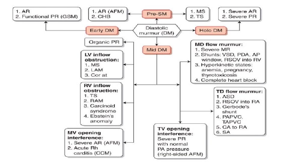
DIASTOLIC MURMUR
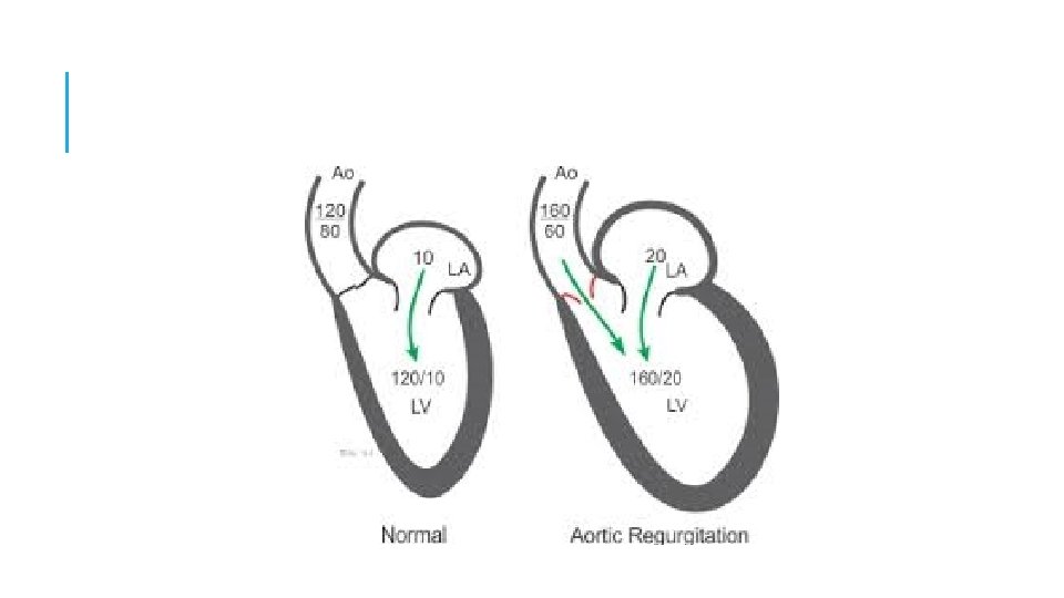
EARLY DIASTOLIC MURMUR AORTIC REGURGITATION: Soft high frequency EDM with pt sitting & leaning forward , breath held in full expiration 3 LICS [2 or 3 RICS in root dilatation Musical quality in eversion Austin flint murmur Cole Cecil murmur-AR murmur in left axilla due to high position of apex
AR Difference between acute and chronic AR A/C AR C/C AR Short mur. -early equalization of diastolic pressures Long mur. Medium n –velocity less rapid and pressure gradient lower High n Associated S 4
HIGH PRESSURE PR High pitched soft blowing decrescendo murmur usually lasts throughout diastole heard in the left upper sternal border Associated with loud P 2 and other features of PAH PR vs. AR Loud P 2, murmur begins after P 2 Normal pulse pressure Clinical setting Squatting and sustained hand grip increases AR
HIGH PRESSURE VS. NORMAL PRESSURE High Pressure Normal pressure Decrescendo Crescendo decrescendo High frequency Medium to low pitched Onset immediately with p 2 Delayed in onset Usu extends throughout diastole Short duration Features of PAH present Usually absent
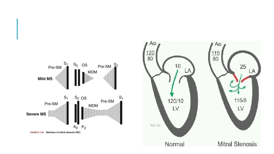
MID DIASTOLIC MURMUR Begins at clear interval after A 2 1. Rapid filling phase: AV valve obstruction- stenosis AV valves, tumors, functional obstruction abnormal pattern of AV flow : increased flow volume , increased flow velocity 2. Incompetent pulmonary valve [PR in normal PA pressure] 3. Atherosclerotic extramural coronary arteries
MID DIASTOLIC MURMUR RV LV OTHERS - TS - MS -Atrial Myxoma ASD - Austin Flint murmur - Carey-Comb's - VSD - PDA - MR
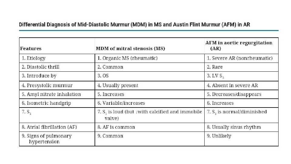
MITRAL STENOSIS Low pitch rough rumbling [sound of distant thunder] MDM Localized to apex, better heard in left lateral position with bell Length - severity Long murmurs up to S 1 even in long cycles of AF- severe MS Late diastolic or Pre systolic accentuation usually seen in pliable valves and in NSR [ sometimes in AF]
TRICUSPID STENOSIS Similar to MS Murmur usually seen associated with AF Diff. from MS Increases during inspiration [Augmentation of RV volume] LLSB
AUSTIN FLINT MURMUR Severe AR regurgitant jet directed toward the AML prevent the latter from opening well during diastole generating turbulent flow Low pitch MDM or late diastolic, best heard at the apex. To differentiate from MS No OS Amyl nitrate inhalation
LATE DIASTOLIC/ PRE-SYSTOLIC MURMURS MS Higher frequency than MDM Sometimes only PSA heard- mild MS Generally absent in calcified valves and most of AF [ may be present during short cycle lengths in AF] Cause. Increased flow during atrial contraction in late systole
OTHER MID DIASTOLIC MURMUR Carey Coomb’s murmurs Acute rheumatic fever- mitral valve structures acutely inflamed with some thickening and edema CAUSES- turbulence of flow during the rapid filling phase. + moderate MR [increased mitral inflow in diastole] Low pitched short MDM. good evidence of active carditis
OTHER DIASTOLIC MURMURS Cabot– Locke Murmur- [Diastolic Flow murmur] The Cabot–Locke murmur is a diastolic murmur that sounds similar to aortic insufficiency but does not have a decrescendo; it is heard best at the left sternal border. [High flow thru coronary vessels, LMCA, LAD] The murmur resolves with treatment of anaemia. Dock’s murmur diastolic crescendo-decrescendo, with late accentuation, [consistent with blood flow through the coronary] in a sharply localized area, 4 cm left of the sternum in the 3 LICS, detectable only when the patient was sitting upright. Due to stenosis of LAD
OTHER DIASTOLIC MURMURS Key–Hodgkin murmur EDM of AR; it has a raspy quality, [sound of a saw cutting through wood]. Hodgkin correlated the murmur with retroversion of the aortic valve leaflets in syphilitic disease. Rytand’s murmur in complete heart block MDM or Late diastolic murmur Atrial contraction coincides with the phase of rapid diastolic filling increased flow short MDM [intermittent]. Another theory- Delayed V. contraction following A. contraction may lead to diastolic MR & TR, because AV valve closure does not occur [unless V. systole supervenes]. When higher V than A pressure during atrial relaxation, an incompletely closed AV valve may lead to a reverse gradient with a considerable regurgitation volume.
CONTINUOUS MURMUR
CONTINUOUS MURMUR Begin in systolic and continue without interruption through the timing of S 2 into all or part of diastole without change in character D/D: to and fro murmurs of VSD + AR , AS + AR , MS + MR
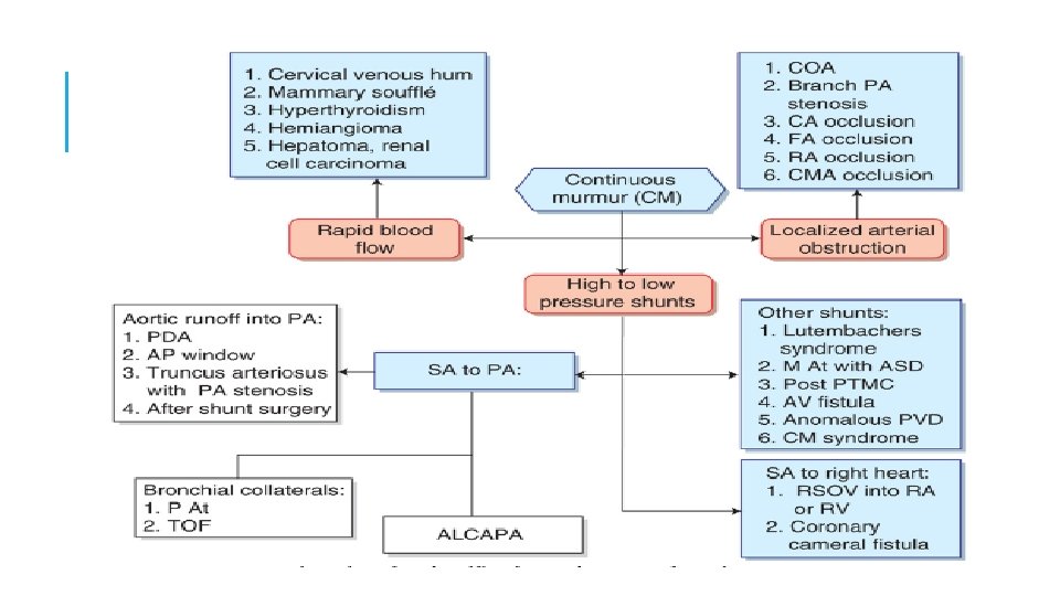
1. CONNECTION B/W HIGH PRESSURE/VESSEL & LOW PRESSURE CHAMBER/VESSELS 1. From the aorta a. persistent ductus arteriosus b. Aorto-pulmonary window c. RSOV 2. From the coronary artery a. coronary arteriovenous fistulae draining into RA, RV, PA b. ALCAPA 3. Other AV communications a. Bronchopulmonary collaterals b. chest wall arteries-pulmonary vessels 3. Peripheral A-V Fistula 4. Others a. lutenbachers syndrome with restricted ASD
PDA Gibsons murmur At 1 or 2 LICS NR- high frequency soft murmur peaks around S 2 Mod R- loud coarse machinery murmur with eddy sounds
PDA WITH NO CONTINUOUS MURMUR Neonates- due to high PVR Very small ductus Very large ductus &large VSD –due to equalization of pulmonary and systemic pressure PAH –first diastolic component goes, then systolic AS, Co. A-due to low aortic pressure
CONTINUOUS MURMURS APW 2 or 3 LICS Usually associated with early development of eisenmenger RSOV No peaking at S 2 seen [ peaks in systolic and diastolic ] To RA- RLSB To RV-LLSB RVOT-3 LICS Lutenbachers syndrome with restricted ASD LLSB (body of RA)
ALCAPA Murmur louder in diastole [LV contraction D/C flow] Do not peak at S 2 Usually LUSB or RUSB
DUE TO RAPID BLOOD FLOW AV fistula Murmur heard in the venous side Due to rapid blood flow cervical venous hum, mammary souffle, hyperthyroidism, hemangioma, hyperemia of neoplasm(HCC, RCC, Pagets disease) Stenosed arteries with inadequate distal collaterals Aortic arch vessels occlusion , atherosclerotic carotids , coarctation of aorta, main pulmonary artery stenosis and peripheral pulmonary artery stenosis
DISTURBANCES IN FLOW PATTERNS IN VEINS VENOUS HUM Healthy children , young healthy adults , pregnancy Sitting , bell, medialaspect of Rt S. Cl fossa , with face pulled leftwards and upwards disappears when returned to normal position. Louder in diastole , +/- high pitched whine Radiation to infraclavicular areas confuse with other murmur. Check by obliteration
APPRECIATING MURMURS IMPORTANT ?
ALL MURMURS TO BE EVALUATED ?
REFERENCES 1. Braunwald 10 th edition , textbook of cardiovascular medicine 2. Synopsis of cardiac physical diagnosis, Abrams, Jonathan 3. Clinical examination in cardiology , 2 nd edition: B. N. Vijay Raghawa Rao
MCQ 1. Effect on SM are similar in HOCM and MVP in all this manuevers EXCEPT A. Amyl nitrate inhalation B. Valsalva C. Handgrip D. Post PVC
2. Murmur increases on hand grip in ………………. A. AS B. PS C. AR D. HOCM E. VSD
3. Brokenbrough Brauwnwalds sign indicates A. Decrease SM in HOCM on handgrip B. Decrease SM in HOCM on amyl nitrate inhalation C. Increase SM in HOCM in PVC D. Increase SM in HOCm on handgrip
4. Complete heart block is associated with: A. ESM B. MSM C. MDM D. EDM
THANK YOU