Cardiovascular System Esraa kiwan Cardiac Cycle Cardiac cycle

 800 msec Diastole Systole 500 msec 300 msec")


 in response to")


 Arterial muscle: increases contractility Ventricular muscle:")


 and a")
 to blood vessel, which is")


- Slides: 35
Cardiovascular System Esraa kiwan
Cardiac Cycle Cardiac cycle (at rest) 800 msec Diastole Systole 500 msec 300 msec
Blood normally flows in a laminar fashion (layer of fluid slide smoothly over each other, doesn't produce sound When blood flow become turbulent, a sound is produced, due to vibrations created in the surrounding structures
Cardiac Output: is the volume of blood pumped by each ventricle per minute
Cardiac Output Cardiac output = Stroke Volume X Heart Rate CO = SV x HR Stroke Volume: volume of blood pumped by each ventricle per beat or stroke (averages 70 ml/beat) Heart Rate: heart beats/min (averages 70 beats/min)
Cardiac Output Stroke Volume: volume of blood pumped per beat or stroke (averages 70 ml/beat) Heart Rate: heart beats/min (averages 70 beats/min) CO = SV x HR CO= 70 ml/beat x 70 beats/min = 4, 900 ml/min ≈ 5 liters/min
Cardiac Output Resting cardiac output ≈ 5 L/min During exercise cardiac output can increase to 20 to 25 L/min Cardiac reserve: the difference between the cardiac output at rest and the maximum volume of blood the heart can pump per minute
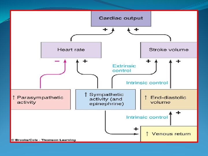
Cardiac Output Cardiac output depends on heart rate and the stroke volume CO = SV x HR ( cardiac output increases or decreases in response to changes in heart rate or stroke volume. i. e. when one of them increase it will increase the cardiac out put)
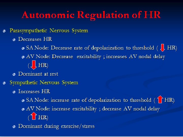
The heart is innervated by both divisions of the autonomic nervous system (sympathetic & parasympathetic), which can modify the rate & strength of contraction. (not initiation of contraction)
Autonomic Regulation of HR
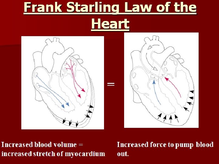
Stroke Volume Control Two types of controls influences stroke volume: Extrinsic control Intrinsic control The extent of venous return The extent of sympathetic stimulation of the heart Both factors increase stroke volume by increasing the strength of contraction of the heart
Stroke Volume Control Intrinsic control: Intrinsic ability to regulate SV (output) in response to changes in venous return (input)
Stroke Volume Control
Stroke Volume Control Extrinsic control: (sympathetic nervous system) Arterial muscle: increases contractility Ventricular muscle: increases contractility Adrenal medulla: increase epinephrine (augments the sympathetic actions on the heart) Veins: increase venous return
Effect Of Sympathetic Stimulation On SV
Shift of the frank starling curve to the left by sympathetic stimulation
Cardiac Output Regulation
Blood Pressure Blood pressure is the force exerted by the blood against a vessel wall Depends on: Volume of blood contained within the vessel Compliance or Distensability of the vessel wall
Blood Pressure During each heartbeat, Blood pressure varies between a maximum (systolic) and a minimum (diastolic) pressure Systolic pressure: The maximum pressure exerted in the arteries when the blood is ejected into them during ventricular systole averages 120 mm. Hg Diastolic pressure: The minimum pressure within the arteries occurs when the blood is draining off into the rest of vessels during ventricular diastole averages 80 mm. Hg
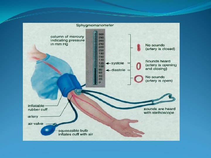
Blood Pressure Measurement Directly: inserting a needle (a canula) to blood vessel, which is linked to a device measure the blood pressure Indirectly: through the use of a sphygmomanometer
Blood Pressure Measurement
Blood Pressure Pulse pressure: is the difference between systolic and diastolic pressure (systolic – diastolic) Blood pressure = 120/80 Pulse pressure 40 mm. Hg Mean arterial pressure: is the average pressure responsible for driving blood forward MAP= diastolic pressure + 1/3 pulse pressure = 80 + (1/3 * 40) = 93 mm. Hg
Regulation of blood pressure Blood pressure is regulated by controlling: Cardiac output Total peripheral resistance Blood volume Blood pressure= cardiac output X peripheral resistance Cardiac output= HR X SV Total peripheral resistance depends on the radius of all arterioles as well as blood viscosity Resistance 1/r� ( r: radius of the vessel)
Regulation of Blood Pressure Cardiac output= HR X SV HR SV
Regulation Of Blood Pressure Total peripheral resistance:
Regulation of Blood Pressure
Regulation of Blood Pressure Baroreceptor:
Regulation of Blood Pressure Baroreceptor:
Regulation of Blood Pressure Baroreceptor:
Regulation of Blood Pressure Blood volume: