Cardiovascular system circulatory system The cardiovascular system consists
 The cardiovascular system consists of the heart and a closed")


 in ELM ERY lumen Pinocytic vesicles Zonula occludens 2 1")


 n lumen")
 n respiratory gasses, nutrients and waste products change between blood")
 n allow the blood cells to pass throughout their wall")


 70 nm , diaphragm (thinner")

 n from 8 to 40 m n n endothelium – fenestra,")


 endothelium +")
 Tunica")
 TI n endothelium n subendothelial connective tissue – thin layer of")

 TA n fibrous connective tissue + smooth muscle cells in veins")
 YES Membrana")
")


 TM: up")


 TM: thin layer of")

 layer Pericardial cavity EPICARDIUM Inner (visceral)")

 Consists of: n Endothelium n Subendothelium –")


 covered with endocardium.")


 region is initially located at the anterior rim of the embryonic")


")


- Slides: 79
Cardiovascular system (circulatory system) The cardiovascular system consists of the heart and a closed system of vessels: the arteries, veins, and capillaries. The heart is the muscular organ that pumps the blood around the circuit of vessels.
Characteristics of c-v system n is an unique and complex hydraulic system n is a closed circle ("circulatory system") n is elastic
Function of c-v system n to maintain homeostasis and an optimal cellular environment. n transport function of oxygen, nutrients and waste products n lining endothelium is important for these functions; is waterproof and incoagulable
Endothelium is a specialized form of mesenchyme-derived epithelium simple squamous epithelium – one layer of flattened cells forms a thin, waterproof and antithrombogenic lining of all blood vessels, heart and lymphatic vessels
Endothelial cells (1, 2) in ELM ERY lumen Pinocytic vesicles Zonula occludens 2 1 bloo d Pinocytic vesicles tissue
Function of endothelium n n n the control of blood pressure by vasoconstriction and vasodilation, blood clotting, formation of new blood vessels (angiogenesis), control of the passage of materials and the transit of white blood cells into and out of the blood, in some organs, there are highly differentiated endothelial cells to perform specialized 'filtering' functions (renal glomerulus in kidney, blood-brain barrier, placental barrier).
Blood vessels are categorized by function n Arteries conduct blood away from the heart and have proportionately more smooth muscle and elastic tissue than veins of comparable size. – Arteries are commonly sub-categorized into elastic arteries (the largest one), muscular arteries (middle-sized), and arterioles. n Veins return blood to the heart. The composition of the wall varies among arteries and veins.
Bloodstream organization veins arteries venules arterioles postcapillaries precapillaries
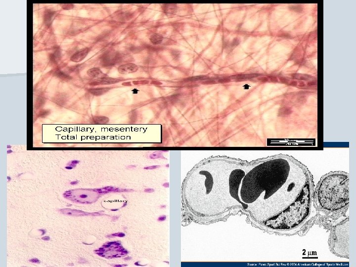
Blood capillaries Network of the smallest, thin- walled vessels, situated between arterial and venous portion of circuit
Blood capillaries n diameter from about 8 µm (to 30 -40 µm) n lumen is lined by 1 -2 endothelial cell n reticular fibers surround the capillaries n capillary bed between arteries and veins n pericytes 3 types of capillaries continuous fenestrated sinusoids
Function of capillaries (1) n respiratory gasses, nutrients and waste products change between blood and tissues The illustration shows satistied cells in well vascularized tissue
Function of capillaries (2) n allow the blood cells to pass throughout their wall into the connective tissue (by diapedesis) Neutrophils microphages Eosinophils Basophils heparinocytes Lymphocytes Monocytes macrophages
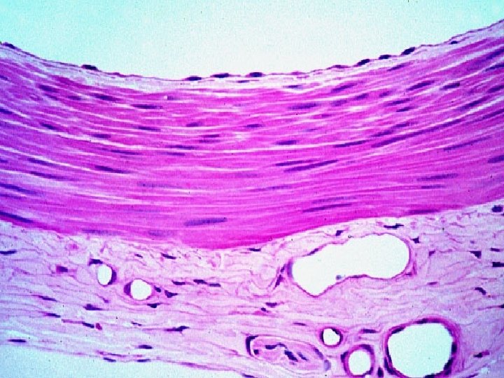
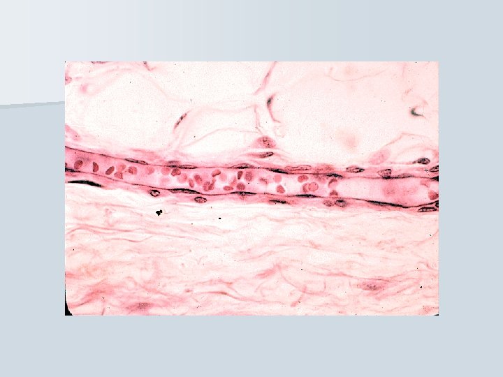
Continuous capillaries The smallest: cca 8 m n The wall: - endothelium – 1 -2 cells (zonulae occludentes and nexuses) - lamina basalis - pericytes - reticular fibers n n only allow small molecules, water and ions to diffuse Example of occurrence: muscle tisue, brain
Capillary
Fenestrated capillaries n n Endothelial cells with fenestra („windows“) 70 nm , diaphragm (thinner than plasma membrane) boards fenestrum continuous basal lamina in the organs with quic and intensive metabolism and substances change allow small molecules and limited amounts of protein to diffuse Exampl of occurrence: intestinal villi, endocrine glands
Capillaries with pores n special type of fenestrated capillaries n not fenestra with diaphragm, but opened pores are in endothelium n in glomeruli of renal corpuscles Pores capillary lumen
Sinusoidal capillaries (sinusoids) n from 8 to 40 m n n endothelium – fenestra, pores and intercellular clefts; some cells are able to phagocyte incomplete basal lamina reticular fibers allow erytrhocytes and serum proteins to enter. Example of occurrence: liver, spleen, bone marrow
Remember! n Sinusoid = type of blood capillary (between arterial and venous part of bloodstream) n Sinus = venous sinus belong to venous, postcapillary part of bloodstream
Pericytes cytoplasmic processes around capillary, n contain actin, myosin, tropomyosin n their own basal lamina fuses together with that one of capillary n
Precapillaries - Postcapillaries 12 – 40 m n endothelium + LB, elastic + collagen fibers, smooth muscle cells n precapillary sphincters n to 200 m n endothelium + LB, smooth muscle cells n
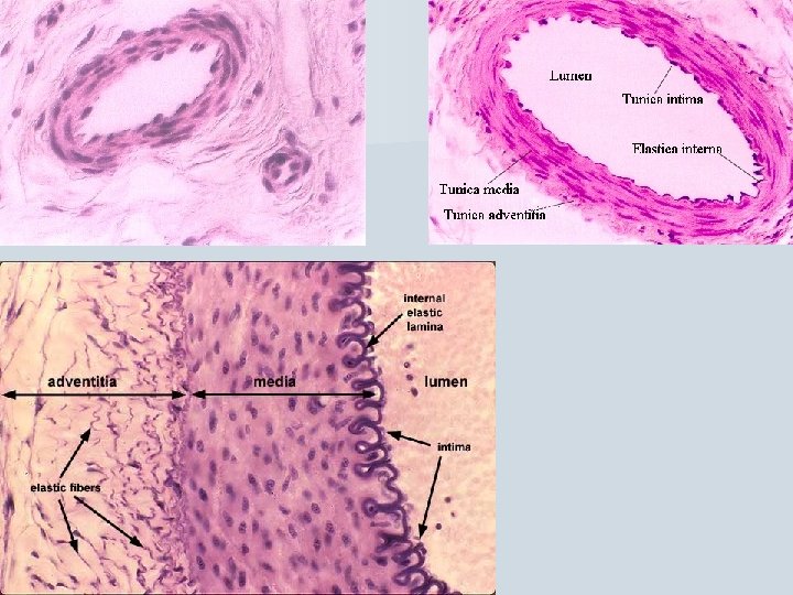
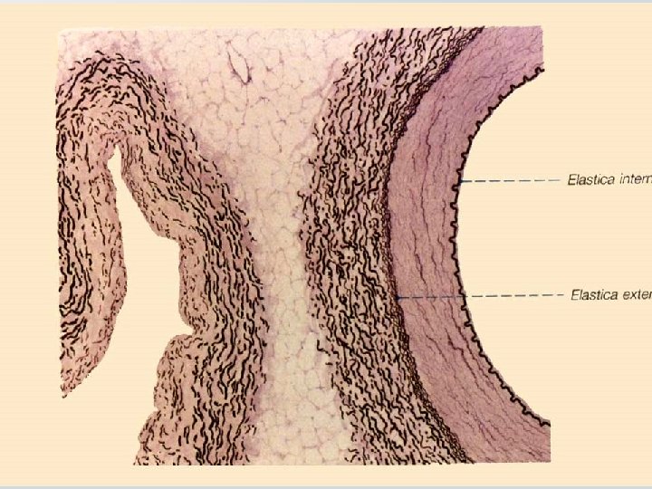
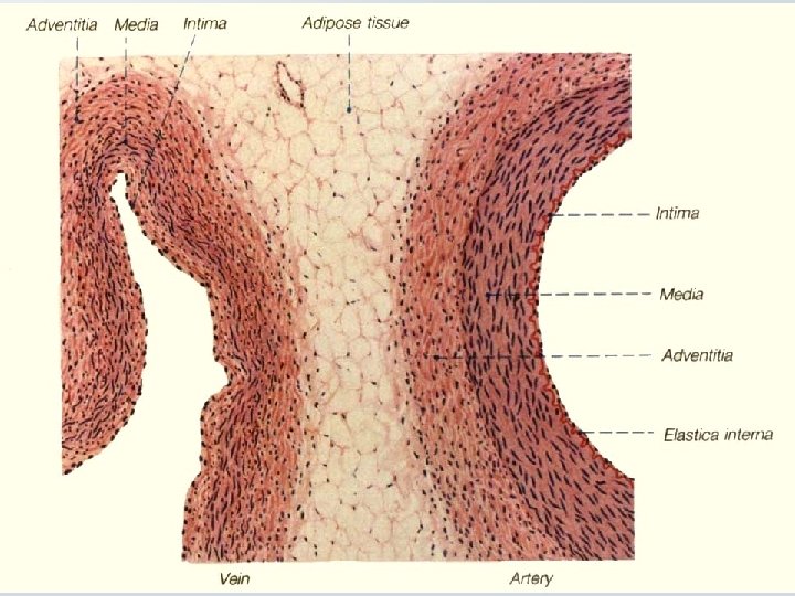
Structure of blood vessel wall – generally – n tunica interna (intima) endothelium + subendothelial connective tissue ____membrana elastica interna_____ n tunica media smooth muscle tissue – circularly oriented ____membrana elastica externa_____ n tunica externa (adventitia) loose connective tissue + nerves + vasa vasorum (+ longitudinal smooth muscle – only in veins)
Endothelium Tunica interna <lungitudinally> Membrana elastica interna Tunica media <circularlly> (Membrana elastica externa) Tunica externa <longitudinally>
Tunica interna (intima) TI n endothelium n subendothelial connective tissue – thin layer of elastic + collagen fibers (longitudinally oriented)
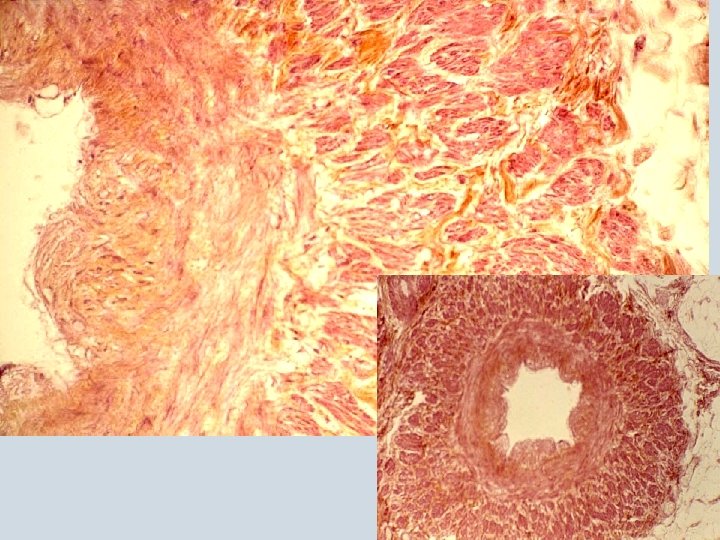
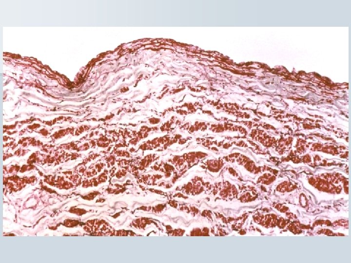
Tunica media TM n consists of smooth muscle cells and elastic membranes in varying proportions (circularly oriented) n is thicker in arteries than in veins Compare aw – vw:
Tunica externa (adventitia) TA n fibrous connective tissue + smooth muscle cells in veins (logitudinally) n is thicker in vein; is the thickest layer in large veins [1] and veins of low limbs [2] n contains vessels and nerves (vasa et nervi vasorum) in large vessels 1 2
Structural differences between arteries and veins – generally: + ++ + (+) YES Membrana elastica int. (YES) thick thin (YES) Membrana elastica ext. NO thin thick
Compare the wall structure of artery and vein adventitia media (+ intima)
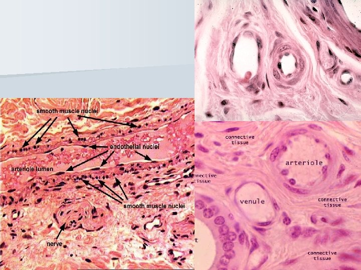
Arterial part of bloodstream According to diameter, morphological differences and ratio of elastic fibers and smooth muscle cells: n Arterioles < 0. 5 mm n Muscular arteries (small and middle-sized) 0. 5 – 1 mm n Elastic arteries (large: aorta and arteries growing from aorta)
Arteriole < 0. 5 mm The wall n TI: endothelium + subendothelium n n membrana elastica int. TM: smooth muscle cells (cca circular 5 layers) n TA: fibrocytes, reticular (+collagen) fibers n
Muscular artery TI: endothelium + subendothelium (with smooth muscle cells (longit. ) TM: up to 40 layers of smooth muscle cells, elastic and collagen fibers n membrana elastica ext. n TA: loose connective tissue TM+TA arrangement is spiral, but … ina n ud membrana elastica int. ng it n l“ „circular“ „lo n
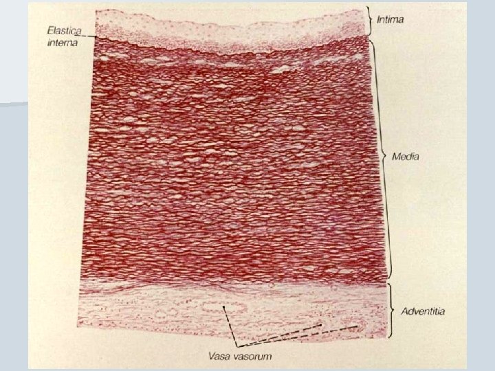
Elastic artery n TI: endothelium + subendothelium (100 m wide layer of connective t. ) TM: up to 40 -60 layers of fenestrated elastic membranes, small amount of smooth muscle cells and reticular fibers n TA: loose connective tissue (+ vasa et nervi vasorum) n
Different types of arteries n Arteriovenous anastomosis (artery contains smooth muscle cells in the wall before vein) n Arteries with intimal pillows (smooth muscle cells form pillows in t. media) – lumen can be closed by their contraction
Portal circulation: arterial or venous n two capillary systems side-by-side 1 2 3 capillaries vessel capillaries WHERE? 1: glomerulus eferent arteriole renal tubules capillaries in KIDNEY 2: GIT organs vena portae hepatic sinusoids in LIVER 3: hypothalamus hypophyseal vein adenohypophysis in HYPOPHYSIS
Venous part of bloodstream n Venules 0. 2 – 1 mm n Small and medium sized veins 1 – 9 mm n Large veins (v. cava inf. et. sup. - the largest vein) n Valves - pocket-like duplication of endothelium scaffolded by elastic c. t. - protection against venous reccurence
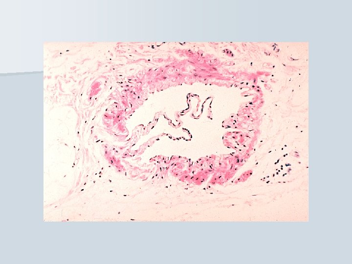
Venule n n n < 0. 2 - 1 mm The wall TI: endothelium only TM: smooth muscle cells (cca circular 1 -3 layers) TA: thick layer of loose connective tissue
Small and medium-sized venules Vein + artery 1 – 9 mm n TI: endothelium + irregular layer of subendothelium + valves n TM: irregular, thin layer of smooth muscle cells, elastic and collagen fibers n TA: thick layer of loose connective tissue with smooth muscle cells n Vein from lower part of body
Large veins n TI: endothelium + subendothelium (+smooth muscle cells) TM: thin layer of connective tissue + reduced amount of smooth muscle cells n TA: longitudinal bundles of smooth muscle cells in loose connective tissue (vasa et nervi vasorum) n
The heart is the hardest working muscle in the human body. Hollow muscular organ – blood pump n Rythmic contraction n Involuntary muscle n
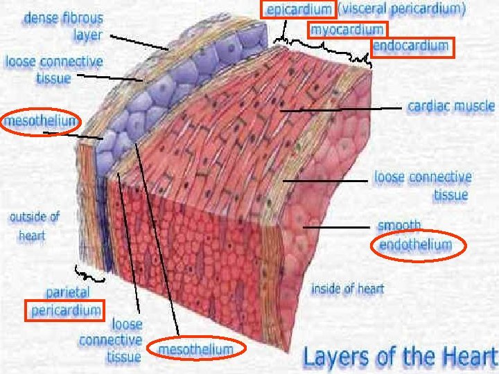
Pericardial sac: pericardium + epicardium PERICARDIUM Outer (parietal) layer Pericardial cavity EPICARDIUM Inner (visceral) layer Pericardial cavity - contains 15 – 50 ml of serous fluid serves as lubricans; - is lined with mesothelium
The wall of heart Epicardium n Myocardium n Endocardium ---------n
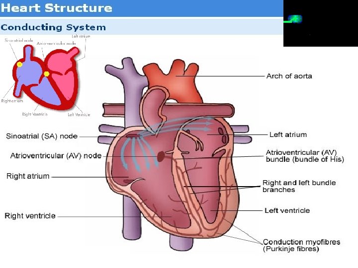
Endocardium (homologous to intima of blood vessels) Consists of: n Endothelium n Subendothelium – thin connective tissue layer Elastic-muscular layer – dense c. t. (elastic fibers, smooth m. cells) n Subendocardium – c. t. + vessels, nerves and distal part of conducting n system (ventricular bundles and Purkinje fibers) Purkinje fibers Purkinje cells
NEXUSES
Myocardium cardiomyocytes „working“ „conducting“ n cells in right ventricle – natriuretic factor (when n n n intravascular volume increases, this factor is released and causes natriuresis and diuresis in kidney) atrial myocardium is thinner than ventricular „left heart“ myocardium is thinner than „right heart“ cords of cardiomyocytes are ended on heart skelston damage of myocardium - infarction low regeneration of myocardium – by scar (decreases function of heart muscle)
Heart skeleton Trigonum fibrosum sin. Trigonum fibrosum dx. Anulus fibrosus sin. Anulus fibrosus dx. Pars membranacea interventricularis
Endocardial valves Plates of dense connective tissue (continuous with heart skeleton) covered with endocardium.
Epicardium Mesothelium lines pericardial space and so it covers outer surface of epicardium and inner surface of pericardiu l sp a i d r a ric Pe ace mesothelium connective tissue subepicardial c. t. myocardium
Practice No. … n n n n Slides: 59. Artery of the muscular type with a vein (HE) 60. Artery of the muscular type with a vein (orcein) 61. Aorta (cross-section, HE) 62. Aorta (cross-section, orcein) 63. Vena cava (HE) 65. Myocardium (Heidenhain´s hematoxylin)
Embryology: Cardiovascular system Development of heart and vessels
Embryological Timetable n Week 3 day 20 – cardiogenic plate n day 21 – endocardial tubes n Week 4 day 22 – fusion into single tube n day 23 – first contraction n day 25 – cardiogenic loop n Week 7 day 49 – 4 -chamber heart
The cardiogenic (heart-forming) region is initially located at the anterior rim of the embryonic disc (rostral to the prechordal plate). As the embryo grows, the developing heart assumes a position ventral to the forming forebrain and foregut.
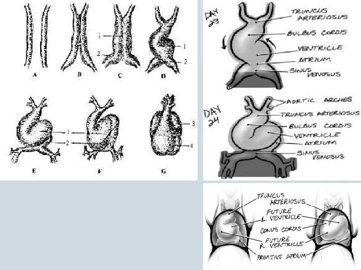
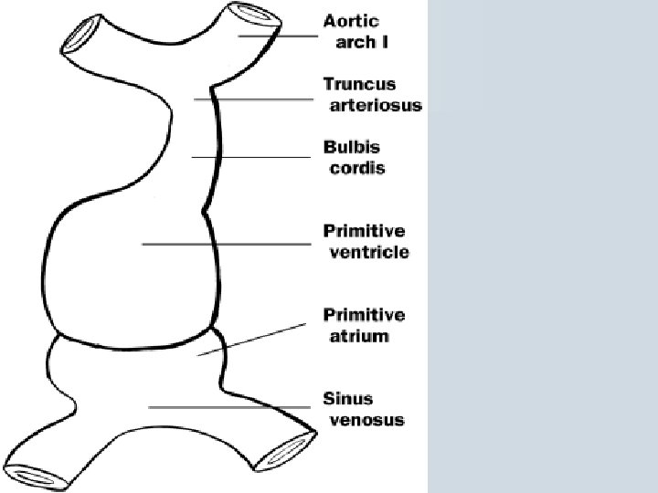
Fusion of the vascular channels in the cardiogenic region results in formation of endothelially-lined heart tube which is surrounded by a layer of splanchnic mesoderm. The intraembryonic coelom in this region becomes the pericardial cavity. the position of the forebrain, foregut and septum transversum relative to the developing heart. septum transversum is located just below the developing heart, which at this stage, begins to beat. As the heart tube elongates and begins to loop, the blood flows into the sinus venosus, then into the primitive atria, ventricles and bulbous cordis before entering the visceral arch vessels.
As the heart tube elongates and bends, the atrial segment assumes a position cranial to the ventricular segment.
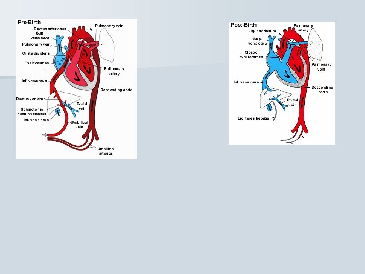
the dorsal mesocardium suspends the heart from the dorsal body Blood flow into the sinus venosus is from three pairs of vessels: the cardinal veins that drain the embryo proper, the vitelline veins from the yolk sac, and the umbilical veins from the placenta.
The umbilical and vitelline veins traverse the liver which forms within the tissue of the septum transversum. Ductus venosus (Arantii) !
Primitive blood circulation ( fetal blood circulation)
the course of blood flow through this part of the heart. The truncus arteriosus will later divide to form the aorta and pulmonary trunk. superior, inferior, right, and left endocardial cushion are around the atrioventricular canal. The superior and inferior cushions must fuse to separate the atrioventricular (AV) canal into a right and left channel. cushions form also to separate the outflow tract of the heart into a pulmonary and aortic trunk.
neural crest cells that form at the level of the 4 th and 6 th aortic arches populate the forming truncal cushions. These cushions must fuse to form the aorticopulmonary septum separating pulmonary and aortic trunks. change in position is indicative of the spiraling of the aorticopulmonary septum, aorta and pulmonary artery
Fusion of the outflow tract cushions results in separation of the blood flow; the blood exits the left ventricle through the aorta and exits the right ventricle through the pulmonary artery. sinoatrial orifice since it is at the junction of the sinus venosus and the primitive atrium. The atrial chambers are divided by septum primum, which grows down to fuse with the endocardial cushions.
An opening, the ostium sccundum, forms in the septum primum. Septum secundum, remains incomplete. The resulting opening is termed the foramen ovale.
The primitive atria form the auricles, the rough-walled portions of the definitive atria. Incorporation of the walls of the sinus venosus on the right and the pulmonary veins on the left forms the smooth-walled portions of the definitive atria.
The aortic vessels, which extend from the aortic sac to the paired dorsal aortae, develop and regress asymmetrically. The left 4 th arch and the left 6 th arch. . .
contribute to the aortic arch and the pulmonary artery and ductus arteriosus