Resting Membrane Potential Esraa kiwan Membrane Potential n

Resting Membrane Potential Esraa kiwan

Membrane Potential n Membrane potential: separation of opposite charges across the plasma membrane n n n difference in the relative number of cations (+ve) & anions (-ve) in the ICF & ECF These separation produce energy differences between inside and outside which called potential This energy is electrical energy and is measured by Millivolts
 and (-ve)")
Membrane Potential n n No membrane potential An equal number of (+ve) and (-ve) charges are on each side of the membrane Membrane potential exist Unequal number of (+ve) and (-ve) charges are on each side of the membrane

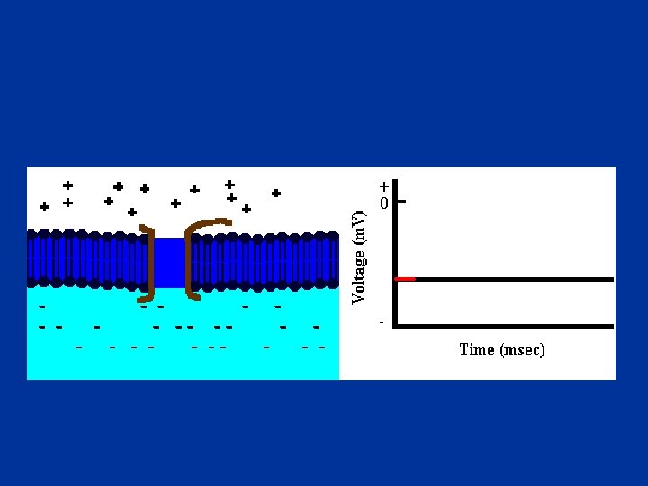
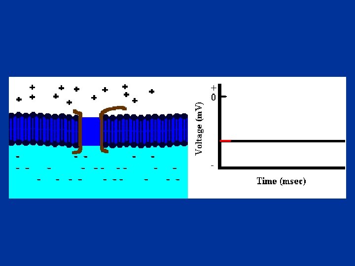
Membrane Potential n Separated charges accumulated in a thin layer along the outer and inner surface of the plasma membrane

Membrane Potential n n The magnitude of the potential depends on the degree of separation of the opposite charges The greater the number of separated charges the larger potential

Resting Membrane Potential n All plasma membrane of all living cells have resting membrane potential: n Varies in value according to the type of the cell: cell -70 m. V n Skeletal and cardiac muscle -80 to -90 m. V n Smooth muscle -60 m. V n Nerve The sign always represent the polarity of the excess charge on the inside of the membrane n Inside is more -ve than outside n

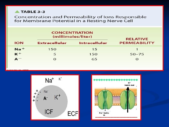
Resting Membrane Potential In the body, the electrical charges are carried by ions n The ions primarily responsible for the generation of resting membrane potential are: n • Na + • K+ • A - (-ve charged intracellular proteins)

Resting Membrane Potential n The electrical prosperities of the cell membrane are due to: Unequal distribution of a key ions across the plasma membrane n Selective movement of these ions through plasma membrane n

Resting membrane potential n Unequal distribution of a key ions between ICF and ECF are maintained by Na+ /K + pump: • Active transport mechanism • Pumps 3 Na+ outside, 2 K+ inside • Maintains high [K+] inside the cell, high [Na+] outside • Unequal transport generates a membrane potential • Outside becoming more +ve than inside (more +ve are transported out than in)

Resting membrane potential n 80% of resting membrane potential is caused by the passive diffusion of K + and Na + down concentration gradient n Na + & K + concentration gradient maintained by Na + /K + pump K + permeability about 50 -75 X than for Na + n K + ions influence the resting membrane potential than Na + n n Na + / K + pump responsible of 20% of resting membrane potential through maintaing concentration gradient for Na +, K +

Resting Membrane Potential n Electrochemical gradient for Na+, K+ in resting state: n Concentration gradient: n Na + inside n K + outside n Electrical gradient ( toward the negative charge) n Na + inside n K + inside
:")
Resting Membrane Potential n K + equilibrium potential (EK+ ):
:")
Resting Membrane Potential n Na + equilibrium potential (ENa+ ):

Resting Membrane Potential n n Effect of both K + and Na + on resting membrane potential: n Na + equilibrium potential (ENa+)= +60 m. V n K + equilibrium potential (EK+ )= -90 m. V K + exerts the dominant effect on the resting membrane potential, because the membrane is more permeable to K + resting membrane potential is much closer to EK+ than to ENa+ which is -70 m. V
 able to")
Changes in Resting Membrane Potential n n Excitable cell (Nerve & muscle) able to undergo transient, repaid changes in their resting membrane potential These changes serve as electrical signal: Nerve cell: use electrical signal to receive, process, initiate, and transmit messages n Muscle: these electrical signals initiate contraction n
")
Changes in Resting Membrane Potential • Polarization: resting state (-ve inside compared with outside) • Depolarization: decrease the negativity of the membrane ( moving toward 0 m. V) • Repolarization: the membrane returns to resting potential • Hyperpolarization: the membrane become more negative than at rest (increase negativity)

Changes in Resting Membrane Potential n Changes in ions movement a cross the membrane (changes in membrane permeability): If net inward of +ve increases compared to the resting state, the membrane become depolarize (decrease -ve inside) n If net outward of +ve increases compared to the resting state, the membrane become hyperpolarized (increase -ve inside) n

Changes in resting membrane potential n Changes in membrane potential could be: Graded potential n Action potential n

Action Potential n n Brief, repaid, large changes in membrane potential The potential actually reverses the inside of the cell become more +ve than outside

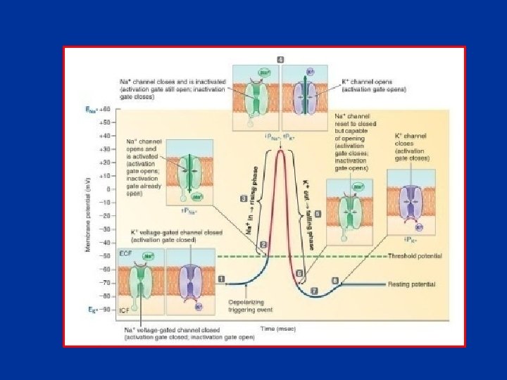
Action Potential Stages of action potential n n n Triggering event causes the membrane to depolarize from resting potential (-70 m. V) Depolarization proceeds slowly at first, until it reaches a critical level, (threshold potential) (-50 _ -55 m. V) At threshold potential, an explosive depolarization takes place: n n n Upward deflection to +30 m. V Reversal of polarity( the inside of the cell becomes more +ve than outside) Then, the membrane repolarizes, dropping back to resting potential

Action Potential n Action Potential is caused by marked changes in membrane permeability of ions: n At rest, membrane permeability to K + is much higher than it for Na + n So, K + makes the greatest contribution to the resting membrane potential. n This will completely change during action potential, due to activation of Voltage. Gated Na + & K + Channels

Action Potential Voltage Gated Channels n n The channels that their state (opening or closure) depends on the voltage changes of the membrane At resting potential, all the Voltage-Gated Channels are closed

Action potential n When a membrane starts to depolarize toward threshold, some of the voltage-gated Na+ channels will open n Then, Na+ will move down its electrochemical gradient into the cell n This influx of Na+ will depolarize the membrane more, & so more channels will open… & so on

Action potential n At threshold, there is an explosive increase in permeability of Na+ (≈600 times more permeable to Na+ than to K+) n The membrane potential will become closer to ENa+= +60 m. V n n The potential will be (+30 m. V) Before reaching the Peak. Potential, Na+ channels will be inactivated, so no more Na+ leak to the inside

Action potential n n Simultaneous with inactivation of Na+ channels, the voltage-gated K+ channels starts to slowly open, with maximum opening occurring at the peak of action potential These channels will greatly increase the K+ permeability (≈300 times more than Na+) n This will lead to marked efflux of K+ out of the cell, down its concentration gradient n This will rapidly restore the negative resting potential
 is caused")
Action potential n n Rising phase: from threshold to (+30 m. V) is caused by Na+ influx Falling phase: from (+30 m. V) to resting potential is caused by K+ efflux Ions distribution is restored by the action of Na+-K+ pump

Action potential Action Potential is an all-or-none response n n n If the membrane is not depolarized to its threshold level, no action potential is produced If the stimulus strength is increased more than what is needed to produce action potential there will be no change in magnitude of action potential (produce same action potential) Strong stimulus produces more action potential (increase in #), and cause more cells to reach their threshold

Action potential Propagation of action potential throughout nerve fiber Once action potential is generated in any part of neuron, it will propagated all the way through the axon to reach axon terminal n Action potential spreads throughout the membrane in undecresing fashion (non-decremental) n

Neuron

Action potential Propagation of action potential throughout nerve fiber n Contiguous conduction in unmyelinated fibers: n Action potential at one point will excited the adjacent point by increasing the permeability of adjacent point to Na+ causing generation of Action Potential

Action potential Propagation of action potential throughout nerve fiber n Saltatory conduction in myelinated fibers

Refractory Period n n A period during which a new action potential cannot be initiated by normal events in a region that has just undergone an action potential Two phases: n Absolute Phase: a second action potential can’t be produced, no matter how strong is the stimulus. The membrane completely unresponsive to second stimulus at all n Relative Phase: during which the second action potential can be produced only by an exceptionally strong triggering

Prosperities of Action Potential n Is an “all or none” event (either pass threshold or dosen’t) n n Is propagated down the axon in nondecremental fashion Has a fixed amplitude ( action potential do NOT change in Height) n Has a refractory period (in which stimulation will not produce an action potential) •

Graded Potential n Is a local change in the membrane n n n potential that occurs in varying grades (changes may be 10 or 20 m. V) The magnitude vary with the strength of the stimulus, the stronger the triggering event the larger the graded potential The magnitude of the graded potential continues to decrease the farther it moves away from the initial active area. (Decremental) Graded potential do not have a refractory period

n A neuron may terminate at: n A muscle n A gland n Another neuron

Synaptic Transmission n n A synapse is the junction between two neurons It involves the junction between an axon terminal of one neuron (presynaptic neuron), & the dendrites or the cell body of a second neuron (postsynaptic neuron)
 crosses the synapse by")
Synaptic Transmission n • The action potential (the electrical signal) crosses the synapse by chemical means to induce electrical changes in other neuron The chemicals released from one neuron to induce electrical changes in next neuron are neurotransmitters n Acetylcholine n Norepinephrine n Histamine n Glycine n Gamma-aminobutyric acid (GABA) Synapses operate in one direction only
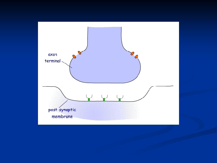

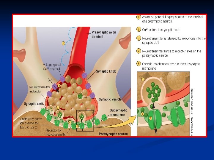
Synaptic Transmission n Events taking place in a synapse: n Propagation of an action potential to the axon terminal of a presynaptic neuron n The arriving action potential produces an influx of calcium ions through voltage-dependent calcium ion channels ([Ca+2] in ECF is much higher than that inside the synaptic knob) n n n Ca+2 induces the release by exocytosis of neurotransmitters from synaptic vesicles into the synaptic cleft After diffusing across the cleft, the neurotransmitter binds with its receptor sites on the subsynaptic membrane This binding triggers the opening of specific ion channels in the subsynaptic membrane. This alters the permeability of the postsynaptic neuron (Chemical messenger-gated channels)

Synaptic Transmission The changes in postsynaptic neuron by neurotransmitter could be excitatory or inhibitory

Types of synapses n Excitatory synapse: n n n the response to the receptor-neurotransmitter combination is an opening of Na+ & K + channels within the subsynaptic membrane. Na+ moves into the postsynaptic neuron & K + out of it, but to a less extent The result is a net movement of positive ions into the cell Small depolarization in the postsynaptic neuron Activation of one excitatory synapse can rarely depolarize the postsynaptic membrane sufficiently to bring it to threshold, but it brings it closer to threshold This postsynaptic potential change is called: Excitatory Post. Synaptic Potential (EPSP)

Types of synapses n Inhibitory synapse: n n The response to the receptorneurotransmitter combination is an opening of K+ & Cl - channels within the subsynaptic membrane The resulting ion movements bring about a small hyperpolarization of the postsynaptic neuron (greater internal negativity) This moves the membrane potential away from threshold This small hyperpolarization of the postsynaptic cell is called: Inhibitory Post. Synaptic Potential (IPSP)

Synaptic Transmission The time needed for action potential to cause the release of the neurotransmitter into synaptic cleft and produce its effect on postsynaptic neuron is called synaptic delay, its about 0. 5 -1 msec

Synaptic Transmission n Fate of neurotransmitter that released from presynaptic membrane: n n Uptake of the neurotransmitter into the cytoplasm of the presynaptic membrane (reused again) Enzymatic deactivation: inactivation of neurotransmitter by certain enzyme
: summation of all potentials ( both")
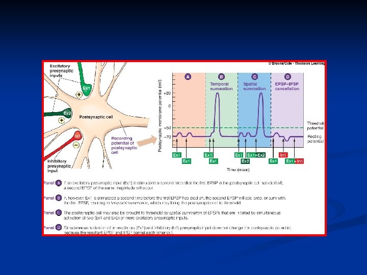
Synaptic Transmission n Grand Post. Synaptic Potential (GPSP): summation of all potentials ( both EPSP + IPSP) that occur at the same time The postsynaptic neuron can be brought to threshold by: n Temporal summation: is the summing of several EPSPs occurring very close together in time because of successive firing of a single presynaptic neuron n Spatial summation: is the summation of EPSPs originating simultaneously from several different presynaptic inputs (from different points in space) If an excitatory & an inhibitory input are simultaneously activated, the concurrent EPSP & IPSP more or less cancel each other out.

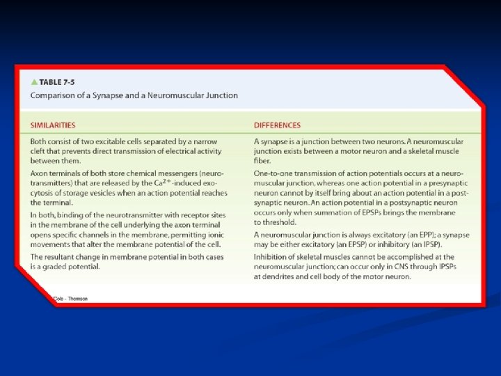
Neuromuscular Junction

Neuromuscular Junction

Neuromuscular Junction

Neuromuscular Junction n n Factors affect transmission of impulse from nerve fiber to muscle fiber at neuromuscular junction: Botulinum toxin: blocks the release of Ach from nerve terminal. Inability of nerve impulses to pass to muscle cause muscle paralysis Venom of black widow spiders: explosive release of Ach at neuromuscular junction causing persistent contraction of muscle Organophosphates: inhibit Ach. E leading to continuous muscle contraction (leading to death because of respiratory failure) Curare: binds with Ach receptors on the motor end plate,
 - controlled")
Muscle 3 Types of Muscles 1. Smooth – involuntary, unstriated (not banded) - controlled by autonomic system 2. Cardiac – involuntary, striated 3. Skeletal – voluntary, striated

Structure of Skeletal Muscle

Structure of Skeletal Muscle n n n A band: consists of thick filaments along with the portions of the thin filaments that overlap on both ends of the thick filaments I band: consists of the remaining portion of the thin filaments that do not project into the A band H zone: The lighter area within the middle of the A band where thin filaments do not reach

Structure of Skeletal Muscle n Z line: Visiblel line in the middle of each I band, flattened disc-like cytoskeletal protein that connects the thin filaments n Sarcomere: The area between two Z lines (functional unit of skeletal muscle) n M line: extends vertically down the middle of the A band within the center of the H zone

Structure of Skeletal Muscle Thick filaments n n n Myosin forms the thick filaments A myosin molecule is a protein consisting of two identical subunits The protein’s tail ends are intertwined around each other, with the two globular heads projecting out at one end These heads form the cross bridges between the thick and thin filaments Each cross bridge has two important sites important to the contractile process: an actin binding site and a myosin ATPase site

Structure of Skeletal Muscle Thin filaments

Structure of Skeletal Muscle Thin filaments n n Composed of 3 proteins: actin, tropomyosin, and troponin Actin : n n n the primary structural proteins of the thin filament (It forms the backbone of the thin filament) Actin spherical in shape. ; joined into two strands and twisted together Each actin molecule has a special binding site for attachment with a myosin cross bridge

Structure of Skeletal Muscle Thin filaments n Tropomyosin: n n Threadlike proteins that lie end to end alongside the groove of the actin spiral Tropomyosin covers the actin sites that bind with the cross bridges thus blocking the interaction that leads to muscle contraction Tropomyosin is stabilized in this blocking position by troponin molecules, which fasten down the ends of each tropomyosin molecule Troponin: n protein complex consisting of 3 units: n Troponin T: that binds to tropomyosin n Troponin I: that binds to actin inhibitin the interaction between Actin & myosin n Troponin C: that can bind with Ca 2+ (When Ca 2+ binds to troponin, the shape of this protein is changed in such a way that tropomyosin is allowed to slide away from its blocking position)

Structure of Skeletal Muscle n Contractile proteins: n n n Actin and Myosin Binding of actin and myosin molecules at the cross bridges results in energy-consuming contraction of the muscle fiber Regulatory proteins: n n n Tropomyosin and Troponin Covering the binding sites for cross-bridge interaction between actin and myosin (preventing contraction) Exposing the binding sites for cross-bridge interaction between actin and myosin (permitting contraction)

Skeletal Muscle Contraction n Sliding filament mechanism: n Contraction is accomplished by the thin filaments sliding closer together between the thick filament

Skeletal Muscle Contraction n Sliding filament mechanism:

Skeletal Muscle Contraction n Cross bridge activity:

Skeletal Muscle Contraction

Skeletal Muscle Contraction
- Slides: 70