Heart Sounds and Murmurs Red very important Green


 or Phonocardiography")


. �")

 • • Systolic between")
")


 Murmur: � Most common kind of heart murmur. Usually Crescendo-decrescendo")


 Systolic murmur Late Holo (Pan) Early Diastolic murmur")


- Slides: 26
Heart Sounds and Murmurs Red: very important. Green: Doctor’s notes. Pink: formulas. Yellow: numbers. Gray: notes and explanation. Physiology Team 436 – Cardiovascular Block Lecture 10 1 Lecture: If work is intended for initial studying. Review: If work is intended for revision.
Objectives � � � Normal heart sounds and its leading causes Causes of abnormal heart sounds Describing abnormal heart sounds Different examples of abnormal heart sounds List the standard positions of stethoscope placement for cardiac auscultation. Distinguish between the 1 st, 2 nd, 3 rd and 4 th heart sounds. Explain physiological splitting of the 2 nd heart sound and depict the pathophysiology of fixed and paradoxical splitting of the 2 nd heart sound. Define and classify cardiac murmurs and list cause of heart murmurs. Outline how heart murmurs are described and graded. Outline the haemodynamic changes and murmurs in conditions of: Aortic stenosis § § § 2 Study Smart: focus on mutual topics. Aortic regurgitation Mitral stenosis Mitral regurgitation Mitral valve prolapse Ventricular septal defect Patent ductus arteriosus
Heart Sounds � Detected over anterior chest wall using either auscultation (Stethoscope) or Phonocardiography (sound recording device) � Best heard at 4 certain areas: (actually we can hear the heart sound anywhere, but these windows provide more obvious sounds) 1. Pulmonary area: 2 nd (2 nd – 3 rd in boys’ slides) left intercostal space 2. Aortic area: 2 nd (2 nd – 3 rd in boys’ slides) right costal cartilage 3. Mitral area: 5 th left intercostal space crossing mid-clavicular line (9 cm (2. 5 -3 in) from sternum) 4. Tricuspid area: lower part of sternum towards right side. Tricuspid area: Left lower sternal border. (in boys’ slides) 4 heart sounds can be detected: 1 st & 2 nd hear sounds are usually audible 3 rd & 4 th heart sounds sometimes detected. � ONLY IN MALES’ SLIDES Phonocardiogram is a graphic recording of the heart sounds. It involves picking up the sonic vibrations from the heart through a highly sensitive microphone. Such waves are then converted into electrical energy and fed into a galvanometer, where they are recorded on paper. • • Generally the sounds produced by each valve is best heard over a particular region of the chest. In general both the first and the second heart sounds can be heard at all areas. 3
Normal Heart Sounds: S 1: � Due to closure of the atrio-ventricular valves. � It marks beginning of ventricular systole. � Recorded at the beginning of the ‘isovolumemetric contraction’ phase. � Long in duration ( 0. 15 sec. ) longest duration of all sounds. � Of low pitch and loud (LUB). � Frequency range (25 -35) (25 -45 in boys' slides) Hz. � Best heard at Mitral & Tricuspid areas. S 1 is soft when the heart rate is low, because the ventricles are well filled with blood and the leaflets of the AV valves float together before systole. S 2 is loud and sharp when the diastolic pressure in the aorta or pulmonary artery is elevated, causing the respective valves to shut briskly at the end of systole. ONLY IN MALES’ SLIDES S 2: � Due to closure of semilunar valves. � Marks the beginning of ventricular diastole and end of ventricular systole. � � Recorded at the beginning of the ‘isometric relaxation’ phase. Short in duration (0. 11 -0. 125 sec. ) (0. 12 sec. in boys' slides). Of high pitch, soft, and sharp (DUB). Frequency : 50 Hz. � Best heard at Aortic & Pulmonary areas. � � 4 Usually the left side closes before the right side, for example semilunar aortic valve closes before semilunar pulmonary valve, but because the duration between them is very short I heard them as one sound (DUB) ventricular systole is between S 1 and S 2, and ventricular diastole is between S 2 and the new S 1
Normal Heart Sounds S 2: � S 2 splits physiologically into 2 sounds during INSPIRATION (physiological splitting) � This splitting occurs due to delay in closure of pulmonary valve S 3: � Recorded during the ‘rapid filling’ phase, due to rush of blood into the ventricle. � Duration 0. 05 sec. � S 3 is usually not audible(soft and very low pitch) � heard in children and young individuals. � Best heard at Mitral area. S 4: � Recorded during ‘atrial systole. ’ � Duration 0. 04 sec. � S 4 is usually not audible (very low pitch) � Usually heard in elderly. � Best heard at Mitral area. 5 ONLY IN MALES’ SLIDES o Normally, only S 1 and S 2 are heard with the stethoscope o S 3 and S 4 are detectable by phonocardiogram. o S 3 coincides with the period of rapid ventricular filling and occurs during transition between rapid filling and slow filling of ventricle, It’s probably due to vibrations set up by the inrush of blood. o S 3 is heard about one third of the way through diastole in children and many normal young individuals. o S 4 is caused by oscillations of the ventricles during atrial contraction. o Occasionally S 4 is heard in normal individuals.
ONLY IN MALES’ SLIDES Splitting of S 2 It has four types: During inspiration the Intra pleural pressure becomes more negative and that triggers stronger blood suction from systemic circulation thus increase Venous return. More venous return will increase blood in RV, RV takes longer time to eject blood (pulmonary valve’s closure is delayed) 1 -Physiological ü ü ü 6 During inspiration, the aortic valve closes before pulmonary valve → reduplication (physiologic splitting of S 2. The increased venous return to the right side of the heart delays closure of the pulmonary valve. The right ventricle has more blood than usual to eject and it thus takes more time. No splitting of the second heart sound is normally seen during expiration. 2 -Fixed ü Splitting of S 2 is heard both during inspiration and expiration, with the aortic valve closing before the pulmonary valve. ü This is heard in cases of ASD.
ONLY IN MALES’ SLIDES 3 -Wide ü A split in the second heart sound during inspiration may become wider and the split may also be seen during expiration if: 1. There is a delay in the closure of the pulmonary valve (as would be seen in right bundle branch block due to delay in right ventricular depolarization and contraction). 2. The aortic valve closes earlier than normal (this is seen with either mitral regurgitation or ventricular septal defect). What causes pulmonary valve to close later ( later than the physiological splitting) • Right bundle branch block Late depolarization of RV pulmonary valve takes more time to close Pulmonary stenosis RV takes more time to eject pulmonary hypertension • • What causes aortic valve to close faster • Mitral regurgitation: during systole of LV, blood is ejected into LA and aorta finishes systole faster aortic closes faster VSD ( ventricular septal defect ) defect between LV and RV, LV started systole blood is ejected into aorta and RV finishes systole faster aortic closes faster • 7 4 -Paradoxical (Reversed) ü Reversed (paradoxical) splitting of the second heart sound is typically heard during expiration, with the pulmonary valve closing before the aortic valve. No splitting is apparent during inspiration, since the pulmonary valve is closing earlier (relative to the aortic valve) than normal. ü This may be caused by the following: ü Delayed onset of left ventricular systole (example: left bundle branch block). ü Prolonged left ventricular systole (examples: aortic stenosis, severe hypertension, left-sided congestive heart failure). ü Early onset of right ventricular systole (example: Wolff. Parkinson White syndrome). Pulmonary valve closes before aortic valve What causes it? What happened to RV? Finished its systole faster, how? Accessory pathway from RA to RV (without passing AV) faster depolarization of RV finishes systole faster pulmonary valve closes faster What happens to LV? Systole takes longer, How? Left bundle branch block depolarization is slower Or aortic stenosis ( LV will take more time to eject aortic valve closure is delayed) / or high aortic pressure
Significance of Heart Sounds � Important for diagnosis of abnormal heart sounds (murmurs). � They are abnormal extra heart sounds heard during heart beat cycle. � They are produced by turbulence (abnormal patterns) of blood flow through the heart and its valves. � Murmurs are longer than heart sounds. � What makes noises in the heart: 1. Valves closing (normal heart sounds) - atrioventricular (S 1) - Semilunar (S 2) 2. Increased intra-cardiac hemodynamics (murmurs): -Blood striking left ventricle (S 3, S 4) -Increased blood flow across normal valves -Turbulent flow through abnormal valves -Turbulent flow through septal defect Problems in the valve: either complete closer of the valve, or stenosis or dilatation of the valve. 8
Causes of Murmurs ONLY IN MALES’ SLIDES Physiological Innocent murmurs: common in children and young adults. Pathological 9 Physiological murmurs(normal murmurs) ● Increased blood flow across normal valves ● Due to pregnancy, fever, anemia, hyperthyroidism, and children ● ﻓﺒﺎﻟﺘﺎﻟﻲ ، ﺑﺎﻟﻘﻠﺐ ﻣﺸﻜﻠﺔ ﻋﻨﺪﻫﻢ ﻣﺎ ﻟﻜﻦ ﻣﺮﻳﻀﻴﻦ ﺻﺢ ﻫﻢ ﻫﻨﺎ ﺍﻟﻤﻘﺼﺪ ﻣﺸﻜﻠﺔ ﻭﺟﻮﺩ ﺑﺴﺒﺐ ﺗﻜﻮﻥ ﻣﺎ ﺑﺄﺴﻤﻌﻬﺎ ﺍﻟﻠﻲ ﺍﻟﻄﺒﻴﻌﻴﺔ ﺍﻟﻐﻴﺮ ﺍﻷﺼﻮﺍﺕ ﺑﺎﻟﻘﻠﺐ Pathological murmurs • Turbulent flow through abnormal valves • Septal defect • Congenital • Tight valves (stenosis) • Leaky valves (regurgitation or insufficiency • Combination of Stenosis and Insufficiency.
How to Describe Heart Murmurs 1 -Timing (systolic or diastolic) • • Systolic between S 1 and S 2 • classified as early, mid, late, and holosystolic (pansystolic) • Diastolic • between S 2 and next S 1 • classified as early, mid, and late. • Continuous ONLY IN MALES’ SLIDES Systolic murmurs: Early very rare ( we can neglect them) mid occurs in rapid ejection phase, mid systolic murmurs = ejection systolic murmurs Pansystolic (holosystolic) heard through out the whole systole ( S 1 -S 2) Diastolic murmurs: Early, Mid, Late. Continuous murmurs: are heard through systole and diastole 10 2 -Shape • • Crescendo (grows louder, increasing intensity) Decrescendo (decreasing intensity) Crescendo-decrescendo (diamond-shaped): (increasing then immediate decreasing intensity). Plateau (uniform): the intensity of the murmur remains uniform throughout.
Cont. ONLY IN MALES’ SLIDES Variation with respiration: If murmur is louder (increases intensity) in inspiration originated from right side (pulmonary or tricuspid). If murmur is louder (increases intensity) in expiration it originated from left side (aortic or mitral). 11
Cont. 8 -Intensity Graded on 6 point scale according to Levine Scale ONLY IN FEMALES' SLIDES ﻣﺸﻜﻠﺔ ﻋﻨﺪﻩ ﻳﺘﻜﻮﻥ ﺑﺪﺍ ﺍﻟﻘﻠﺐ ﺇﻥ ﻣﻌﻨﺎﺗﻪ ﻫﺬﺍ ﻃﺒﻴﻌﻴﺔ ﻏﻴﺮ ﺃﺼﻮﺍﺕ ﻳﻄﻠﻊ ﻭﺑﺪﺍ A thrill is a slight palpable vibration felt by the hand over the chest wall. ONLY IN MALES’ SLIDES ﺃﺤﺲ ﺑﺎﻟﻤﺮﻣﺮ ﺑﻴﺪﻱ ﻛﺄﻨﻪ ﺧﺮﺑﺸﺔ ﺟﺎﻳﻪ ﻋﻠﻰ ﻳﺪﻱ ﺃﺴﻤﻊ ﺃﻘﺪﺭ ( )ﻣﺎﻳﻠﻪ ﺍﻟﺼﺪﺭ ﻋﻠﻰ ﻣﻨﻬﺎ ﺟﺰﺀ ﺍﻟﺴﻤﺎﻋﺎﺕ ﺣﻄﻴﺖ ﻟﻮ ﻳﻌﻨﻲ 12 Grade 1: Grade I/VI Grade 2: Grade II/VI Grade 3: Grade III/VI Grade 4: Grade IV/VI Grade 5: Grade V/VI Grade 6: Grade VI/VI
Heart Murmur Intensity ONLY IN FEMALES' SLIDES 13 ONLY IN MALES’ SLIDES
ONLY IN FEMALES' SLIDES Common Systolic Murmurs and Timing Aortic Stenosis ejection murmur May cause S 2 to become quieter or absent Pulmonary Stenosis ejection murmur Splitting of S 2 Mitral/tricuspid holosystoli c regurgitation Ventricular Septal Defect (VSD) Mitral valve prolapse 15 holosystoli c Mid-late systole You will hear click first For further information to help you understand heart murmurs and their various causes click here.
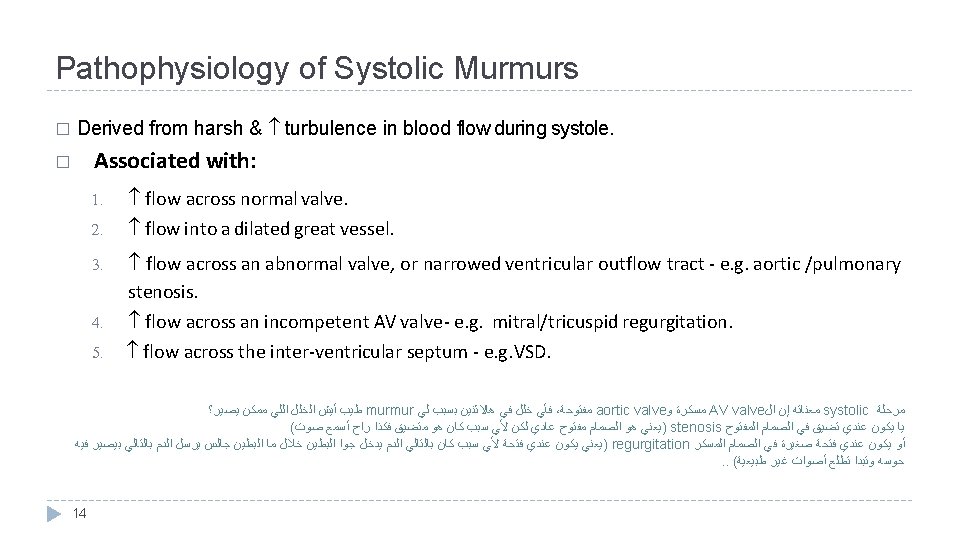
Systolic Murmurs Ejection (mid-systolic) Murmur: � Most common kind of heart murmur. Usually Crescendo-decrescendo (diamond shaped). � It may be: 1. Innocent: as in children and young adults. 2. Physiological: in hyper-dynamic states (increased intra-cardiac hemodynamics) such as anemia, pregnancy, fever, and hyperthyroidism. 3. Pathological: secondary to structural CV abnormalities such as aortic/pulmonary stenosis or hypertrophic cardiomyopathy, and mitral prolapse. � Pan-Systolic (Holosystolic) Murmurs: • Pathological. • Begins with S 1 immediately and continues until S 2. • Heard with: Mitral/tricuspid regurgitation and ventricular septal defect (VSD). 16
Cont. Aortic Stenosis: � Narrowing of aortic outflow tract causing obstruction of flow from left ventricle into ascending aorta. � Timing: mid-systolic (ejection) murmur. � Location: best heard at aortic area and it radiates along carotid arteries (going to neck). � Character: harsh, loud, may have associated thrill, ejection click. � Association: older age as a result of wear and tear of the aortic valve, bicuspid aortic valve, Scarring of the aortic valve due to rheumatic fever as a child or young adult. � The ESM of aortic stenosis has a crescendo-decrescendo contour and a gap between the end of the audible sound and S 2 (Splitting of S 2). ONLY IN MALES’ SLIDES • • • The narrow orifice of the aortic valve increases resistance of the aortic valve and slows the rate at which SV (stoke volume) is ejected. Ventricular systolic pressure increases to overcome the increased resistance of the aortic valve. Thus, there is a pressure gradient between the left ventricle and aorta during ejection. AS may result in concentric hypertrophy of the LV. Aortic stenosis causes reversed splitting of S 2 sound 17
Mitral Prolapse & Regurgitation Mitral Prolapse: � Bulging of one or both mitral valve leaflets in left atrium during left ventricular systole ﻓﺘﺮﺓ ﻓﻲ ﺇﻥ ﻗﻠﻨﺎ ﻛﻨﺎ cardiac cycle ﻣﺤﺎﺿﺮﺓ ﻣﻦ � Timing: mid-late systolic murmur � Location: best heard at the apex region � Character: mid systolic click � Association: 5% normal population, asymptomatic, sudden death Bulging ﻋﻨﺪﻱ ﺑﻴﻜﻮﻥ isometric contraction ﻟﻜﻦ prolapse ﻋﻨﺪﻱ ﺻﺎﻳﺮﺓ ﻛﺄﻨﻪ ﻳﻌﻨﻲ AV ﺑﺠﻬﺔ ﻓﺘﺮﺓ ﺑﺲ ﻷﻨﻬﺎ ، ﻣﺮﺽ ﺃﻲ ﻓﻴﻬﺎ ﻭﻣﺎ ﻃﺒﻴﻌﻴﺔ ﺣﺎﻟﺔ ﻫﺬﻱ ﺑﺘﻨﺘﻬﻲ ﻭﺧﻼﺹ isometric contraction Mitral Regurgitation: Retrograde flow from left ventricle to left atrium through an incompetent mitral valve. Timing: holosystolic murmur. Location: best heard at apex radiates to left axilla. Character: soft, high pitched, blowing. Association: Mitral valve prolapse, mitral valve myxomatous degeneration*, myocardial infarction, rheumatic heart disease, cardiomyopathy, endocarditis. The sound is of reasonably constant intensity throughout the ejection period. ﺇﻟﻰ ﻭﻳﺆﺪﻱ ﺍﻟﺪﻡ ﻣﻊ ﻭﻳﺘﺤﺮﻙ ﻣﻨﻪ ﺟﺰﺀ ﻳﻨﻘﻄﻊ ﻣﻤﻜﻦ ﺍﻟﺘﺠﻤﻊ ﻫﺬﺍ ، ﺍﻟﻤﺘﻀﺮﺭ ﺍﻟﺼﻤﺎﻡ ﻣﻨﻄﻘﺔ ﻓﻲ platelets ﻭ ﻟﻠﺒﻼﺯﻣﺎ ﺗﺠﻤﻊ ﻋﻨﺪﻱ ﻳﺘﻜﻮﻥ *ﻳﻌﻨﻲ stroke in the brain 18 ONLY IN MALES’ SLIDES 1. 2. Left atrial volume and pressure are increased during ventricular systole. Left ventricular volume and pressure are increased during diastole, but there is NO pressure gradient between the LA and the LV.
Diastolic Murmurs Almost always indicates heart disease � Two basic types: � Early decrescendo diastolic murmurs • Signify regurgitation flow through an incompetent semilunar valve • i. e. aortic/pulmonary regurgitation 19 Rumbling diastolic murmurs in mid or late diastole • Suggests stenosis of AV valve • i. e. mitral/tricuspid stenosis ONLY IN FEMALES' SLIDES Ø Common murmurs and timing: soft, blowing, gurgle 1. Aortic regurgitation: early diastole 2. Mitral stenosis: mid to late (presystolic) diastole
Cont. Aortic regurgitation: � Retrograde flow from aorta into LV through incompetent aortic cusps � Timing: diastolic (early) murmur � Location: best heard at 2 nd – 4 th left intercostal spaces with the patient sitting up, leaning forward, at end expiration. � Character: high pitched, loud blowing, decrescendo, it wanes with time as aortic pressure falls. � Association: aortic root degeneration, rheumatic heart disease, VSD w/aortic valve prolapse (kids. ) • • semilunar ﺍﻝ ﺇﻥ ﻣﻌﻨﺎﺗﻪ diastole ﻣﺮﺣﻠﺔ ﻓﺄﻲ ، ﻣﻔﺘﻮﺣﺔ AV valve ﻭ ﻣﺴﻜﺮﺓ valve ﻃﻴﺐ murmur ﻟﻲ ﺑﻴﺴﺒﺐ ﻫﺎﻻﺛﻨﻴﻦ ﻓﻲ ﺧﻠﻞ ﻳﺼﻴﺮ؟ ﻣﻤﻜﻦ ﺍﻟﻠﻲ ﺍﻟﺨﻠﻞ ﺃﻴﺶ ﺍﻟﻤﻔﺘﻮﺡ ﺍﻟﺼﻤﺎﻡ ﻓﻲ ﺗﻀﻴﻖ ﻋﻨﺪﻱ ﻳﻜﻮﻥ ﻳﺎ ﻋﺎﺩﻱ ﻣﻔﺘﻮﺡ ﺍﻟﺼﻤﺎﻡ ﻫﻮ )ﻳﻌﻨﻲ stenosis ﺃﺴﻤﻊ ﺭﺍﺡ ﻓﻜﺬﺍ ﻣﺘﻀﻴﻖ ﻫﻮ ﻛﺎﻥ ﺳﺒﺐ ﻷﻲ ﻟﻜﻦ ( ﺻﻮﺕ ﺍﻟﻤﺴﻜﺮ ﺍﻟﺼﻤﺎﻡ ﻓﻲ ﺻﻐﻴﺮﺓ ﻓﺘﺤﺔ ﻋﻨﺪﻱ ﻳﻜﻮﻥ ﺃﻮ ﻷﻲ ﻓﺘﺤﺔ ﻋﻨﺪﻱ ﻳﻜﻮﻥ )ﻳﻌﻨﻲ regurgitation ﺧﻼﻝ ﺍﻟﺒﻄﻴﻦ ﺟﻮﺍ ﻳﺪﺧﻞ ﺍﻟﺪﻡ ﺑﺎﻟﺘﺎﻟﻲ ﻛﺎﻥ ﺳﺒﺐ ﻓﻴﻪ ﺑﻴﺼﻴﺮ ﺑﺎﻟﺘﺎﻟﻲ ﺍﻟﺪﻡ ﻳﺮﺳﻞ ﺟﺎﻟﺲ ﺍﻟﺒﻄﻴﻦ ﻣﺎ ( ﻃﺒﻴﻌﻴﺔ ﻏﻴﺮ ﺃﺼﻮﺍﺕ ﺗﻄﻠﻊ ﻭﺗﺒﺪﺍ ﺣﻮﺳﻪ The aortic valve does not close properly at the beginning of diastole. ONLY IN MALES’ SLIDES As a result, there is retrograde flow of blood from the aorta into the ventricle during diastole. The amount of the blood regurgitated into the left ventricle may be as much as 60 -70% of the amount ejected during systole. Thus, there is: • Decreased aortic diastolic pressure. • increased left ventricular and aortic systolic pressures. • Increased aortic pulse pressure. 20
Cont. Mitral Stenosis: � Obstruction of flow from LA to LV during diastole because of a narrowed mitral orifice (Valve becomes thickened & calcified). � Timing: diastolic (mid-diastolic or presystolic) murmur with (opening snap) after closure of aortic valve. � Location: best heard at apex. � Characteristic: low pitched (heard with bell of stethoscope). � Association: Rheumatic fever. • Narrowing of the mitral valve orifice impairs emptying of the left atrium into the left ventricle during diastole. ONLY IN MALES’ SLIDES • Left atrial pressure greatly exceeds left ventricular pressure when the stenotic valve is open. This generates a pressure gradient between the left atrium and the left ventricle during filling. • Thus, pressure and volume can be dramatically elevated in the left atrium. • However, in most cases of MS, LV pressure curve is normal, and similarly, the aortic pressure curve is also normal. • The murmur is often heard with an opening snap (OS) This gives the murmur a decreschendo-creschendo profile. • It begins early after the OS. Its timing is thus mid-diastolic or pre-systolic. • It is a low-frequency (low pitched) blowing sound and thus heard with the bell of the stethoscope. 21
Continuous Murmurs murmur ﺃﺴﻤﻌﻪ ﺍﻟﻠﻲ ﻛﻞ DUB ﻭﻻ LUB ﺃﺴﻤﻊ ﺭﺍﺡ ﻣﺎ ﺑﺎﻟﺤﺎﻟﺔ Begin in systole, peak near S 2 & continue into all or part of diastole. � Heard with: � 1. Patent ductus arteriosus (PDA) 2. Ventricular septal defect (VSD) � Common murmurs and timing: 1. Patent ductus arteriosus (PDA) 2. Ventricular Septal defect (VSD) Patent Ductus Arteriosus: Failure of closure of the duct between pulmonary artery & aorta. � Timing: continuous murmur. � Location: best heard at upper left sternal border. � Characteristics: machine-like. � Association: left to right shunt, cyanosis. � 22
ONLY IN MALES’ SLIDES Pressures In Valvular Abnormalities In AS: � LV systolic pressure increases Aortic systolic pressure decreases ( less SV) � There’s a pressure gradient between LV and aorta which isn’t normal during systole � In MR � Systole LA pressure increased. � Diastole LV pressure increased � 23 (no pressure gradient). In AR: � LV diastolic and systolic pressure increases. � Aortic diastolic pressure decreases (blood is flowing into LV). � Aortic systolic pressure increases (increased SV). In MS: � LA pressure increases. � LV pressure and aortic pressure are normal.
Heart Murmurs - Early Mid (Ejection) Systolic murmur Late Holo (Pan) Early Diastolic murmur Mid 24 Location Aortic stenosis: Narrowing of aortic outflow Best heard at aortic area, tract causing obstruction of radiates along flow from left ventricle into Carotid arteries ascending aorta Character Association Harsh , loud, may have associated thrill, “ejection click. ” Older age, bicuspid aortic valve, rheumatic fever. Mid systolic click 5% normal population, asymptomatic, sudden death Mitral Prolapse: Bulging of one or both mitral valve leaflets in left atrium during left ventricular systole (it’s midlate systole) Best heard at the apex region Mitral Regurgitation: Retrograde flow from left ventricle to left atrium through an incompetent mitral valve Best heard at apex radiates Soft, high pitched, blowing to left axilla Mitral valve prolapse, mitral valve myxomatous degeneration, myocardial infarction, rheumatic heart disease, cardiomyopathy, endocarditis. Aortic Regurgitation: Retrograde flow from aorta into LV through incompetent aortic cusps Best heard at 2 nd – 4 th left intercostal spaces High pitched, blowing, decrescendo Aortic root degeneration, rheumatic heart disease, VSD w/aortic valve prolapse (kids. ) Mitral Stenosis: Best heard at apex Low pitched (heard with bell of stethoscope), opening snap after closure of aortic valve Rheumatic fever Obstruction of flow from LA to LV because of a narrowed mitral orifice (Valve becomes thickened & calcified) (it’s mid-late systole) - Late Patent Ductus
Quiz � https: //www. onlineexambuilder. com/heart-murmurs/exam-141803 Link to Editing File (Please be sure to check this file frequently for any edits or updates on all of our lectures. ) References: • Girls’ and boys’ slides. • Guyton and Hall Textbook of Medical Physiology (Thirteenth Edition. ) 25
Thank you!. ﻋﻤﻼ ﺃﺤﺴﻦ ﻣﻦ ﺃﺠﺮ ﻳﻀﻴﻊ ﻻ ﺍﻟﻠﻪ ﺃﻦ ﺗﻌﻠﻢ ﺃﻨﺖ ﻭ ﺍﻋﻤﻞ ، ﺩﻣﻌﺔ ﻟﺘﻤﺴﺢ ﺍﻋﻤﻞ ، ﺑﺴﻤﺔ ﻟﺘﺮﺳﻢ ﺍﻋﻤﻞ The Physiology 436 Team: Female Members: Amal Alshaibi Allulu Alsulayhem Najd Al. Theeb Ghada Alskait Heba Alnasser 26 Male Members: Mohammad Almutlaq Team Leaders: Qaiss Almuhaideb Lulwah Alshiha Contact us: Physiology 436@gmail. com @Physiology 436