Atlantooccipital dislocation Findings anterior and superior displacement of


 • • Findings: – intra-articular radial styloid fracture – mild")


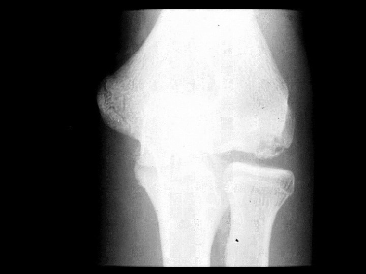
 • Findings: – small avulsion of the medial")


- Slides: 62
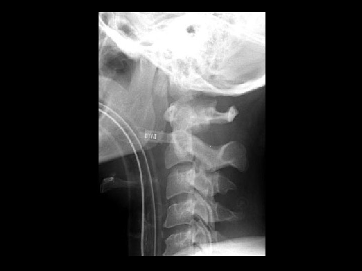
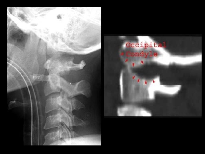
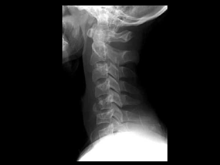
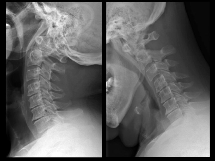
Atlanto-occipital dislocation • Findings: – anterior and superior displacement of occipital condyles in relation to C 1 – pre-vertebral soft tissue swelling – mechanism: hyperflexion or hyperextension – UNSTABLE
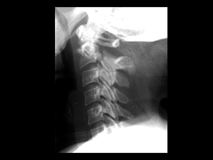
Bilateral jumped facets • Findings: – complete anterior dislocation of C 5 on C 6 – locked bilateral facets – disruption of ALL & PLL – “bat wing” appearance – mechanism: extreme hyperflexion – UNSTABLE
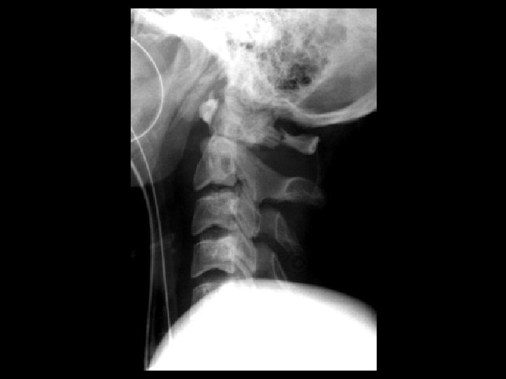
Unilateral jumped facet • Findings: – anterolisthesis of C 5 on C 6, less than 50% – discordant rotation (C 25 bodies appear smaller than C 6 -7) – C 5 inferior facet perched on C 6 superior facet – mechanism: flexion and rotation – STABLE
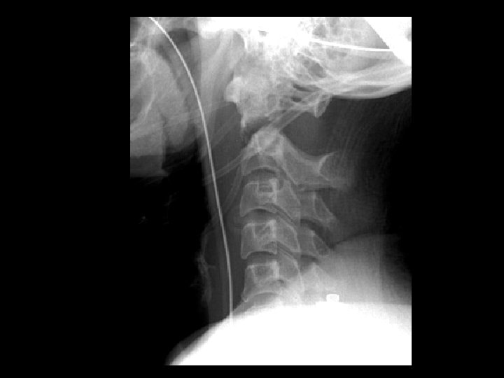
Flexion teardrop fracture • Findings: – anterior teardrop fragment of C 5 – prevertebral soft tissue swelling – widening of C 5 -C 6 interspinous distance – mechanism: hyperflexion and compression (diving accident) – UNSTABLE
Hangman’s fracture • Findings: – bilateral C 2 pars fractires – anterolisthesis and forward tilting of the C 2 body – prevertebral soft tissue swelling – may see avulsion of anterior inferior C 2 – mechanism: hyperextension – UNSTABLE
Jefferson fracture • Findings: – burst fracture of C 1 – lateral view: lateral displacement of lateral masses > 2 mm – mechanism: axial load injury – UNSTABLE
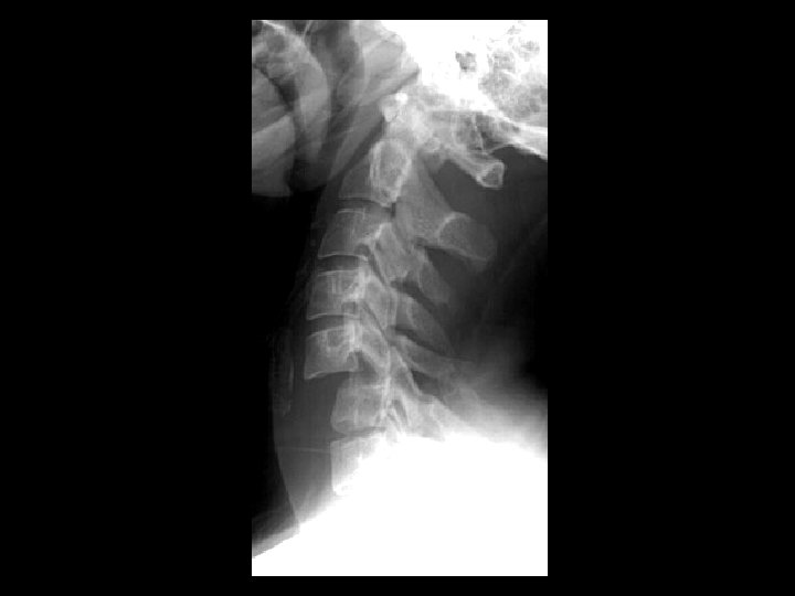
Odontoid fracture • Findings: – base of dens fracture – anterior and superior distraction of dens and atlas – prevertebral soft tissue swelling – mechanism: hyperflexion – UNSTABLE
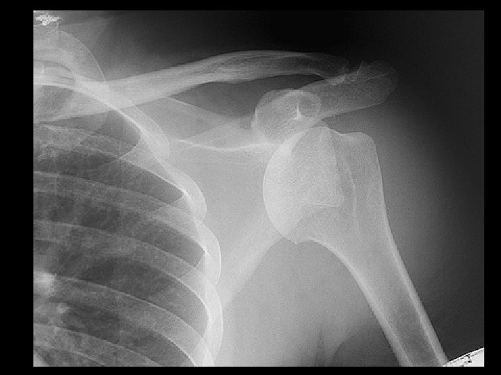
Anterior shoulder dislocation • Findings: – inferomedial, subcoracoid position of the humeral head – too much overlap of humeral head and glenoid – axillary view shows Hill Sachs - Bankhart configuration • ddx: – NONE! – This is an Aunt Minnie!
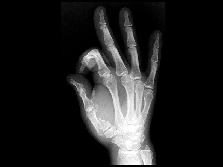
Bennett Fracture • Findings: – Vertical oblique intraarticular fracture at the base of the thumb MC – Radial subluxation of distal fragment – must be fixed surgically • ddx: – NONE! – This is an Aunt Minnie!
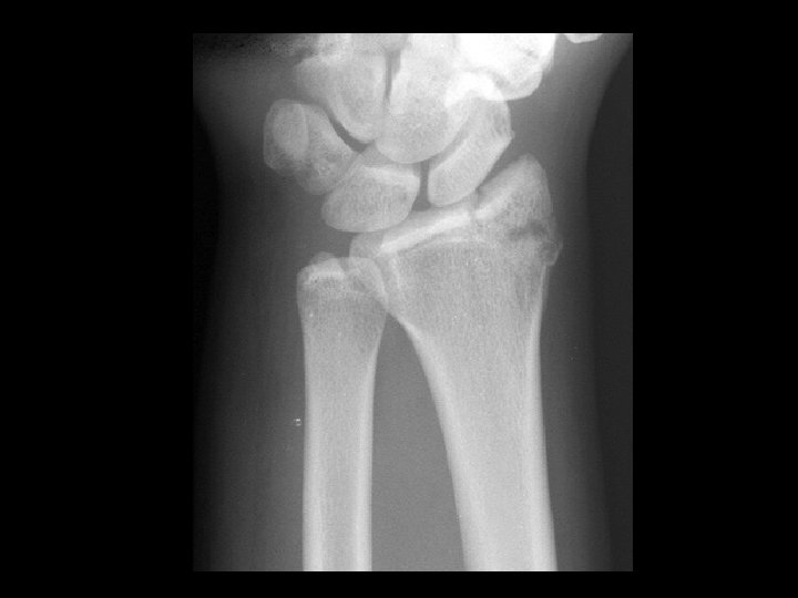
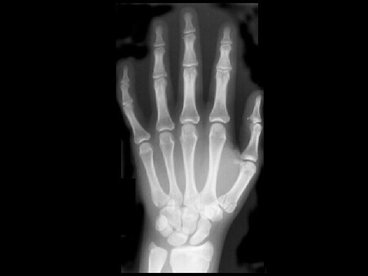
Chauffer’s fracture (Hutchinson fracture) • • Findings: – intra-articular radial styloid fracture – mild widening of SL distance – direct injury to thenar side of wrist ddx: – NONE! – This is an Aunt Minnie!
Radial head fracture • Findings: – Visualization of both the anterior and posterior fat – “spinnaker sign” – most common adult elbow fracture – assume radial head fracture even if not clearly seen • ddx: – NONE! – This is an Aunt Minnie!
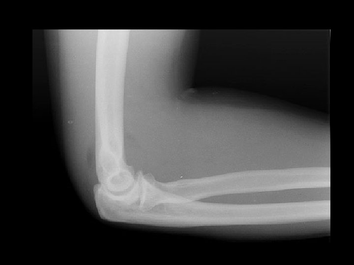
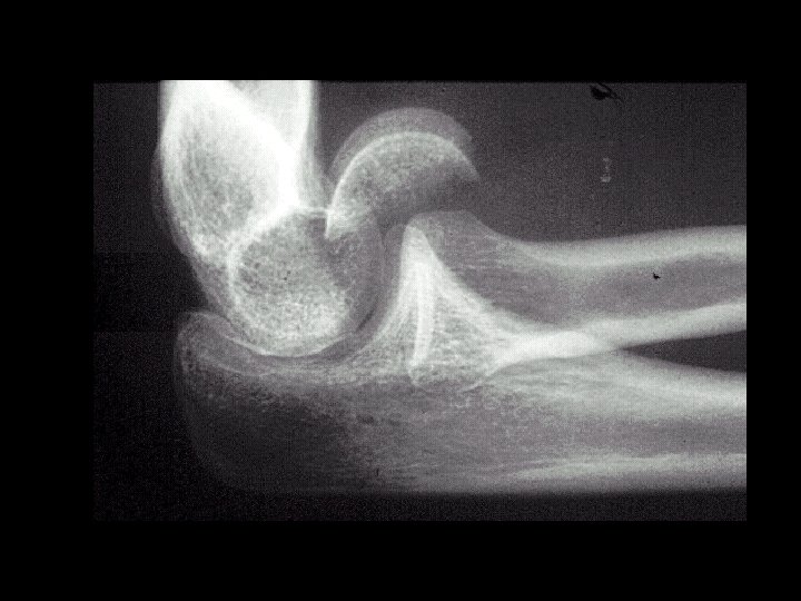
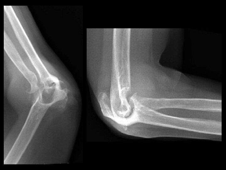
Capitellum fracturedislocation • Findings: – fracture-dislocation of the capitellum – due to direct trauma – second most common site in kids (most common is suprcondylar) • ddx: – NONE! – This is an Aunt Minnie!
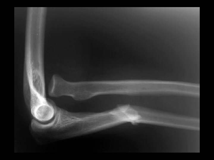
Medial epicondyle fracture (Little Leaguer’s elbow) • Findings: – small avulsion of the medial epicondyle – due to large valgus forces – little league pitchers • ddx: – NONE! – This is an Aunt Minnie!
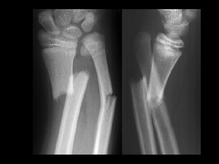
Galeazzi fracture-dislocation • Findings: – Angulated distal radius and ulna fractures – Distal ulnar dislocation • ddx: – NONE! – This is an Aunt Minnie!
Monteggia fracturedislocation • Findings: – Overlapping, angulated fracture of the proximal ulna – radial head dislocation • ddx: – NONE! – This is an Aunt Minnie!
Rolando fracture • Findings: – comminuted intraarticular fracture of the thumb MC • ddx: – NONE! – This is an Aunt Minnie!
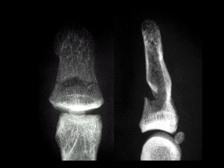
Nail bed injury • Findings: – incomplete fracture of the dorsal distal phalanx – involves the nail bed – treat as open fracture – high risk of osteomyelitis • ddx: – NONE! – This is an Aunt Minnie!
Dialysis arthropathy • Findings: – Multiple erosions on both sides of multiple PIP joints – Osteopenia, fuzzy trabecula, and dialysis graft = evidence of ESRD • ddx: – Inflammatory arthritis
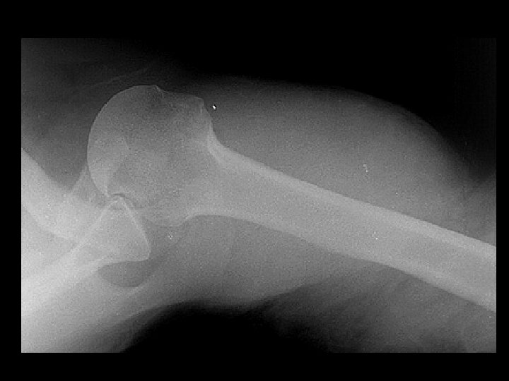
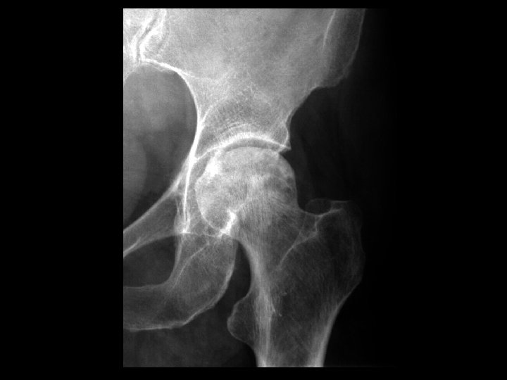
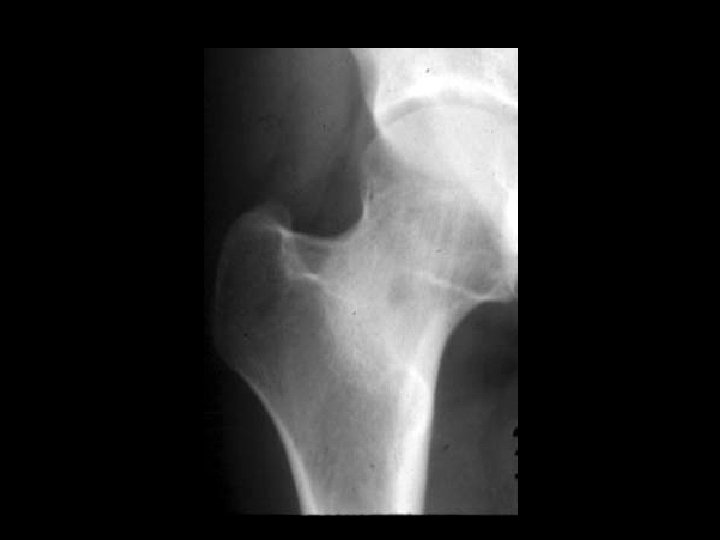
Femoral Head AVN with collapse • Findings: – Mixed sclerotic and lytic appearance of the femoral head – 2 mm collapse of the superior articular surface – Joint space still preserved • ddx: – NONE! – This is an Aunt Minnie!
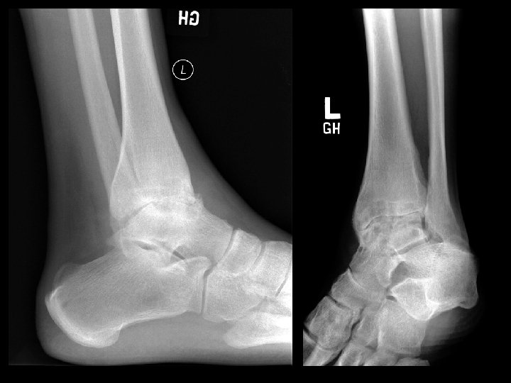
Hemophilia • Findings: – Severe secondary arthritis of the ankle • ddx: – Inflammatory – Post-infectious
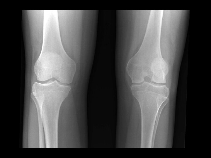
Juvenile Rheumatoid Arthritis • Findings: – Flared metaphyses – Gracile diaphyses – Joint space narrowing – Abnormal patellae • ddx: – NONE! – This is an Aunt Minnie!
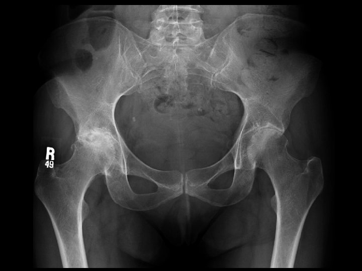
Inflammatory Arthritis • • Findings: – Severe joint space narrowing – Sub-chondral cystic changes and sclerosis – No significant osteophyte formation ddx: – Post-infectious
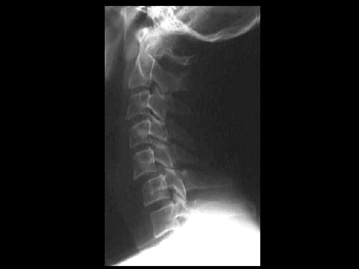
Odontoid fracture with C 1 subluxation • Findings: – 5 mm anterior subluxation of C 1 relative to C 2 with flexion • ddx: – Destroyed dens • Rheumatoid • Metastasis
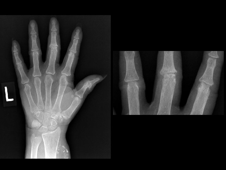
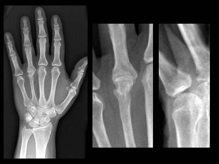
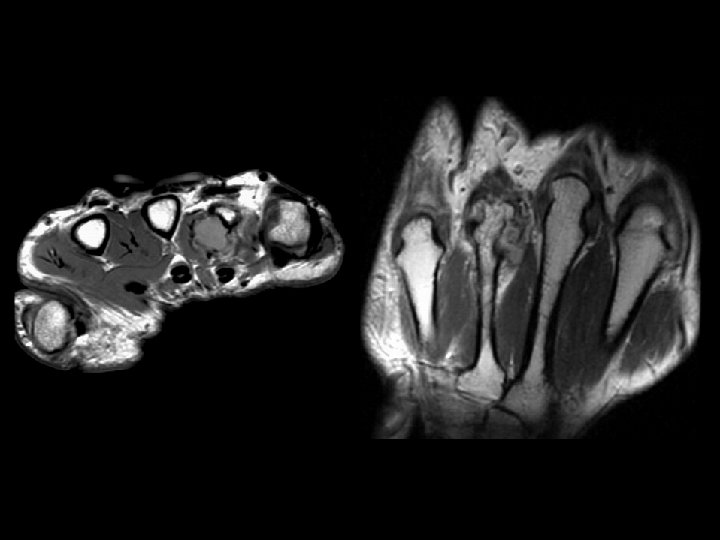
Synovial chondromatosis • Findings: – Smoothly marginated erosions on both sides of the 4 th MCP joint – Soft tissue mass causing narrowing of the 4 th MC – Single joint involvement • ddx: – Rheumatoid – PVNS
Hyperparthyroidism • Findings: – Subperiosteal absorption along the medial aspect of the proximal tibial metadiaphysis – Subligamentous resorption at the attachments of patellar ligament • ddx: – NONE! – This is an Aunt Minnie!
Rheumatoid Arthritis • Findings: – Deformed elbow joint due to complete loss of articular cartilage and large erosions – Soft tissue pannus within the joint • ddx: – NONE! – This is an Aunt Minnie!
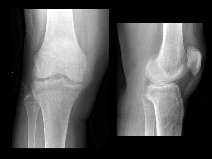
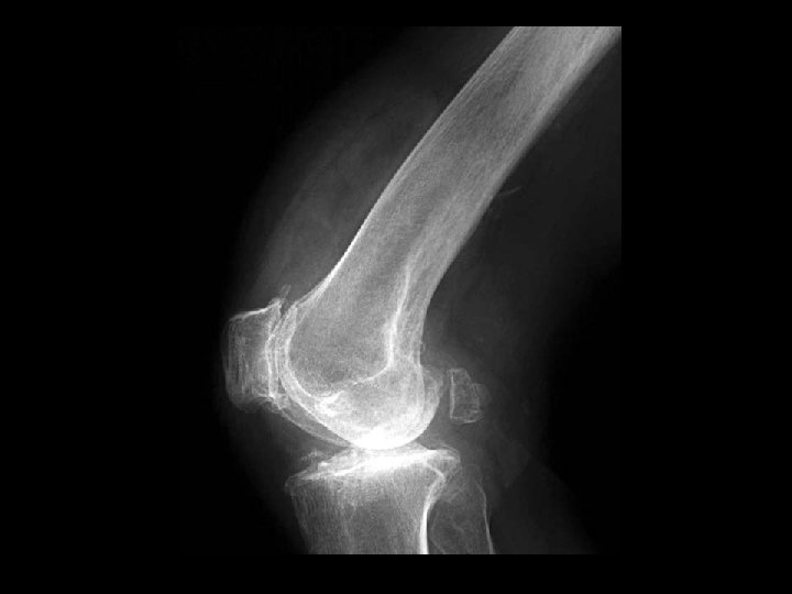
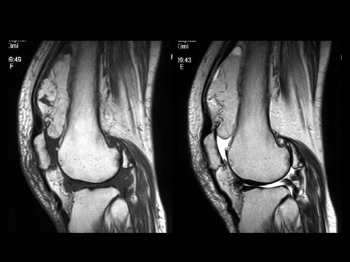
Lipoma arborescens • Findings: – Fatty lesion within the suprapatellar pouch • ddx: – NONE! – This is an Aunt Minnie!
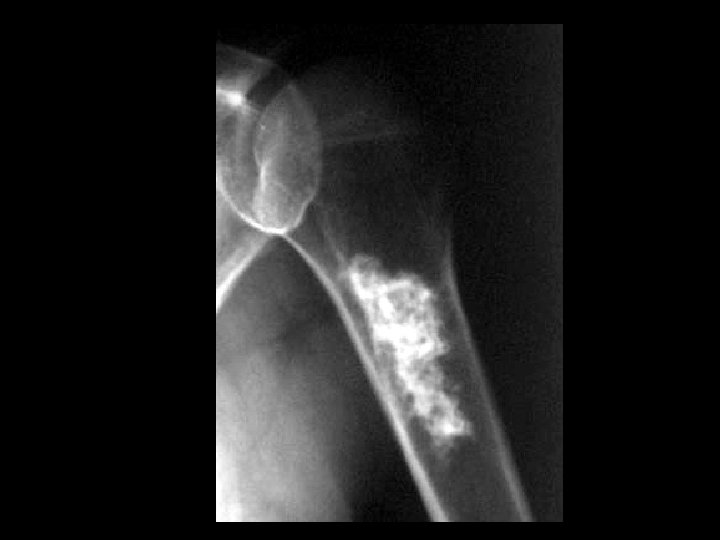
Enchondroma • Findings: – Bone forming lesion in the medullary space of the humeral metadiaphysis – Rings and arcs calcification • ddx: – Bone infarction
Osteoid osteoma • Findings: – Small lucent lesion with surrounding sclerosis within femoral neck • ddx: – Brodie’s abscess – EG