Bones of the Hip Ilium Iliac fossa Iliac
![Bones of the Hip Bones of the Hip](https://slidetodoc.com/presentation_image/5300bd76c00603884f0d7b3040795288/image-1.jpg)
Bones of the Hip
![Ilium • Iliac fossa • Iliac ala • Arcuate line – True pelvis – Ilium • Iliac fossa • Iliac ala • Arcuate line – True pelvis –](http://slidetodoc.com/presentation_image/5300bd76c00603884f0d7b3040795288/image-2.jpg)
Ilium • Iliac fossa • Iliac ala • Arcuate line – True pelvis – False pelvis • Acetabulum
![Male and female pelvis Male and female pelvis](http://slidetodoc.com/presentation_image/5300bd76c00603884f0d7b3040795288/image-3.jpg)
Male and female pelvis
![Differences • Angle of pubic arch – female wider • Pelvic inlet – female Differences • Angle of pubic arch – female wider • Pelvic inlet – female](http://slidetodoc.com/presentation_image/5300bd76c00603884f0d7b3040795288/image-4.jpg)
Differences • Angle of pubic arch – female wider • Pelvic inlet – female larger • Sacrum – female less curved • Obturator foramen – female oval, male round • Iliac ala – female wider and shallower
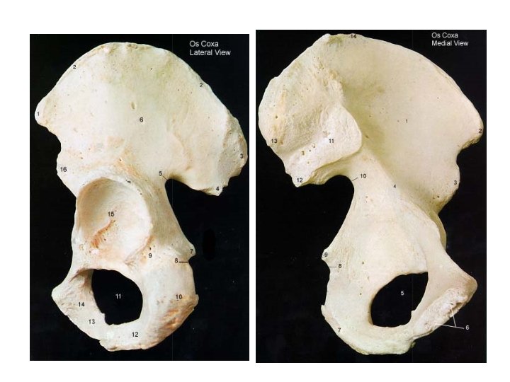
![figure figure](http://slidetodoc.com/presentation_image/5300bd76c00603884f0d7b3040795288/image-5.jpg)
figure
![Iliac crest • Thicker posteriorly • Highest point palpated posteriorly is at level of Iliac crest • Thicker posteriorly • Highest point palpated posteriorly is at level of](http://slidetodoc.com/presentation_image/5300bd76c00603884f0d7b3040795288/image-6.jpg)
Iliac crest • Thicker posteriorly • Highest point palpated posteriorly is at level of L 4 • Lips of iliac crest – Internal lip – Intermediate lip – External lip
![L 4 is colored L 4 is colored](http://slidetodoc.com/presentation_image/5300bd76c00603884f0d7b3040795288/image-7.jpg)
L 4 is colored
![Attachments to lips • Internal lip – Anterior lip - Transversus abdominis muscle – Attachments to lips • Internal lip – Anterior lip - Transversus abdominis muscle –](http://slidetodoc.com/presentation_image/5300bd76c00603884f0d7b3040795288/image-8.jpg)
Attachments to lips • Internal lip – Anterior lip - Transversus abdominis muscle – Middle lip - Quadratus lumborum muscle – Posterior lip - Sacrospinalis muscle (erector spinae muscles of back) • Intermediate lip – Anterior lip – internal abdominal oblique muscle
![• External lip – Anterior lip – external abdominal oblique muscle – Anterior • External lip – Anterior lip – external abdominal oblique muscle – Anterior](http://slidetodoc.com/presentation_image/5300bd76c00603884f0d7b3040795288/image-9.jpg)
• External lip – Anterior lip – external abdominal oblique muscle – Anterior lip – origin of tensor fascia latae muscle – Posterior lip – origin of latissimus dorsi muscle – Whole lip – fascia lata (deep fascia of the thigh)
![](http://slidetodoc.com/presentation_image/5300bd76c00603884f0d7b3040795288/image-10.jpg)
![Other Structures on Iliac Crest • Anterior Superior Iliac Spine (ASIS) – Sartorius muscle Other Structures on Iliac Crest • Anterior Superior Iliac Spine (ASIS) – Sartorius muscle](http://slidetodoc.com/presentation_image/5300bd76c00603884f0d7b3040795288/image-11.jpg)
Other Structures on Iliac Crest • Anterior Superior Iliac Spine (ASIS) – Sartorius muscle – Tensor fascia latae • Posterior Superior Iliac Spine (PSIS) – Skin dimples on back – Level of S 2 vertebrae
![Other Structures on the Ilium • Anterior Inferior Iliac Spine – Origin of part Other Structures on the Ilium • Anterior Inferior Iliac Spine – Origin of part](http://slidetodoc.com/presentation_image/5300bd76c00603884f0d7b3040795288/image-12.jpg)
Other Structures on the Ilium • Anterior Inferior Iliac Spine – Origin of part of rectus femoris muscle – Attachment for iliofemoral ligament • Posterior Inferior Iliac Spine – Posterior part of the surface that articulates with the sacrum
![• Greater Sciatic Notch – Posterior part of ilium – Just inferior to • Greater Sciatic Notch – Posterior part of ilium – Just inferior to](http://slidetodoc.com/presentation_image/5300bd76c00603884f0d7b3040795288/image-13.jpg)
• Greater Sciatic Notch – Posterior part of ilium – Just inferior to the PIIS – Next slide – structures that pass through the greater sciatic notch
![• • • Superior gluteal nerve and vessels Piriformis muscle Inferior gluteal nerve • • • Superior gluteal nerve and vessels Piriformis muscle Inferior gluteal nerve](http://slidetodoc.com/presentation_image/5300bd76c00603884f0d7b3040795288/image-14.jpg)
• • • Superior gluteal nerve and vessels Piriformis muscle Inferior gluteal nerve and vessels Sciatic nerve Nerve to superior gemellus and obturator internus muscles • Nerve to inferior gemellus and quadratus femoris muscles • Pudendal nerve and vessels • Posterior femoral cutaneous nerve • Note structures which are above and below the piriformis muscle
![Post hip fig Post hip fig](http://slidetodoc.com/presentation_image/5300bd76c00603884f0d7b3040795288/image-15.jpg)
Post hip fig
![Post hip 2 Post hip 2](http://slidetodoc.com/presentation_image/5300bd76c00603884f0d7b3040795288/image-16.jpg)
Post hip 2
![Structures on the external surface of the ilium • Three gluteal lines – Posterior Structures on the external surface of the ilium • Three gluteal lines – Posterior](http://slidetodoc.com/presentation_image/5300bd76c00603884f0d7b3040795288/image-17.jpg)
Structures on the external surface of the ilium • Three gluteal lines – Posterior Gluteal line – Anterior Gluteal line – Inferior Gluteal line – Rough area above acetabulum • Origin of reflected head of the rectus femoris muscle
![Gluteal lines on ADAM • Origins of: – Gluteus maximus – Gluteus medius – Gluteal lines on ADAM • Origins of: – Gluteus maximus – Gluteus medius –](http://slidetodoc.com/presentation_image/5300bd76c00603884f0d7b3040795288/image-18.jpg)
Gluteal lines on ADAM • Origins of: – Gluteus maximus – Gluteus medius – Gluteus minimus
![The ISCHIUM • Body of the ischium – External surface is the articular and The ISCHIUM • Body of the ischium – External surface is the articular and](http://slidetodoc.com/presentation_image/5300bd76c00603884f0d7b3040795288/image-19.jpg)
The ISCHIUM • Body of the ischium – External surface is the articular and nonarticular surface of the acetabulum • Acetabulum – Articular surface – lunate surface which articulates with the head of the femur – Non-articular surface – acetabular fossa
![• Internal surface of ischium – Forms part of the wall of the • Internal surface of ischium – Forms part of the wall of the](http://slidetodoc.com/presentation_image/5300bd76c00603884f0d7b3040795288/image-20.jpg)
• Internal surface of ischium – Forms part of the wall of the true pelvis
![Structures on posterior border of ischium • Ischial spine – Separates the greater and Structures on posterior border of ischium • Ischial spine – Separates the greater and](http://slidetodoc.com/presentation_image/5300bd76c00603884f0d7b3040795288/image-21.jpg)
Structures on posterior border of ischium • Ischial spine – Separates the greater and the lesser sciatic notches – Origin of superior gemellus muscle – Attachment site for the sacrospinous ligament • Ligament helps convert the greater sciatic notch into the greater sciatic foramen • Part of sacroischial joint
![Post hip lig Post hip lig](http://slidetodoc.com/presentation_image/5300bd76c00603884f0d7b3040795288/image-22.jpg)
Post hip lig
![• Lesser Sciatic notch – Passageway for: • Obturator internus muscle • Nerve • Lesser Sciatic notch – Passageway for: • Obturator internus muscle • Nerve](http://slidetodoc.com/presentation_image/5300bd76c00603884f0d7b3040795288/image-23.jpg)
• Lesser Sciatic notch – Passageway for: • Obturator internus muscle • Nerve to obturator internus muscle • Pudendal nerve and vessles – Note these also travel through the greater sciatic notch
![• Ischial tuberosity – Attachment of hamstring muscles • Long head of the • Ischial tuberosity – Attachment of hamstring muscles • Long head of the](http://slidetodoc.com/presentation_image/5300bd76c00603884f0d7b3040795288/image-24.jpg)
• Ischial tuberosity – Attachment of hamstring muscles • Long head of the biceps femoris muscle • Semitendinosus muscle • Semimembranosus muscle – Origin of Inferior Gemellus muscle – Attachment of sacrotuberous ligament • Part of sacroischial joint • Helps convert both the greater and lesser sciatic notches into foramen
![• Superior ischial ramus – External or lateral wall • Origin of quadratus • Superior ischial ramus – External or lateral wall • Origin of quadratus](http://slidetodoc.com/presentation_image/5300bd76c00603884f0d7b3040795288/image-25.jpg)
• Superior ischial ramus – External or lateral wall • Origin of quadratus femoris muscle – On ridge between the superior ischial ramus and the ischial tuberosity – Lower part is part of the origin of the obturator externus muscle
![The PUBIS • Body of the pubis – External surface - acetabulum • Articular The PUBIS • Body of the pubis – External surface - acetabulum • Articular](http://slidetodoc.com/presentation_image/5300bd76c00603884f0d7b3040795288/image-26.jpg)
The PUBIS • Body of the pubis – External surface - acetabulum • Articular surface • Non-articular surface – acetabular fossa – Internal surface • Forms part of wall of true pelvis
![Superior Ramus of the Pubis • External surface – Origin of adductor longus muscle Superior Ramus of the Pubis • External surface – Origin of adductor longus muscle](http://slidetodoc.com/presentation_image/5300bd76c00603884f0d7b3040795288/image-27.jpg)
Superior Ramus of the Pubis • External surface – Origin of adductor longus muscle • Just below pubic crest • Internal surface – part of wall of true pelvis
![• Superior border of superior ramus (medial part) – Pubic crest – RA • Superior border of superior ramus (medial part) – Pubic crest – RA](http://slidetodoc.com/presentation_image/5300bd76c00603884f0d7b3040795288/image-28.jpg)
• Superior border of superior ramus (medial part) – Pubic crest – RA and pyramidalis – Pubic tubercle – inguinal ligament – Medial border – articular surface for attachment of fibrocartilage of pubic symphysis – Lateral part – part of edge of obturator foramen
![• Superior border of superior ramus (lateral part) – Pectineal line – origin • Superior border of superior ramus (lateral part) – Pectineal line – origin](http://slidetodoc.com/presentation_image/5300bd76c00603884f0d7b3040795288/image-29.jpg)
• Superior border of superior ramus (lateral part) – Pectineal line – origin of pectineus muscle – Iliopubic eminence • Junction of iliac and pubic bones • Insertion of psoas minor muscle
![• Inferior surface of superior pubic ramus – Obturator crest – part of • Inferior surface of superior pubic ramus – Obturator crest – part of](http://slidetodoc.com/presentation_image/5300bd76c00603884f0d7b3040795288/image-30.jpg)
• Inferior surface of superior pubic ramus – Obturator crest – part of margin of obturator foramen – Obturator “groove” – passageway for obturator nerve and vessels as they pass through the obturator foramen – Posterior surface – part of origin of obturator internus muscle
![Inferior pubic ramus • External surface – Origin of adductor brevis muscle – Origin Inferior pubic ramus • External surface – Origin of adductor brevis muscle – Origin](http://slidetodoc.com/presentation_image/5300bd76c00603884f0d7b3040795288/image-31.jpg)
Inferior pubic ramus • External surface – Origin of adductor brevis muscle – Origin of gracilis muscle – Origin of adductor magnus muscle (lateral) – Origin of obturator externus muscle (bone near foramen)
![Structures common to more than one part of the innominate bone • Acetabulum • Structures common to more than one part of the innominate bone • Acetabulum •](http://slidetodoc.com/presentation_image/5300bd76c00603884f0d7b3040795288/image-32.jpg)
Structures common to more than one part of the innominate bone • Acetabulum • Obturator foramen
![Acetabulum • Lateral innominate bone • Surfaces – Lunate surface – articulating with head Acetabulum • Lateral innominate bone • Surfaces – Lunate surface – articulating with head](http://slidetodoc.com/presentation_image/5300bd76c00603884f0d7b3040795288/image-33.jpg)
Acetabulum • Lateral innominate bone • Surfaces – Lunate surface – articulating with head of femur – Non-articulating surface – acetabular fossa • Contains a fat pad • Acetabular notch – gap in wall of acetabulum – Attachment of ligamentum teres (ligament of the head of the femur) – Transverse ligament attaches to the edges of the notch and completes the articular surface
![Obturator foramen • Covered by obturator membrane • Origin of obturator internus – from Obturator foramen • Covered by obturator membrane • Origin of obturator internus – from](http://slidetodoc.com/presentation_image/5300bd76c00603884f0d7b3040795288/image-34.jpg)
Obturator foramen • Covered by obturator membrane • Origin of obturator internus – from inner (posterior) surface of the membrane and the surrounding bone • Origin of obturator externus – from the external (anterior) surface of the membrane and the surrounding bone • Passageway through the membrane for obturator nerve and vessels to the medial thigh
![Passages • Structures that enter the lower extremity must travel through the pelvis • Passages • Structures that enter the lower extremity must travel through the pelvis •](http://slidetodoc.com/presentation_image/5300bd76c00603884f0d7b3040795288/image-35.jpg)
Passages • Structures that enter the lower extremity must travel through the pelvis • There are several different ways that structures (nerves, blood vessels, lymph vessels, muscles and tendons) can travel through or around the pelvis
![• Greater Sciatic Notch – Many structures – Sciatic nerve – Superior and • Greater Sciatic Notch – Many structures – Sciatic nerve – Superior and](http://slidetodoc.com/presentation_image/5300bd76c00603884f0d7b3040795288/image-36.jpg)
• Greater Sciatic Notch – Many structures – Sciatic nerve – Superior and inferior gluteal nerves and vessels – Piriformis muscle • Lesser Sciatic Notch – Pudendal nerve and vessels travels back through – Obturator internus muscle and its nerve
![Obturator foramen • Obturator nerve and vessels through an opening in the obturator membrane Obturator foramen • Obturator nerve and vessels through an opening in the obturator membrane](http://slidetodoc.com/presentation_image/5300bd76c00603884f0d7b3040795288/image-37.jpg)
Obturator foramen • Obturator nerve and vessels through an opening in the obturator membrane at the obturator groove
![Structures which pass over the pelvic brim to lower extremity • • • Tendon Structures which pass over the pelvic brim to lower extremity • • • Tendon](http://slidetodoc.com/presentation_image/5300bd76c00603884f0d7b3040795288/image-38.jpg)
Structures which pass over the pelvic brim to lower extremity • • • Tendon of the iliacus muscle Tendon of the psoas major muscle Femoral Nerve, Artery and Vein Lateral femoral cutaneous nerve Lymph vessels
![Femoral triangle fig Femoral triangle fig](http://slidetodoc.com/presentation_image/5300bd76c00603884f0d7b3040795288/image-39.jpg)
Femoral triangle fig
![Post hip lig Post hip lig](http://slidetodoc.com/presentation_image/5300bd76c00603884f0d7b3040795288/image-40.jpg)
Post hip lig
![Post hip 2 Post hip 2](http://slidetodoc.com/presentation_image/5300bd76c00603884f0d7b3040795288/image-41.jpg)
Post hip 2
![Quiz on bony landmarks • http: //www. mhhe. com/biosci/ap/saladin 2 e/ graphics/saladin 02 ap/ch Quiz on bony landmarks • http: //www. mhhe. com/biosci/ap/saladin 2 e/ graphics/saladin 02 ap/ch](http://slidetodoc.com/presentation_image/5300bd76c00603884f0d7b3040795288/image-42.jpg)
Quiz on bony landmarks • http: //www. mhhe. com/biosci/ap/saladin 2 e/ graphics/saladin 02 ap/ch 09/others/chap 09 l abeling 09. html • http: //www. mhhe. com/biosci/ap/saladin 2 e/ graphics/saladin 02 ap/ch 09/others/chap 09 l abeling 08. html
- Slides: 42