Vesseles of post abdominal wall Asst prof Dr
![Vesseles of post. abdominal wall Asst. prof. Dr. Alaa Jamel Cabs, rcsi, mbchb Vesseles of post. abdominal wall Asst. prof. Dr. Alaa Jamel Cabs, rcsi, mbchb](https://slidetodoc.com/presentation_image_h/000bdabdb5df6c366061ec1782a8b85f/image-1.jpg)
Vesseles of post. abdominal wall Asst. prof. Dr. Alaa Jamel Cabs, rcsi, mbchb
![Abdominal aorta Beginning; diaphragmatic opening at lower border of T 12 as a continuation Abdominal aorta Beginning; diaphragmatic opening at lower border of T 12 as a continuation](http://slidetodoc.com/presentation_image_h/000bdabdb5df6c366061ec1782a8b85f/image-2.jpg)
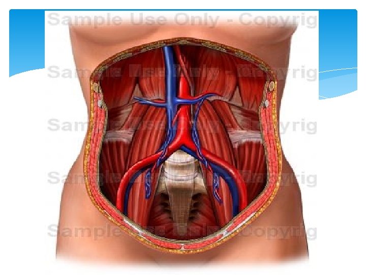
Abdominal aorta Beginning; diaphragmatic opening at lower border of T 12 as a continuation of descending thoracic aorta. Course; descend infront of upper 4 lumber vertebra. Termination; lower border of L 4 BY dividing to rt & lf common iliac art.
![](http://slidetodoc.com/presentation_image_h/000bdabdb5df6c366061ec1782a8b85f/image-3.jpg)
![](http://slidetodoc.com/presentation_image_h/000bdabdb5df6c366061ec1782a8b85f/image-4.jpg)
![Relations; Anterior; from above downward; 1 - coeliac trunk 2 - body of pancreas Relations; Anterior; from above downward; 1 - coeliac trunk 2 - body of pancreas](http://slidetodoc.com/presentation_image_h/000bdabdb5df6c366061ec1782a8b85f/image-5.jpg)
Relations; Anterior; from above downward; 1 - coeliac trunk 2 - body of pancreas and 3 structures behind to it *splenic v * lf renal v* origin of sup. Mesenteric art. 3 - 3 rd part of duod. & origin of inf. mes. art. 4 -upper part of root of mesentery 5 -paraital peritoneum separating it from coil of small intestine.
![Posterioly ; 1 - upper 4 lumber vertebra & intervertebral disc 2 -ant. longitudinal Posterioly ; 1 - upper 4 lumber vertebra & intervertebral disc 2 -ant. longitudinal](http://slidetodoc.com/presentation_image_h/000bdabdb5df6c366061ec1782a8b85f/image-6.jpg)
Posterioly ; 1 - upper 4 lumber vertebra & intervertebral disc 2 -ant. longitudinal ligament 3 - 3 rd and 4 th lumber veins
![Rt and lf relation of a. a On rt ; cisterna chyli & thorasic Rt and lf relation of a. a On rt ; cisterna chyli & thorasic](http://slidetodoc.com/presentation_image_h/000bdabdb5df6c366061ec1782a8b85f/image-7.jpg)
Rt and lf relation of a. a On rt ; cisterna chyli & thorasic duct , lower part of RT crus of diaphragm azygous vein , RT coeliac ganglion , & IVC On lf --- lf crus of diaphragm , lf coeliac ganglia, lf sympathetic chain, body of pancreas, inf. mesenteric v.
![BRANCHEAS OF A. A; Single branches 1 - coeliac trunk upper of L 1 BRANCHEAS OF A. A; Single branches 1 - coeliac trunk upper of L 1](http://slidetodoc.com/presentation_image_h/000bdabdb5df6c366061ec1782a8b85f/image-8.jpg)
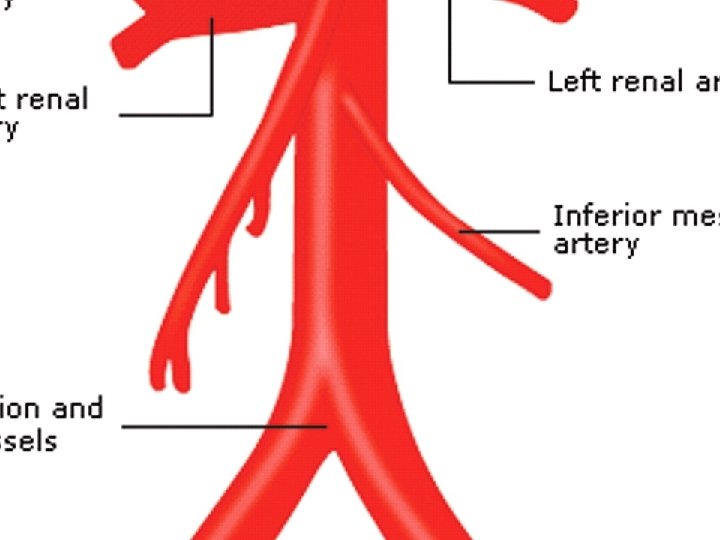
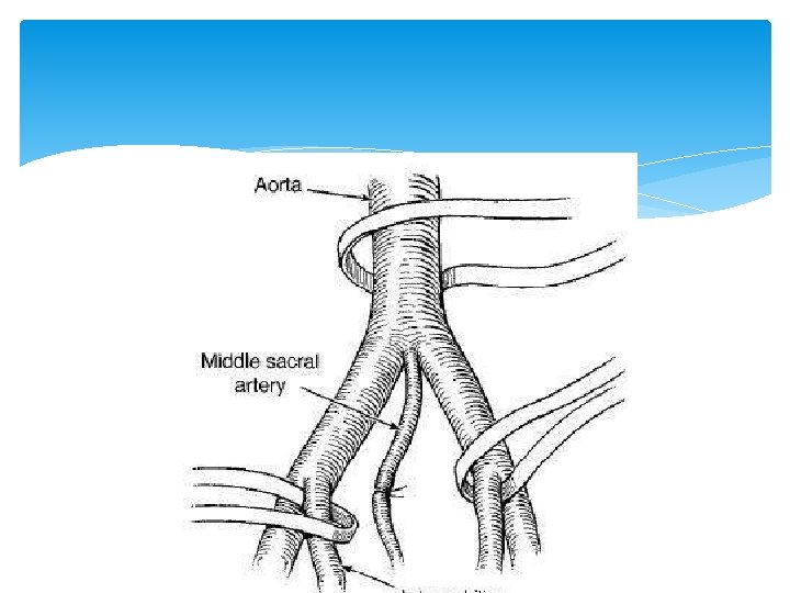
BRANCHEAS OF A. A; Single branches 1 - coeliac trunk upper of L 1 2 - sup. Mesenteric A lower of L 1 3 - inf. Mesenteric A. L 3 4 - median sacral art. From back of aorta as a continuation of A.
![](http://slidetodoc.com/presentation_image_h/000bdabdb5df6c366061ec1782a8b85f/image-9.jpg)
![Paired branches; 1 -Rt & lf inferior phrenic A. ; upper L 1 2 Paired branches; 1 -Rt & lf inferior phrenic A. ; upper L 1 2](http://slidetodoc.com/presentation_image_h/000bdabdb5df6c366061ec1782a8b85f/image-10.jpg)
Paired branches; 1 -Rt & lf inferior phrenic A. ; upper L 1 2 - rt&lf middle supra renal A. lower. L 1 3 - rt&lf renal art. L 2 4 - rt& lf gonodal art. L 3 5 - rt& lf common iliac A. lower L 4. + 4 pairs of lumber arteries from back of A. from l 1 to l 4
![MEDIAN SACRAL ART. It arise from the back aorta, just above its bifurcation. descend MEDIAN SACRAL ART. It arise from the back aorta, just above its bifurcation. descend](http://slidetodoc.com/presentation_image_h/000bdabdb5df6c366061ec1782a8b85f/image-11.jpg)
MEDIAN SACRAL ART. It arise from the back aorta, just above its bifurcation. descend in front of L 4&L 5. It enter the pelvis in front of sacral promontory. Branches; 1 - 5 th pair of lumber arteries 2 - twigs to the sacral canal 3 - twigs to the rectum
![](http://slidetodoc.com/presentation_image_h/000bdabdb5df6c366061ec1782a8b85f/image-12.jpg)
![](http://slidetodoc.com/presentation_image_h/000bdabdb5df6c366061ec1782a8b85f/image-13.jpg)
![Inf. phrenic arteries rt&lf; Arise from side of aa at level of L 1 Inf. phrenic arteries rt&lf; Arise from side of aa at level of L 1](http://slidetodoc.com/presentation_image_h/000bdabdb5df6c366061ec1782a8b85f/image-14.jpg)
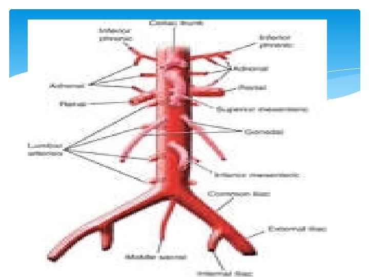
Inf. phrenic arteries rt&lf; Arise from side of aa at level of L 1 upper border Ascend upward to diaphragm Rt A pass behind ivc Lf pass behind the oesophagus Each artery give suprarenal art.
![Middle suprarenal art rt&lf Arise; from the side of AA at lower border of Middle suprarenal art rt&lf Arise; from the side of AA at lower border of](http://slidetodoc.com/presentation_image_h/000bdabdb5df6c366061ec1782a8b85f/image-15.jpg)
Middle suprarenal art rt&lf Arise; from the side of AA at lower border of L 1. then go to supplying the suprarenal glands Renal arteries rt &lf; Origin; side of AA at L 2 level Reach kidney infront of renal pelvis and ureter Rt one is longer and pass behind ivc, rt renal vein& head of pancreas Lf one shorter pass behind the lf renal vein &and body of pancreas
![Branches; 1 - inf suprarenal art, 2 - terminal branches 5 3 - ureteric Branches; 1 - inf suprarenal art, 2 - terminal branches 5 3 - ureteric](http://slidetodoc.com/presentation_image_h/000bdabdb5df6c366061ec1782a8b85f/image-16.jpg)
Branches; 1 - inf suprarenal art, 2 - terminal branches 5 3 - ureteric branches to the upper end of ureter
![Gonodal arteries ; They are testicular or ovarian art. they arise from anterio lateral Gonodal arteries ; They are testicular or ovarian art. they arise from anterio lateral](http://slidetodoc.com/presentation_image_h/000bdabdb5df6c366061ec1782a8b85f/image-17.jpg)
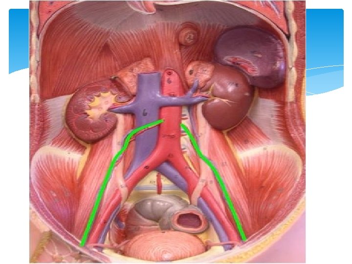
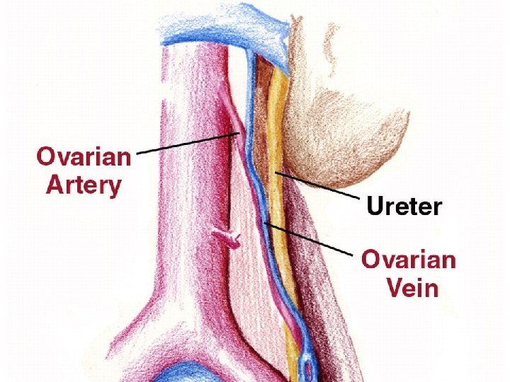
Gonodal arteries ; They are testicular or ovarian art. they arise from anterio lateral aspect of AA. At level of L 3. TERMINATION; Testicular a. reach deep ing. ring then superfascial ing. ring pass through ing. canal as a constitution of spermatic cord to reach the testis.
![Ovarian art. it cross the ext. ilic vessels to enter the suspensory lig. of Ovarian art. it cross the ext. ilic vessels to enter the suspensory lig. of](http://slidetodoc.com/presentation_image_h/000bdabdb5df6c366061ec1782a8b85f/image-18.jpg)
Ovarian art. it cross the ext. ilic vessels to enter the suspensory lig. of ovary to reach the broad ligament then reach the ovary via mesoovarium.
![](http://slidetodoc.com/presentation_image_h/000bdabdb5df6c366061ec1782a8b85f/image-19.jpg)
![](http://slidetodoc.com/presentation_image_h/000bdabdb5df6c366061ec1782a8b85f/image-20.jpg)
![Lumber arteries 4 pairs arise from back of AA. They supply ; muscles of Lumber arteries 4 pairs arise from back of AA. They supply ; muscles of](http://slidetodoc.com/presentation_image_h/000bdabdb5df6c366061ec1782a8b85f/image-21.jpg)
Lumber arteries 4 pairs arise from back of AA. They supply ; muscles of post abdominal wall Muscles of lat. abdominal wall Spinal cord 5 th paird arise from median sacral a.
![Inferior vena cava Beginning; in front body of vertebra L 5 (AORTA L 4) Inferior vena cava Beginning; in front body of vertebra L 5 (AORTA L 4)](http://slidetodoc.com/presentation_image_h/000bdabdb5df6c366061ec1782a8b85f/image-22.jpg)
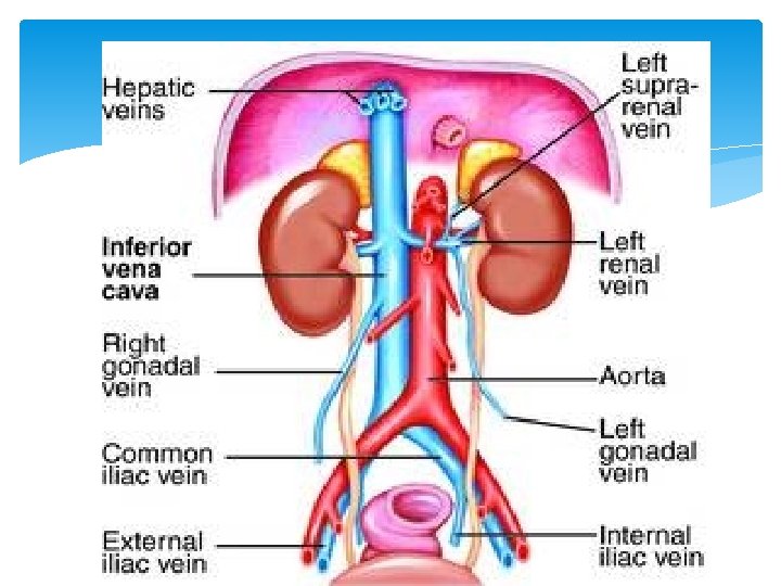
Inferior vena cava Beginning; in front body of vertebra L 5 (AORTA L 4) by union rt&lf common iliac v. Course ascend up rt to the aorta and pierce diaphragm at level of t 8 Termination; open to the lower part of rt atrium.
![](http://slidetodoc.com/presentation_image_h/000bdabdb5df6c366061ec1782a8b85f/image-23.jpg)
![](http://slidetodoc.com/presentation_image_h/000bdabdb5df6c366061ec1782a8b85f/image-24.jpg)
![Relations s. Anterioly; Below the duodenum; 1 - RT common iliac art. 2 - Relations s. Anterioly; Below the duodenum; 1 - RT common iliac art. 2 -](http://slidetodoc.com/presentation_image_h/000bdabdb5df6c366061ec1782a8b85f/image-25.jpg)
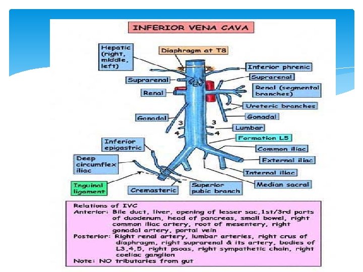
Relations s. Anterioly; Below the duodenum; 1 - RT common iliac art. 2 - root of mesentery and sup. mesentric vessels 3 - RT gonodal vessel At the duodenum 1 - 3 rd part of duod. 2 -head of pancreas and cbd 3 - 1 st part of duod. which separate from ivc by cbd , gastroduod. art. &portal vein
![Above the duodenum; 1 -The epiploic foramen ; which separate the ivc from the Above the duodenum; 1 -The epiploic foramen ; which separate the ivc from the](http://slidetodoc.com/presentation_image_h/000bdabdb5df6c366061ec1782a8b85f/image-26.jpg)
Above the duodenum; 1 -The epiploic foramen ; which separate the ivc from the margin of lesser omentum and its contents( portal v, hepatic art. &cbd) 2 -Post. surface of rt lobe of liver Post relation; A- the lower part of ivc is related to 1 - bodies of lower 3 l. vertbra 2 -Rt psoas major m 3 -Rt sympathetic chain
![Upper part of ivc is related to ; 1 - RT crus of diaphragm Upper part of ivc is related to ; 1 - RT crus of diaphragm](http://slidetodoc.com/presentation_image_h/000bdabdb5df6c366061ec1782a8b85f/image-27.jpg)
Upper part of ivc is related to ; 1 - RT crus of diaphragm but separated from it by ; a- RT coeliac ganglion B- 3 arteries; *RT inf phrenic art *RT middle supra renal art *RT renal art.
![Rt side relation to ivc; * rt lobe of the liver(in the upper part) Rt side relation to ivc; * rt lobe of the liver(in the upper part)](http://slidetodoc.com/presentation_image_h/000bdabdb5df6c366061ec1782a8b85f/image-28.jpg)
Rt side relation to ivc; * rt lobe of the liver(in the upper part) *medial border of rt kidney( in middle part) *rt ureter ( in the lower part) Lf side related to *caudate of the liver( in upper part) *RT crus of diaphragm (in middle part) *abdominal aorta( in the lower part)
![Tributaries of ivc 1 - 2 common iliac v. 22 pair of lumber veins Tributaries of ivc 1 - 2 common iliac v. 22 pair of lumber veins](http://slidetodoc.com/presentation_image_h/000bdabdb5df6c366061ec1782a8b85f/image-29.jpg)
Tributaries of ivc 1 - 2 common iliac v. 22 pair of lumber veins ( 3 rd &4 th) 32 renal v (rt&lf) 42 rt side v. (rt suprarenal & rt gonodal) 52 inf phrenic v. 62 hepatic veins
![Renal veins 2 large vein open to the ivc Rt – short and lies Renal veins 2 large vein open to the ivc Rt – short and lies](http://slidetodoc.com/presentation_image_h/000bdabdb5df6c366061ec1782a8b85f/image-30.jpg)
Renal veins 2 large vein open to the ivc Rt – short and lies behind 2 nd part of duod. Lf vein-- longer in 3 times, pass infront of aorta and behind body of pancreas , splenic vein It receive 2 tributaries 1 - lf gonodal vein ( LF VARICOCELE MORE COMMON) 2 - lf supra renal vein
![Collateral venous anastemosis between svc & ivc Sites ; A- in the ant abdominal Collateral venous anastemosis between svc & ivc Sites ; A- in the ant abdominal](http://slidetodoc.com/presentation_image_h/000bdabdb5df6c366061ec1782a8b85f/image-31.jpg)
Collateral venous anastemosis between svc & ivc Sites ; A- in the ant abdominal wall; 1 - the thoroco- epigastric v; long vein it connect a-lat thoracic v of axillary v(svc) B-superfascial epigastric v of femoral v (ivc) 2 - sup. epigastric v of internal mammary v. (svc) anastemose inside the rectus sheath with inf epigastric v of ext. iliac v. (ivc)
![B- in the post abdominal wall 1 - the azygous vein. from ivc to B- in the post abdominal wall 1 - the azygous vein. from ivc to](http://slidetodoc.com/presentation_image_h/000bdabdb5df6c366061ec1782a8b85f/image-32.jpg)
B- in the post abdominal wall 1 - the azygous vein. from ivc to svc direct connection. 2 - inf hemiazygous v. 3 - the vertebral plexus of veins; Present inside vertebral canal it connect A-lumber veins (ivc) B- post. intercostal v (tributaries of azygous and hemiazygous v tri. Of svc in the thorax)
![Applied anatomy why the varicocele more common on the left side? Which is more Applied anatomy why the varicocele more common on the left side? Which is more](http://slidetodoc.com/presentation_image_h/000bdabdb5df6c366061ec1782a8b85f/image-33.jpg)
Applied anatomy why the varicocele more common on the left side? Which is more dangerous anterior aortic aneurysm rapture or posterior aneurysm?
![Thank you Thank you](http://slidetodoc.com/presentation_image_h/000bdabdb5df6c366061ec1782a8b85f/image-34.jpg)
Thank you
- Slides: 34